. 2023 Sep 21;18(9):e0291927.
doi: 10.1371/journal.pone.0291927. eCollection 2023.

Peptide immunization against the C-terminal of alpha-synuclein reduces locomotor activity in mice overexpressing alpha-synuclein

Affiliations

Peptide immunization against the C-terminal of alpha-synuclein reduces locomotor activity in mice overexpressing alpha-synuclein

Yu-Sung Chiu et al. PLoS One. .

Abstract

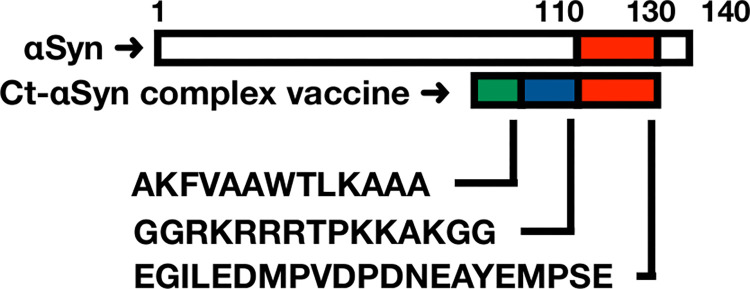
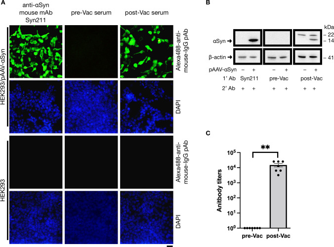
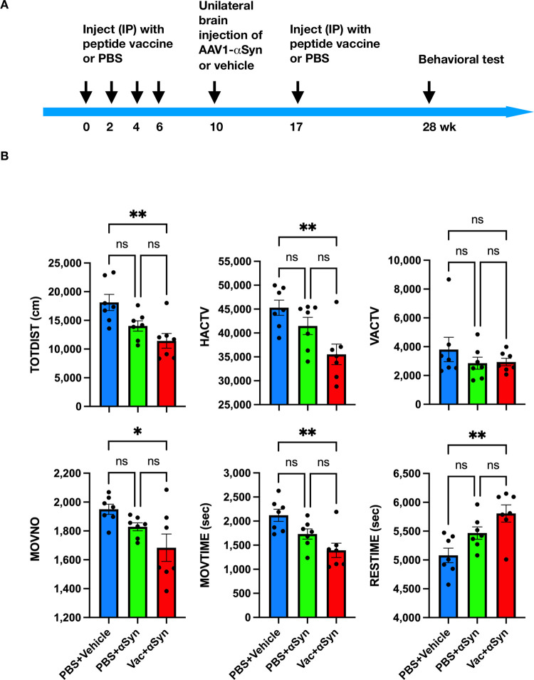
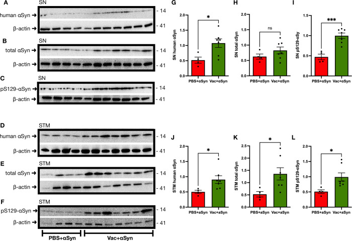
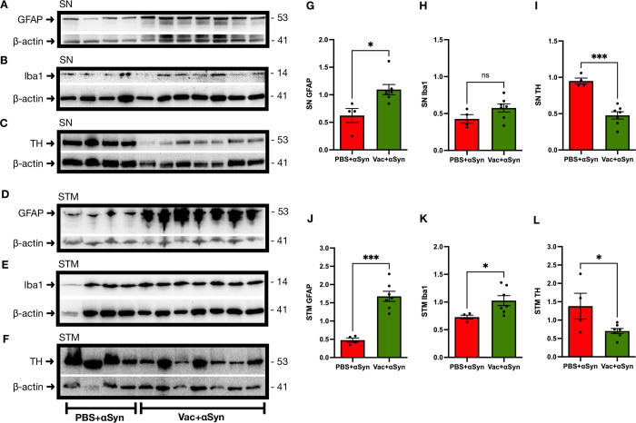
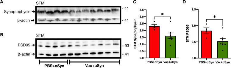
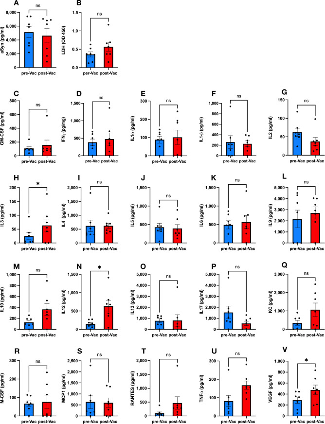
Abnormal accumulation of alpha-synuclein (αSyn) in the remaining nigra dopaminergic neurons is a common neuropathological feature found in patients with Parkinson's disease (PD). Antibody-based immunotherapy has been considered a potential approach for PD treatment. This study aims to investigate the effectiveness of active immunization against αSyn in a mouse model of PD. Adult mice were immunized with or without a synthetic peptide containing the C-terminal residues of human αSyn and activation epitopes, followed by an intranigral injection of adeno-associated virus vectors for overexpressing human αSyn. Upon the peptide injection, αSyn-specific antibodies were raised, accompanied by degeneration of dopaminergic neurons and motor deficits. Furthermore, the induction of neuroinflammation was postulated by the elevation of astroglial and microglial markers in the immunized mice. Instead of lessening αSyn toxicity, this peptide vaccine caused an increase in the pathogenic species of αSyn. Our data demonstrated the potential adverse effects of active immunization to raise antibodies against the C-terminal fragment of αSyn. This drawback highlights the need for further investigation to weigh the pros and cons of immunotherapy in PD. Applying the αSyn C-terminal peptide vaccine for PD treatment should be cautiously exercised. This study provides valuable insights into the intricate interplay among immune intervention, αSyn accumulation, and neurodegeneration.


Conflict of interest statement

The authors have declared that no competing interests exist.

Figures

Fig 1
Fig 1. Schematic illustration of the Ct-αSyn complex vaccine.
A linear peptide consists of a pan DR T helper epitope (AKFVAAWTLKAAA; green), an arginine-rich motif (GGRKRRRTPKKAKGG; blue) derived from the Tat protein of simian immunodeficiency virus, and a peptide corresponding to amino acid residues 110–130 of human αSyn (EGILEDMPVDPDNEAYEMPSE; red). This combination peptide was adjuvanted with alum salts and used as a Ct-αSyn complex vaccine to immunize mice in this study.
Fig 2
Fig 2. Ct-αSyn complex vaccine induces antibodies to recognize αSyn.
Adult mice (n = 7) were intraperitoneally injected with Ct-αSyn complex vaccine four times at 2-week intervals. Sera were collected from tail veins before vaccination (pre-Vac) and after the fourth vaccination (post-Vac). Pooled sera and an αSyn-specific mouse monoclonal antibody (Syn211) were used as primary antibodies (1’ Ab) to probe αSyn in HEK293 cells transfected with or without pAAV-αSyn plasmids (encoding human αSyn) by (A) immunofluorescence staining and (B) Western blot analyses. In the immunofluorescence staining, Alexa488-conjugated polyclonal antibodies were used as secondary antibodies (green); cellular nuclei were labeled with DAPI (blue); scale bar = 20 μm. In the Western blot analysis, HRP-conjugated anti-mouse IgG antibodies were used as secondary antibodies (2’ Ab); the migration molecular weights of αSyn (14 kDa) and β-actin (41 kDa) were indicated. (C) All serum samples were 2-fold serially diluted for antibody titration by enzyme-linked immunosorbent assay conducted in microplates coated with αSyn110-130 peptide. The antibody titer was defined as the reciprocal of the highest serum dilution that gives an OD450 value 3-fold higher than the background value. Data are expressed as mean values ± SEM. Significant differences between groups are indicated (**p < 0.01; Student’s t-test).
Fig 3
Fig 3. αSyn C-terminal peptides disrupt the interaction between vaccine-induced antibodies and αSyn.
Adult mice (n = 7) were administered with Ct-αSyn complex vaccine via intraperitoneal injection four times at 2-week intervals. Sera were collected from tail veins after the fourth vaccination. By immunofluorescence staining analysis, pooled sera were examined for their specificity to αSyn in cells (HEK293/pAAV-αSyn) transfected with pAAV-αSyn plasmids (encoding human αSyn) and naive cells (HEK293) in the presence of αSyn110–130 or Spike-binder peptides at different concentrations (0, 0.7, 0.07 μg/μl). Cellular nuclei were labeled with DAPI (blue); Alexa488-conjugated polyclonal antibodies were applied to detect mouse IgG (green); scale bar = 20 μm.
Fig 4
Fig 4. Ct-αSyn complex vaccine deteriorates motor function in mice overexpressing αSyn in the substantia nigra.
(A) Experiment timeline. Adult mice were randomly divided into three groups and intraperitoneally injected with PBS (group 1: n = 7, blue, PBS + vehicle; group 2: n = 7, green, PBS + αSyn) or Ct-αSyn complex vaccine (group 3: n = 7, red, Vac + αSyn) on weeks 0, 2, 4, 6, and 17. On week 10, mice received a unilateral brain injection of AAV1-αSyn (groups 2 and 3) or vehicle (group 1) in the right substantia nigra. The behavioral test was performed in the activity chamber for 2 h on week 28. (B) Six parameters of locomotor activity were recorded, including total distance traveled (TOTDIST), horizontal activity (HACTV), vertical activity (VACTV), movement number (MOVNO), movement time (MOVTIME), and rest time (RESTIME). Data are expressed as mean ± SEM, and differences between groups were determined by ordinary ANOVA followed by Tukey’s multiple comparison tests (*p < 0.05, **p < 0.01, ns: not significant).
Fig 5
Fig 5. Ct-αSyn complex vaccine enhances the accumulation of αSyn and phosphorylated αSyn in the brain.
Vaccinated mice (n = 7; Vac + αSyn) and non-vaccinated mice (n = 4; PBS + αSyn) were intracranially injected with the viral vector AAV1-αSyn in the right substantia nigra for overexpressing human αSyn. (A—F) Twenty weeks later, substantia nigra (SN) and striatum (STM) tissues of the right hemisphere brain were collected for Western blot analysis to probe human αSyn (by anti-V5 tag antibody), total αSyn (by BD clone-42 antibody), phosphorylated αSyn (pS129-αSyn), and β-actin. The molecular weight (kDa) and migration location of protein markers are indicated. (G—L) The levels of detected proteins were quantified by densitometric analysis and normalized to β-actin levels. Data are expressed as mean values ± SEM. Significant differences between groups are indicated (*p < 0.05, ***p < 0.001, ns: not significant; Student’s t-test).
Fig 6
Fig 6. Ct-αSyn complex vaccine induces neuroinflammation and reduces TH levels.
Vaccinated mice (n = 7; Vac + αSyn) and non-vaccinated mice (n = 4; PBS + αSyn) received an intracranial injection of the viral vector AAV1-αSyn in the right substantia nigra for overexpressing human αSyn. (A—F) Twenty weeks later, substantia nigra (SN) and striatum (STM) tissues of the right hemisphere brain were subjected to Western blot analysis to detect glial fibrillary acidic protein (GFAP), ionized calcium-binding adaptor molecule-1 (Iba1), tyrosine hydroxylase (TH), and β-actin. The molecular weight (kDa) and migration location of protein markers are indicated. (G—L) The protein levels of GFAP, Iba1, and TH were quantified by densitometric analysis and normalized to β-actin levels. Data are expressed as mean values ± SEM. Significant differences between groups are indicated (*p < 0.05, ***p < 0.001, ns: not significant; Student’s t-test).
Fig 7
Fig 7. Ct-αSyn complex vaccine downregulates synaptic markers.
Vaccinated mice (n = 7; Vac + αSyn) and non-vaccinated mice (n = 4; PBS + αSyn) were administered with the viral vector AAV1-αSyn in the right substantia nigra for overexpressing human αSyn. (A, B) Twenty weeks later, striatum (STM) tissues of the right hemisphere brain were examined by western blot analysis to probe synaptophysin, postsynaptic density protein-95 (PSD95), and β-actin. The molecular weight (kDa) and migration location of protein markers are indicated. (C, D) The protein levels of synaptophysin and PSD95 were measured and normalized to β-actin by densitometric analysis. Data are expressed as mean values ± SEM, and significant differences between groups are indicated (*p < 0.05; Student’s t-test).
Fig 8
Fig 8. Peripheral levels of αSyn, LDH, and cytokines in the mice vaccinated with the Ct-αSyn complex.
Mice were intraperitoneally injected with the Ct-αSyn complex (n = 7) on weeks 0, 2, 4, 6, and 17. On week 10, mice received a unilateral brain injection of AAV1-αSyn in the right substantia nigra and were subjected to behavioral tests on week 28. Peripheral blood sera were collected on days -2 (pre-Vac; 2 days before the first vaccination) and 60 (post-Vac: 18 days after the fourth vaccination) for the measurement of αSyn (A), lactate dehydrogenase (B) and 20 inflammation-related cytokines (C—V). Data were expressed as mean concentrations (pg/ml or OD450) ± SEM, and differences between groups were determined by the paired, two-tailed Student’s t-test (*p < 0.05, ns: not significant).

Similar articles

Cited by

References

    1. de Lau LM, Breteler MM. Epidemiology of Parkinson’s disease. Lancet Neurol. 2006;5(6):525–35. doi: 10.1016/s1474-4422(06)70471-9 . - DOI - PubMed
    1. Spillantini MG, Schmidt ML, Lee VM, Trojanowski JQ, Jakes R, Goedert M. Alpha-synuclein in Lewy bodies. Nature. 1997;388(6645):839–40. doi: 10.1038/42166 . - DOI - PubMed
    1. Armstrong MJ, Okun MS. Diagnosis and Treatment of Parkinson Disease: A Review. Jama. 2020;323(6):548–60. doi: 10.1001/jama.2019.22360 . - DOI - PubMed
    1. Kahle PJ, Neumann M, Ozmen L, Muller V, Jacobsen H, Schindzielorz A, et al.. Subcellular localization of wild-type and Parkinson’s disease-associated mutant alpha -synuclein in human and transgenic mouse brain. J Neurosci. 2000;20(17):6365–73. Epub 2000/08/31. doi: 10.1523/JNEUROSCI.20-17-06365.2000 ; PubMed Central PMCID: PMC6772969. - DOI - PMC - PubMed
    1. Burré J. The Synaptic Function of α-Synuclein. Journal of Parkinson’s disease. 2015;5(4):699–713. Epub 2015/09/26. doi: 10.3233/jpd-150642 ; PubMed Central PMCID: PMC4927875. - DOI - PMC - PubMed

Publication types