. 2023 Sep 26;120(39):e2308435120.
doi: 10.1073/pnas.2308435120. Epub 2023 Sep 21.

Cryo-EM structures of human GPR34 enable the identification of selective antagonists

Affiliations

Cryo-EM structures of human GPR34 enable the identification of selective antagonists

Anjie Xia et al. Proc Natl Acad Sci U S A. .

Abstract

GPR34 is a functional G-protein-coupled receptor of Lysophosphatidylserine (LysoPS), and has pathogenic roles in numerous diseases, yet remains poorly targeted. We herein report a cryo-electron microscopy (cryo-EM) structure of GPR34 bound with LysoPS (18:1) and Gi protein, revealing a unique ligand recognition mode with the negatively charged head group of LysoPS occupying a polar cavity formed by TM3, 6 and 7, and the hydrophobic tail of LysoPS residing in a lateral open hydrophobic groove formed by TM3-5. Virtual screening and subsequent structural optimization led to the identification of a highly potent and selective antagonist (YL-365). Design of fusion proteins allowed successful determination of the challenging cryo-EM structure of the inactive GPR34 complexed with YL-365, which revealed the competitive binding of YL-365 in a portion of the orthosteric binding pocket of GPR34 and the antagonist-binding-induced allostery in the receptor, implicating the inhibition mechanism of YL-365. Moreover, YL-365 displayed excellent activity in a neuropathic pain model without obvious toxicity. Collectively, this study offers mechanistic insights into the endogenous agonist recognition and antagonist inhibition of GPR34, and provides proof of concept that targeting GPR34 represents a promising strategy for disease treatment.

Keywords: GPCR; GPR34; antagonist; cryo-EM.


Conflict of interest statement

Sichuan University has applied for Chinese patents covering YL-365 and related compounds. Other authors declared no competing interests.

Figures
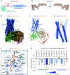
Fig. 1.
Fig. 1.
Cryo-EM structure of GPR34-Gi in complex with LysoPS (18:1). (A) Chemical structure of LysoPS (18:1). The hydrophilic head, glycerol linker, and hydrophobic tail of LysoPS (18:1) are highlighted with cadet blue, pink, and cornflower blue shading, respectively. (B and C) The cryo-EM density map (B) and model (C) of LysoPS (18:1)-bound GPR34-Gi1 complex. Cornflower blue, GPR34; pale green, Gαi; tan, Gβ; pink, Gγ; dark gray, scFv16; bright orange, LysoPS (18:1). (D) Cryo-EM density map shown as dark gray meshes allowed unambiguous identification of LysoPS (18:1). The density map of LysoPS (18:1) are depicted at contour level of 0.397. (E) Orthogonal view of cartoon model of LysoPS (18:1)-bound GPR34. The hydrophobic tail of LysoPS (18:1) extended toward TM3-TM5. The helical bundle of GPR34 is presented as cylindrical helices. (F) Key residues involved in LysoPS (18:1) recognition. The residues in contact with the hydrophilic head of LysoPS (18:1) are colored cadet blue, and those engaged with the hydrophobic tail are colored cornflower blue. Polar interactions are highlighted as gray dashed lines. (G) Mutagenesis effects of orthosteric-site residues of GPR34 on their activities in response to LysoPS (18:1) stimulation examined by Gαi1-Gγ2 dissociation assay. Bars represent differences in calculated potency (ΔpEC50) for each mutant shown as percentage of the maximum in wild type (WT). The ΔpEC50 values were derived from the dose-dependent curves in SI Appendix, Fig. S3 G and H. Statistical differences between WT and mutants were determined by two-sided, one-way ANOVA with Tukey’s test. *P < 0.05; **P < 0.01; ***P < 0.001; ND, not detectable due to low signal; NS, no significant difference. Data are presented as the mean ± SEM of three independent experiments performed in triplicate.
Fig. 2.
Fig. 2.
Hit identification and structural optimization. (A) Flowchart for the screening process of hit compound (Hit-1). (B) Structural optimization of Hit-1 led to the identification of a potent GPR34 antagonist YL-365. Bioactivities of compounds were measured by the Tango assay. Dose–response curves of Hit-1 and YL-365 are shown. IC50 values are mean ± SD from three independent experiments.
Fig. 3.
Fig. 3.
Overall architecture of the inactive GPR34-YL-365 complex. (A) Overall cryo-EM structure of GPR34 (light coral)-YL-365(bremen blue) complex. YL-365 is shown as bremen blue stick. (B) Cartoon representation of GPR34. YL-365 is shown as bremen blue spheres. (C) Top views of GPR34-YL-365. The disulfide bond formed between C1273.25 and C204ECL2 is shown as brilliant yellow stick. (D) Overall structure of hydrophobic-binding pocket viewed from the membrane plane, showing interactions between the bound YL-365 and residues in the pocket. Key residues are highlighted as sky blue. (E) Chemical structure of the antagonist YL-365 showing the R1, R2 and R3 groups. The key residues forming the hydrophobic interactions between GPR34 and YL-365 are marked with orange and blue circles, respectively. (F) Mutagenic effects of the binding pocket residues to YL-365-induced receptor inhibition examined by Gαi1-Gγ2 dissociation assay. Bars represent differences in calculated potency (ΔpEC50) for each mutant shown as percentage of the maximum in WT. The ΔpEC50 values were derived from the dose–dependent curves in SI Appendix, Fig. S7E. Statistical differences between WT and mutants were determined by two-sided, one-way ANOVA with Tukey’s test. *P < 0.05; **P < 0.01; ***P < 0.001; ND, not detectable due to low signal; NS, no significant difference. Data are presented as the mean ± SEM of three independent experiments performed in triplicate.
Fig. 4.
Fig. 4.
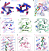
Structural comparison of GPR34 in active state and inactive state. (A) Structural superposition of active and inactive GPR34 from the extracellular view (Left) and intracellular view (Right). Significant conformational changes of TM4, TM5, TM6, and ECL2 in the extracellular domain, as well as TM5, TM6, and TM7 in the intracellular domain were observed. The helical bundles were presented as cylindrical helices. (B) Structural superposition of LysoPS (18:1) (bright orange) bound and YL-365 (deep teal) bound GPR34 indicated that polar interactions between hydrophilic head of LysoPS (18:1) and GPR34 connected the core helical bundle TM3, TM5, and TM6. The conformational changes from active to inactive state are indicated as red arrows, and the polar interactions are highlighted with grey dashed lines. (C) The F6.48 together with I5.50-I3.40-F6.44 (I-I-F) motif in GPR34 form a special core quaternary motif. The inactive μOR (PDB: 4DKL), active μOR (PDB: 7T2G), inactive β2AR (PDB: 3NY8) and active β2AR (PDB: 3SN6) are used for comparison. (D) YL-365 binding-induced conformational change of M1894.60, F2195.39, L2235.43, and M2265.46 in the OBP and formation of an ion lock between E2165.36 and K196ECL2. (E) F2275.47 contacts with the F6.48-I-I-F core quaternary motif in the inactive GPR34. (F) 4-benzyloxyphenyl moiety of YL-365 engaged with an extended binding pocket (EBP) formed by I1433.41, F1473.45, I1774.48, and L1814.52.
Fig. 5.
Fig. 5.
Pharmacological and toxicological effects of YL-365 in vivo. (A) The flow diagram of the in vivo antinociceptive experiments of YL-365 in the mouse model of neuropathic pain. (B) Body weight variation over time of C57BL/6J mice treated with vehicle or YL-365 (100 mg/kg, i.p., b.i.d.). Data are presented as averages ± SEM of five biological replicates. (CG) Complete blood count analyses (white blood cells, neutrophils, lymphocytes, red blood cells, and platelets) and of peripheral blood samples harvested from mice at the end of 14-d treatment with vehicle or YL-365 (100 mg/kg, i.p., b.i.d.). Data are presented as averages ± SEM of five biological replicates. P values were analyzed with paired t tests. (H) Antinociceptive effects of YL-365 in the mouse model of neuropathic pain. Mechanical allodynia was assessed on day 3 in sham-operated (sham) (n = 6) or SNI (spinal nerve injury) mice treated with vehicle or YL-365 (i.p., b.i.d.) (n = 8 to 10). Data are mean ± SEM. The P values were analyzed using one-way ANOVA compared with the vehicle group, *P < 0.05, ***P < 0.001. (I and J) The gene expression level of proinflammatory genes (I) and anti-inflammatory genes (J) in L4 spinal cord were assessed by qRT-PCR. Data are mean ± SEM (n = 7 for sham group, n = 13 for vehicle and YL-365 (20 mg/kg, i.p., b.i.d.) treatment group). The P values were analyzed using one-way ANOVA compared with the vehicle group, *P < 0.05, ***P < 0.001.

References

    1. Makide K., Kitamura H., Sato Y., Okutani M., Aoki J., Emerging lysophospholipid mediators, lysophosphatidylserine, lysophosphatidylthreonine, lysophosphatidylethanolamine and lysophosphatidylglycerol. Prostaglandins Other Lipid Mediat. 89, 135–139 (2009). - PubMed
    1. Bellini F., Bruni A., Role of a serum phospholipase A1 in the phosphatidylserine-induced T cell inhibition. FEBS Lett. 316, 1–4 (1993). - PubMed
    1. Wang X., et al. , GPR34-mediated sensing of lysophosphatidylserine released by apoptotic neutrophils activates type 3 innate lymphoid cells to mediate tissue repair. Immunity 54, 1123–1136.e8 (2021). - PubMed
    1. Lou L., et al. , Mafba and Mafbb regulate microglial colonization of zebrafish brain via controlling chemotaxis receptor expression. Proc. Natl. Acad. Sci. U.S.A. 119, e2203273119 (2022). - PMC - PubMed
    1. Sayo A., et al. , GPR34 in spinal microglia exacerbates neuropathic pain in mice. J. Neuroinflammation 16, 1–11 (2019). - PMC - PubMed

Publication types

LinkOut - more resources