. 2023 Sep 21;21(9):e3002280.
doi: 10.1371/journal.pbio.3002280. eCollection 2023 Sep.

Inhibitory feedback from the motor circuit gates mechanosensory processing in Caenorhabditis elegans

Affiliations

Inhibitory feedback from the motor circuit gates mechanosensory processing in Caenorhabditis elegans

Sandeep Kumar et al. PLoS Biol. .

Abstract

Animals must integrate sensory cues with their current behavioral context to generate a suitable response. How this integration occurs is poorly understood. Previously, we developed high-throughput methods to probe neural activity in populations of Caenorhabditis elegans and discovered that the animal's mechanosensory processing is rapidly modulated by the animal's locomotion. Specifically, we found that when the worm turns it suppresses its mechanosensory-evoked reversal response. Here, we report that C. elegans use inhibitory feedback from turning-associated neurons to provide this rapid modulation of mechanosensory processing. By performing high-throughput optogenetic perturbations triggered on behavior, we show that turning-associated neurons SAA, RIV, and/or SMB suppress mechanosensory-evoked reversals during turns. We find that activation of the gentle-touch mechanosensory neurons or of any of the interneurons AIZ, RIM, AIB, and AVE during a turn is less likely to evoke a reversal than activation during forward movement. Inhibiting neurons SAA, RIV, and SMB during a turn restores the likelihood with which mechanosensory activation evokes reversals. Separately, activation of premotor interneuron AVA evokes reversals regardless of whether the animal is turning or moving forward. We therefore propose that inhibitory signals from SAA, RIV, and/or SMB gate mechanosensory signals upstream of neuron AVA. We conclude that C. elegans rely on inhibitory feedback from the motor circuit to modulate its response to sensory stimuli on fast timescales. This need for motor signals in sensory processing may explain the ubiquity in many organisms of motor-related neural activity patterns seen across the brain, including in sensory processing areas.


Conflict of interest statement

The authors have declared that no competing interests exist.

Figures
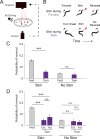
Fig 1
Fig 1. Turns decrease the likelihood of mechanosensory-evoked reversals.
(A) Closed-loop optogenetic stimulation is delivered to animals as they crawl based on their current behavior. (B) Optogenetic stimulation is delivered to gentle-touch mechanosensory neurons in worms that are either moving forward (top row) or turning (bottom row). (C) The probability of a reversal is shown in response to stimulation during forward movement or turn. Responses are also shown for a low-light no-stimulation control. This figure only is a reanalysis of recordings from [19]. The number of stimulation events, from left to right: 6,002, 1,114, 5,996, and 1,050. (D) The probability of reversal in response to stimulation during turning is shown broken down further by turn subtype: escape-like turns “Esc” and isolated turns “Iso.” N = 6,002, 602, 512, 5,996, 599, and 451 stim events, from left to right. The number of plates for forward and turn context are 29 and 47, respectively. The 95% confidence intervals for population proportions are reported; *** indicates p<0.001, “n.s.” indicates p>0.05 via two proportion Z-test. Exact p values for all the statistical tests are listed in S1 Table. All data underlying this figure can be found at https://doi.org/10.25452/figshare.plus.23903202.
Fig 2
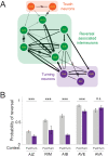
Fig 2. Turns decrease the likelihood of interneuron evoked reversals, except for AVA.
(A) Anatomical connectivity showing chemical (arrows) and electrical (resistor symbol) synapses among the anterior mechanosensory neurons, downstream interneurons, and turning-associated neurons. (B) Probability of a reversal response is shown for 3 s optogenetic stimulation to the listed neurons either during forward movement or immediately after the onset of turning. Strains are listed in Table 1. Illumination was 80 μW/mm2 red light to activate Chrimson in AVE or AVA, 300 μW/mm2 blue light to activate ChR2 in RIM or AIB, and 340 μW/mm2 to activate ChR2 in AIZ. Error bars indicate 95% confidence intervals for population proportions; *** indicates p<0.001, “n.s.” indicates p>0.05 via two-proportion Z-test, and p value for AVA stimulation group is 0.125. Exact p values for all the statistical tests are listed in S1 Table. N = 2,612, 601, 883, 107, 880, 511, 1,007, 342, 409, and 191 stimulus events, from left-to-right, measured across the following number of plates: 16, 27, 12, 19, 4, 24, 8, 16, 8, and 20. All data underlying this figure can be found at https://doi.org/10.25452/figshare.plus.23903202.
Fig 3
Fig 3. RIV, SMB, and SAA neurons influence reversal duration.
Neurons RIV, SMB, and SAA were optogenetically inhibited when worms spontaneously reversed. The time spent going backwards is reported in a 10-s window coinciding with optogenetic inhibition upon reversal onset. Worms expressed the inhibitory opsin gtACR2 in neurons RIV, SMB, and SAA under the lim-4 promoter. Illumination intensity of either 180 μW/mm2 (“+”) or 2 μW/mm2 (“o” control) was delivered. Worms spent more time reversing when these neurons were inhibited than in the control. Error bars represent 95% confidence intervals; p value via two-proportion Z-test is 1.93E−09. N = 612 and 695 stimulus events for “o” and “+” conditions, respectively, across 14 plates. All data underlying this figure can be found at https://doi.org/10.25452/figshare.plus.23903202.
Fig 4
Fig 4. Optogenetic inhibition of neurons RIV, SAA, and SMB during turns restore mechanosensory-evoked reversal response.
Probability of reversals when touch neurons are activated or when touch neurons are activated and RIV, SMB, and SAA are inhibited simultaneously, during either forward movement or turn onset. Touch neurons express Chrimson and are activated with red light. RIV, SMB, and SAA express gtACR2 and are inhibited with blue light. Strains are listed in Table 1. The 95% confidence intervals for population proportions are reported; *** indicates p<0.001, “n.s.” indicates p>0.05 via two-proportion Z-test. Exact p values for all the statistical tests are listed in S1 Table. N = 5,381, 1,525, and 1,115 stimulation events from left to right. The number of plates from left to right bars are: N = 8, 16, and 16. Additional controls are shown in S7 Fig. All data underlying this figure can be found at https://doi.org/10.25452/figshare.plus.23903202.
Fig 5
Fig 5. Putative circuit mechanism.
In response to gentle-touch, mechanosensory neurons propagate signals downstream through the network and reach neuron AVA to evoke a reversal. But during turning, neurons RIV, SMB, and/or SAA send inhibitory signals that disrupt sensory-related signals before they reach AVA, thus gating the likelihood of a reversal.

Update of

Similar articles

Cited by

References

    1. Mante V, Sussillo D, Shenoy KV, Newsome WT. Context-dependent computation by recurrent dynamics in prefrontal cortex. Nature. 2013;503(7474):78–84. doi: 10.1038/nature12742 - DOI - PMC - PubMed
    1. Remington ED, Narain D, Hosseini EA, Jazayeri M. Flexible Sensorimotor Computations through Rapid Reconfiguration of Cortical Dynamics. Neuron. 2018;98(5):1005–1019.e5. doi: 10.1016/j.neuron.2018.05.020 - DOI - PMC - PubMed
    1. Zhang SX, Rogulja D, Crickmore MA. Dopaminergic Circuitry Underlying Mating Drive. Neuron. 2016;91(1):168–181. doi: 10.1016/j.neuron.2016.05.020 - DOI - PubMed
    1. Ghosh DD, Sanders T, Hong S, McCurdy LY, Chase DL, Cohen N, et al. Neural Architecture of Hunger-Dependent Multisensory Decision Making in C. elegans. Neuron. 2016;92(5):1049–1062. doi: 10.1016/j.neuron.2016.10.030 - DOI - PMC - PubMed
    1. Raizen DM, Zimmerman JE, Maycock MH, Ta UD, Yj Y, Sundaram MV, et al. Lethargus is a Caenorhabditis elegans sleep-like state. Nature. 2008;451(7178):569–572. doi: 10.1038/nature06535 - DOI - PubMed