. 2023 Sep 21;19(9):e1011446.
doi: 10.1371/journal.ppat.1011446. eCollection 2023 Sep.

Susceptibility to Zika virus in a Collaborative Cross mouse strain is induced by Irf3 deficiency in vitro but requires other variants in vivo

Affiliations

Susceptibility to Zika virus in a Collaborative Cross mouse strain is induced by Irf3 deficiency in vitro but requires other variants in vivo

Marie Bourdon et al. PLoS Pathog. .

Abstract

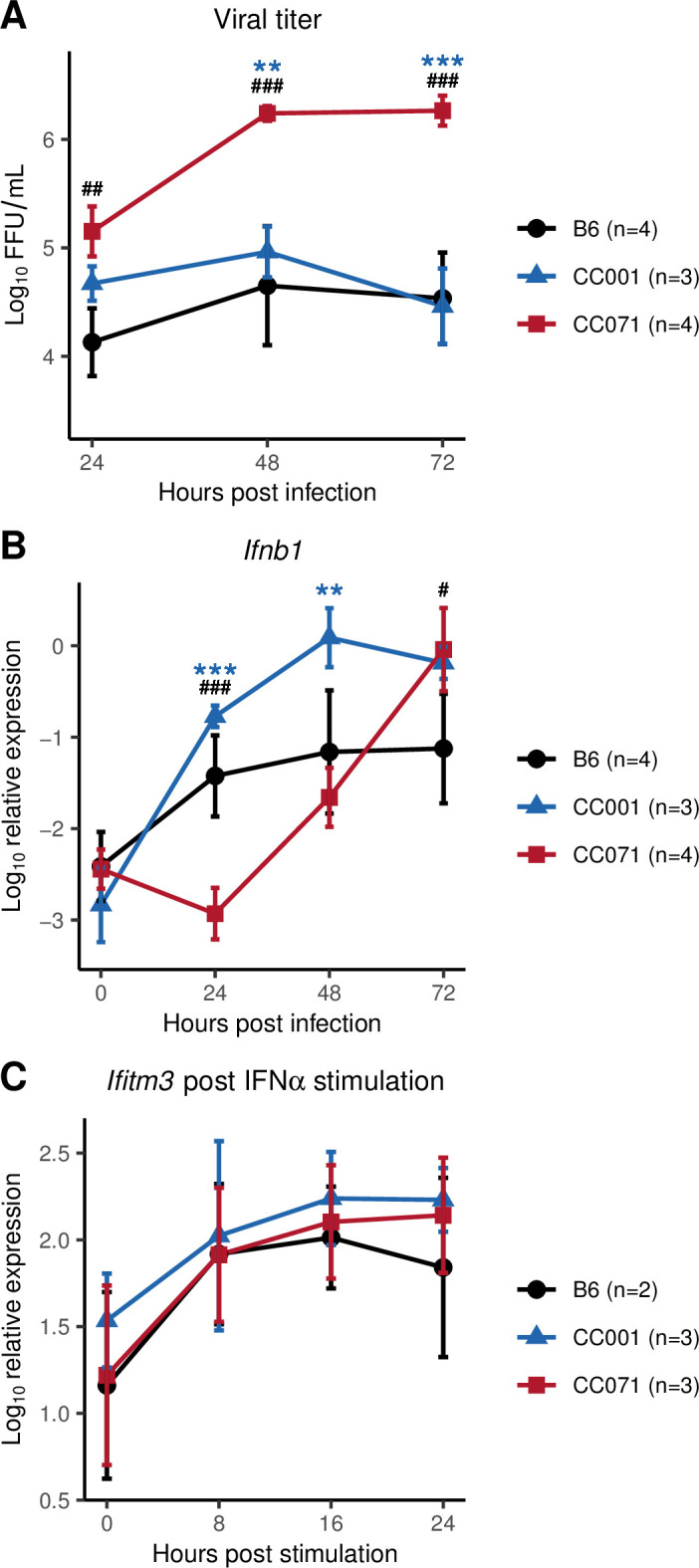
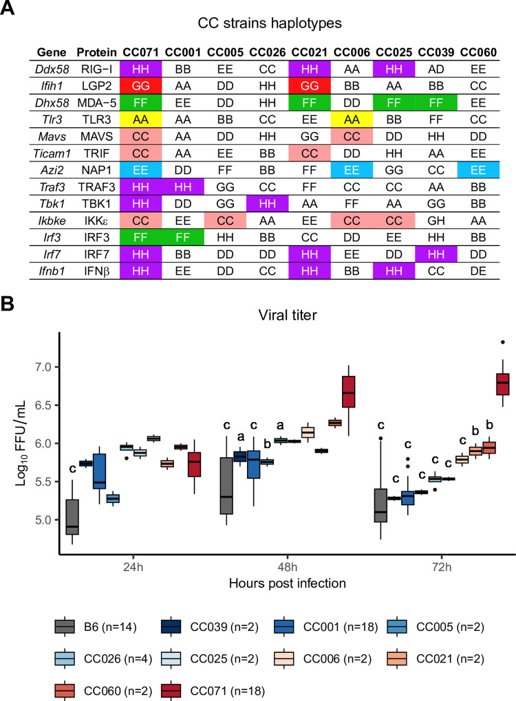
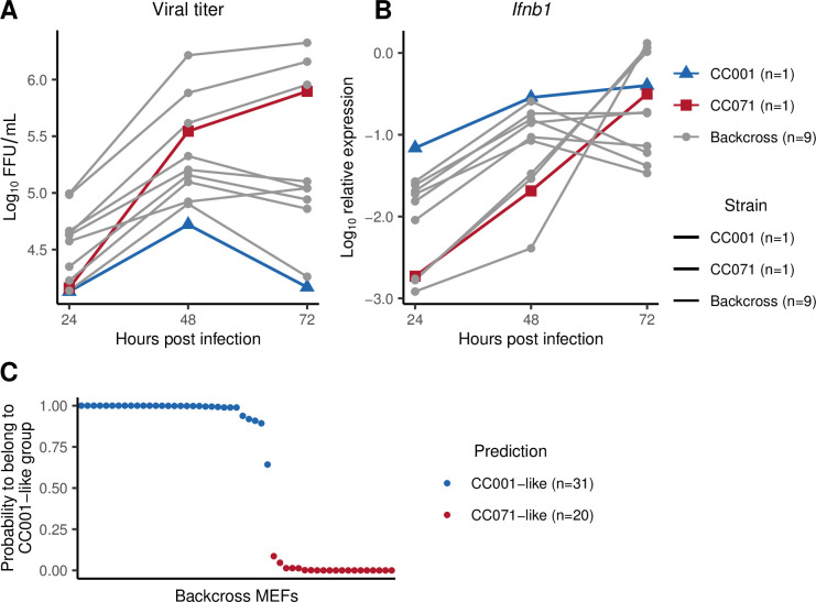
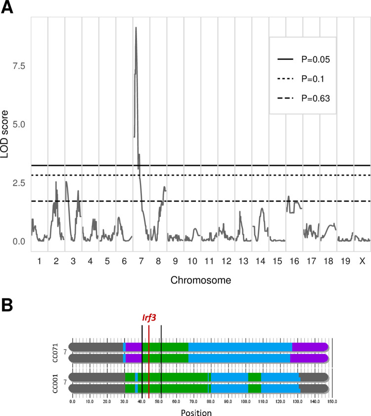
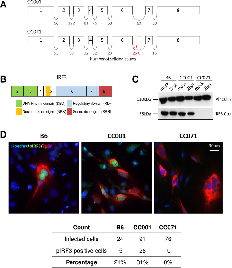
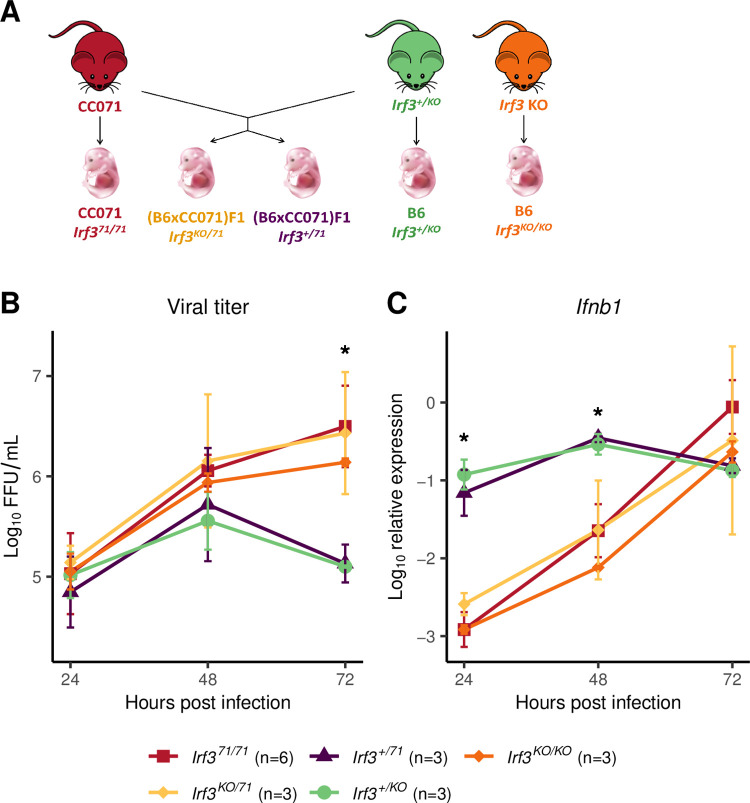
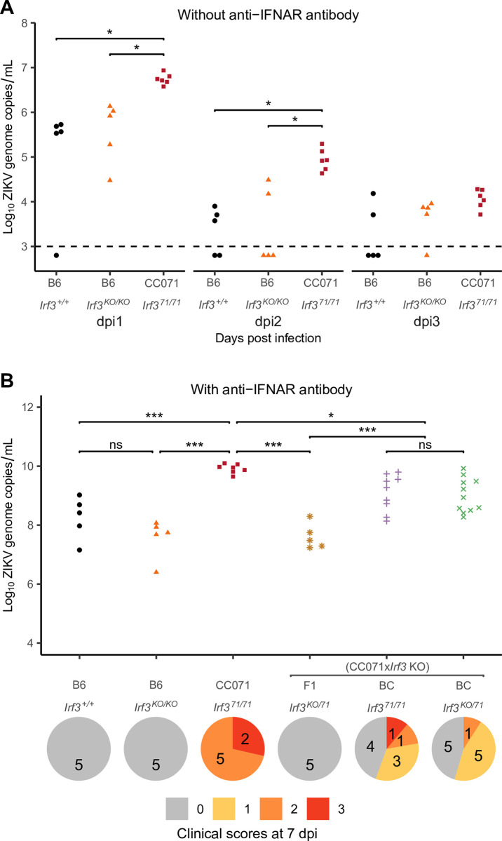
Zika virus (ZIKV) is a Flavivirus responsible for recent epidemics in Pacific Islands and in the Americas. In humans, the consequences of ZIKV infection range from asymptomatic infection to severe neurological disease such as Guillain-Barré syndrome or fetal neurodevelopmental defects, suggesting, among other factors, the influence of host genetic variants. We previously reported similar diverse outcomes of ZIKV infection in mice of the Collaborative Cross (CC), a collection of inbred strains with large genetic diversity. CC071/TauUnc (CC071) was the most susceptible CC strain with severe symptoms and lethality. Notably, CC071 has been recently reported to be also susceptible to other flaviviruses including dengue virus, Powassan virus, West Nile virus, and to Rift Valley fever virus. To identify the genetic origin of this broad susceptibility, we investigated ZIKV replication in mouse embryonic fibroblasts (MEFs) from CC071 and two resistant strains. CC071 showed uncontrolled ZIKV replication associated with delayed induction of type-I interferons (IFN-I). Genetic analysis identified a mutation in the Irf3 gene specific to the CC071 strain which prevents the protein phosphorylation required to activate interferon beta transcription. We demonstrated that this mutation induces the same defective IFN-I response and uncontrolled viral replication in MEFs as an Irf3 knock-out allele. By contrast, we also showed that Irf3 deficiency did not induce the high plasma viral load and clinical severity observed in CC071 mice and that susceptibility alleles at other genes, not associated with the IFN-I response, are required. Our results provide new insight into the in vitro and in vivo roles of Irf3, and into the genetic complexity of host responses to flaviviruses.


Conflict of interest statement

The authors have declared that no competing interests exist.

Figures

Fig 1
Fig 1. CC071 MEFs fail to control viral replication, with delayed Ifnb1 expression but normal response to IFN-I.
(A, B) MEFs derived from B6 (black circles), CC001 (blue triangles) and CC071 (red squares) were infected with ZIKV at a MOI of 5 and analyzed 24, 48 and 72 hpi. (A) Viral titer in supernatants was quantified by FFA. (B) Ifnb1 expression was normalized to Tbp reference gene. Data are mean +/- sem from 3 to 4 biological replicates per strain (MEFs derived from individual embryos). (C) MEFs were stimulated with recombinant IFNα. Ifitm3 relative expression normalized to Tbp reference gene is shown as an example of ISG. Data are mean +/- sem from 2 to 3 biological replicates. Blue asterisks and black hashes show statistical significance of CC071 compared to CC001 and to B6, respectively (ANOVA followed by Tukey HSD, */# p < 0.05, **/## p < 0.01, ***/### p < 0.001).
Fig 2
Fig 2. Haplotype analysis fails to identify a gene from the Ifnb1 induction pathway associated with uncontrolled viral replication.
(A) Identification of CC strains which carry the same ancestral haplotype as CC071 at the genes involved in the pathway leading to Ifnb1 expression. Colored boxes indicate matched haplotypes between CC071 and other CC strains. Letters and colors designate the eight CC founder strains. A: A/J (yellow); B: C57BL/6J; C: 129S1/SvImJ (pink); D: NOD/ShiLtJ; E: NZO/HILtJ (light blue); F: CAST/EiJ (green); G: PWK/PhJ (red); H: WSB/EiJ (purple). Doubled letters (eg AA) indicate homozygous genotypes. Heterozygous genotypes are indicated by the two corresponding letters (eg AD). (B) Kinetics of viral titer in MEFs from CC071 and the 8 CC strains shown in (A). Experimental conditions were as in Fig 1A n: number of technical replicates for each strain. Data are mean +/- sem from the technical replicates. Letters show statistical significance between CC071 and other strains. (ANOVA followed by Tukey HSD, a: p < 0.05, b: p < 0.01, c: p < 0.001).
Fig 3
Fig 3. Backcross MEFs display either a CC001-like or a CC071-like phenotype.
MEFs derived from CC001 (blue triangles), CC071 (red squares) and backcross (gray circles) embryos were infected with ZIKV at a MOI of 5. (A) Viral titer and (B) Ifnb1 expression in ZIKV-infected MEFs from 9 backcross embryos. Experimental conditions were as in Fig 1A and 1B. Red and blue curves show the results for CC071 and CC001 MEFs, respectively, from the same infection experiment. (C) Results of LDA on the backcross MEFs. LDA coefficients were calculated from Ifnb1 expression data in CC001 and CC071 infected MEFs, and applied to backcross MEFs. The graph shows the probability of each BC MEF to belong to the "CC001-like" group, resulting in two distinct populations shown in blue ("CC001-like") and in red ("CC071-like"). n: number of BC MEFs assigned to each group.
Fig 4
Fig 4. Genetic analysis of Ifnb1 expression in backcross MEFs identifies a major determinant mapping to the Irf3 locus.
(A) Genome-wide linkage analysis of the LDA classification of backcross MEFs performed with R/qtl. X-axis: genomic location. Y-axis: LOD score of the phenotype-genotype association. Genome-wide significance thresholds (P = 0.05, P = 0.1 and P = 0.63) were computed from 1000 data permutation. The chromosome 7 peak has a LOD score of 9.138. (B) Schematic representation of CC001 and CC071 chromosome 7 haplotypes, from https://csbio.unc.edu/CCstatus/CCGenomes/. Colors represent the CC ancestral haplotypes (same colors as in Fig 2A). Thick vertical black lines show the peak’s 95% Bayesian confidence interval (25.9–31.3 cM, corresponding to 40.1–50.6 Mb). The red line shows the position of Irf3 gene.
Fig 5
Fig 5. CC071 Irf3 mRNAs show abnormal splicing, resulting in defective IRF3 protein.
(A) Schematic representation of the exons of the Irf3 gene with the number of reads spanning successive exons in the CC001 and CC071 RNAseq data (one sample of each strain). The red box between CC071’s exons 6 and 7 depicts a novel exon resulting from abnormal splicing. (B) Schematic representation of the IRF3 protein structural domains (exon 1 is untranslated). Exon 8 encodes the serine rich region containing the phosphorylation sites for IRF3 activation. (C) Western blot using an anti-C-terminal IRF3 antibody from mock-infected and ZIKV-infected B6, CC001 and CC071 MEFs at 2 hpi, showing the absence of full-length IRF3 in CC071 MEFs. Vinculin was used as a loading control. (D) Immunofluorescence using an anti-phosphorylated IRF3 (pIRF3, green) in ZIKV-infected B6, CC001 and CC071 MEFs at 24 hpi, showing the absence of pIRF3 in the nucleus of CC071 MEFs upon infection. Red-labeled 4G2 antibody labels ZIKV-infected cells. Cell nuclear DNA labeled by Hoechst (blue). Quantification of the number of infected and pIRF3 positive cells is presented in the table. Proportions were established on 420, 428 and 551 cells for CC001, CC071 and B6, respectively.
Fig 6
Fig 6. Quantitative complementation test confirms that Irf3 LOF in CC071 is responsible for uncontrolled viral replication and delayed Ifnb1 expression.
(A) Mice heterozygous for an inactivated Irf3 allele (B6-Irf3+/KO) were mated with CC071 mice to produce four types of embryos from which MEFs were derived. The genetic background and Irf3 genotype is shown below each type of embryos. (B) Viral titer and (C) Ifnb1 expression in ZIKV-infected MEFs. Experimental conditions were as in Fig 1A and 1B. Data are mean +/- sem from 6 biological replicates for CC071 and 3 for the other groups. The asterisks represent the results of ANOVA test between all groups (* p < 0.05). Results of the Tukey HSD post-hoc are as follows. Viral titer at 72h: CC071 vs Irf3+/71 and vs Irf3+/KO: p < 0.001. Irf3KO/71 vs Irf3+/71 and vs Irf3+/KO: p < 0.01. Irf3KO/KO vs Irf3+/71 and vs Irf3+/KO: p < 0.05. Ifnb1 expression at 24hpi: CC071 vs Irf3+/71 and vs Irf3+/KO, Irf3KO/71 vs Irf3+/71 and vs Irf3+/KO, Irf3KO/KO vs Irf3+/71 and vs Irf3+/KO: p < 0.001. Ifnb1 expression at 48hpi: CC071 vs Irf3+/71and vs Irf3+/KO, Irf3KO/71 vs Irf3+/71 and vs Irf3+/KO, Irf3KO/KO vs Irf3+/71 and vs Irf3+/KO: p < 0.01. Fig 6 includes a drawing of a mouse which was taken from https://publicdomainvectors.org/fr/gratuitement-des-vecteurs/Dessin-de-souris-de-dessin-anim%C3%A9-avec-longue-moustache-vectoriel/22268.html and a picture of a mouse embryon taken from https://commons.wikimedia.org/wiki/File:201309_mouse_embryo.png.
Fig 7
Fig 7. Irf3 LOF is not sufficient to explain CC071 susceptibility to ZIKV infection in vivo.
(A) B6, B6-Irf3 KO and CC071 mice were infected IP with 107 FFUs of ZIKV and monitored for 7 days, without prior IP injection of MAR1-5A3 IFNAR-blocking monoclonal antibody. Plasma viral load was quantified at days 1 to 3 p.i. by RT-qPCR. The two lines of the group legend (X-axis) indicate the genetic background of the mice and their genotype at the Irf3 locus, respectively. Each dot represents one mouse. Groups were compared by Kruskal-Wallis followed by Wilcoxon test with Holm correction for multiple testing (* p < 0.05). (B) B6, B6-Irf3 KO, CC071, (CC071 x Irf3 KO) F1 and (CC071 x Irf3 KO) x CC071 BC mice were infected IP with 107 FFUs of ZIKV after IP injection of 2 mg of MAR1-5A3 IFNAR-blocking monoclonal antibody 24 h before infection, and monitored for 7 days. The graph shows plasma viral loads quantified at day 2 p.i. by RT-qPCR. The two lines of the group legend (X-axis) indicate the genetic background of the mice and their genotype at the Irf3 locus, respectively. BC mice are separated into two groups depending on the genotype at the Irf3 gene: homozygous for the CC071 mutant allele (Irf371/71), or heterozygous for the CC071 and the KO alleles (Irf3KO/71). Irf3 genotyping results are presented in S5 Fig. Each dot represents one mouse. Groups were compared by ANOVA followed by Tukey HSD (* p < 0.05, ** p < 0.01, *** p < 0.001). Below the graph is shown the distribution of clinical scores at day 7 p.i. in the same groups of mice as above (0, no symptoms; 1, slight hunched posture; 2, ruffled fur, hunched posture and/or mild ataxia; 3: prostration, ataxia, partial paralysis).

Similar articles

Cited by

References

    1. Cao-Lormeau VM, Blake A, Mons S, Lastère S, Roche C, Vanhomwegen J, et al.. Guillain-Barré Syndrome outbreak associated with Zika virus infection in French Polynesia: a case-control study. Lancet. 9 avr 2016;387(10027):1531–9. - PMC - PubMed
    1. Carod-Artal FJ. Neurological complications of Zika virus infection. Expert Review of Anti-infective Therapy. 4 mai 2018;16(5):399–410. doi: 10.1080/14787210.2018.1466702 - DOI - PubMed
    1. Oliveira Melo AS, Malinger G, Ximenes R, Szejnfeld PO, Alves Sampaio S, Bispo de Filippis AM. Zika virus intrauterine infection causes fetal brain abnormality and microcephaly: tip of the iceberg?: Physician Alert. Ultrasound Obstet Gynecol. janv 2016;47(1):6–7. - PubMed
    1. Paixao ES, Cardim LL, Costa MCN, Brickley EB, de Carvalho-Sauer RCO, Carmo EH, et al.. Mortality from Congenital Zika Syndrome—Nationwide Cohort Study in Brazil. N Engl J Med. 24 févr 2022;386(8):757–67. doi: 10.1056/NEJMoa2101195 - DOI - PMC - PubMed
    1. Rossi SL, Ebel GD, Shan C, Shi PY, Vasilakis N. Did Zika Virus Mutate to Cause Severe Outbreaks? Trends in Microbiology. 1 oct 2018;26(10):877–85. doi: 10.1016/j.tim.2018.05.007 - DOI - PMC - PubMed

Publication types