. 2023 Sep 22;381(6664):1324-1330.
doi: 10.1126/science.adf9325. Epub 2023 Sep 21.

Reproductive outcomes after pregnancy-induced displacement of preexisting microchimeric cells

Affiliations

Reproductive outcomes after pregnancy-induced displacement of preexisting microchimeric cells

Tzu-Yu Shao et al. Science. .

Abstract

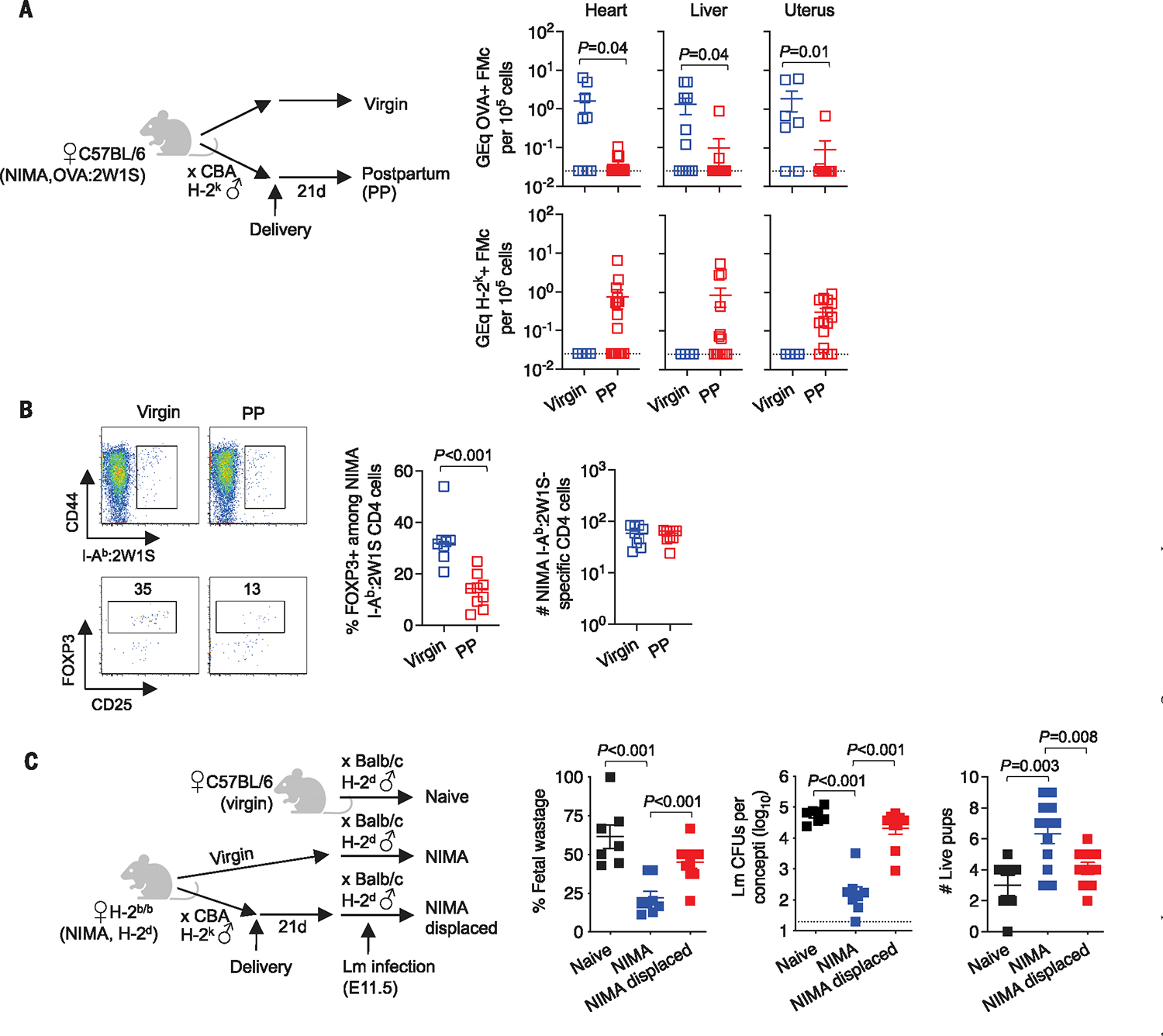
Pregnancy confers partner-specific protection against complications in future pregnancy that parallel persistence of fetal microchimeric cells (FMcs) in mothers after parturition. We show that preexisting FMcs become displaced by new FMcs during pregnancy and that FMc tonic stimulation is essential for expansion of protective fetal-specific forkhead box P3 (FOXP3)-positive regulatory T cells (Treg cells). Maternal microchimeric cells and accumulation of Treg cells with noninherited maternal antigen (NIMA) specificity are similarly overturned in daughters after pregnancy, highlighting a fixed microchimeric cell niche. Whereas NIMA-specific tolerance is functionally erased by pregnancy, partner-specific resiliency against pregnancy complications persists in mothers despite paternity changes in intervening pregnancy. Persistent fetal tolerance reflects FOXP3 expression plasticity, which allows mothers to more durably remember their babies, whereas daughters forget their mothers with new pregnancy-imprinted immunological memories.


Conflict of interest statement

Competing interests: The authors declare that they have no competing interests.

Figures

Fig. 1.
Fig. 1.. MMcs and NIMA-specific tolerance displaced after pregnancy.
(A) Levels of ovalbumin (OVA) DNA specific to OVA:2W1S+ MMcs or H-2k DNA specific to H-2k+ FMcs in each tissue among OVA:2W1S NIMA mice 21 days postpartum (PP) after allogeneic pregnancy sired by H-2k+ CBA males (red) or age-matched 12- to 14-week-old virgin OVA:2W1S-NIMA mice (blue). GEq, genome equivalents. (B) Representative plots and composite data showing FOXP3+ among CD4 cells with I-Ab :2W1S surrogate NIMA specificity and numbers of I-Ab :2W1S+ CD4 cells in pooled secondary lymphoid organs for the mice described in (A). (C) Percent fetal wastage, average recoverable colony-forming units (CFUs) from concepti in each litter, and numbers of live pups per litter 5 days after maternal Lm infection midgestation [embryonic day 11.5 (E11.5)] for naïve H-2b C57BL/6 mice during primary pregnancy sired by H-2d BALB/c males (black), compared with genetically identical H-2d NIMA mice undergoing primary pregnancy sired by H-2d BALB/c males (blue) or H-2d NIMA mice during secondary pregnancy by H-2d BALB/c males with prior pregnancy by H-2k CBA males (red). Each point indicates the data from an individual mouse and is representative of at least three independent experiments, each with similar results. Bar, mean ± standard error.
Fig. 2.
Fig. 2.. Pregnancy-induced FMc displacement does not override fetal tolerance.
(A) Levels of OVA DNA specific to OVA:2W1S+ FMcs from primary pregnancy or H-2k DNA specific to H-2k+ FMcs from secondary pregnancy in each tissue among C57BL/6 female mice 9 to 10 weeks after a single pregnancy sired by OVA:2W1S+ H-2d BALB/c males (blue) compared with mice 3 weeks after second pregnancy sired by H-2k+ CBA males (red). (B) Representative plots and composite data showing percent FOXP3+ among CD4 cells with I-Ab :2W1S surrogate fetal specificity and numbers of I-Ab :2W1S+ CD4 cells in pooled secondary lymphoid organs for the mice described in (A). (C) Percent fetal wastage, average recoverable CFUs from concepti in each litter, and number of live pups per litter 5 days after maternal Lm infection at midgestation (E11.5) for naïve mice during primary pregnancy sired by H-2d BALB/c males (black), compared with mice during secondary pregnancy sired by H-2d BALB/c males (blue) or during tertiary pregnancy sired by H-2d BALB/c males with intervening secondary pregnancy sired by H-2k CBA males (red). Each point indicates the data from an individual mouse and is representative of at least three independent experiments, each with similar results. Bar, mean ± standard error.
Fig. 3.
Fig. 3.. Resiliency against fetal wastage despite FMc depletion.
(A) Levels of OVA DNA specific to OVA:2W1S+ FMcs in each tissue of mice 30 days PP from primary pregnancy sired by OVA:2W1S+ H-2d BALB/c males treated with α-OVA IgG on days 2 and 16 after parturition (red) or rabbit IgG (blue), or virgin controls (black). (B) Percent FOXP3+ among CD4 cells with I-Ab:2W1S surrogate fetal specificity and numbers of I-Ab:2W1S+ CD4 cells in pooled secondary lymphoid organs for the mice described in (A). (C) Percent fetal wastage, average recoverable CFUs from concepti in each litter, and number of live pups per litter 5 days after maternal Lm infection midgestation (E11.5) for mice during primary pregnancy sired by OVA:2W1S+ H-2d BALB/c males (black), compared with during secondary pregnancy sired by OVA:2W1S+ H-2d BALB/c males in mice treated with rabbit IgG (blue) or anti-OVA IgG (red) PP on days 2 and 16 after primary pregnancy. (D) Percent FOXP3+ among CD4 cells with I-Ab:2W1S surrogate fetal specificity, number of I-Ab:2W1S+ FOXP3+ CD4 cells in pooled secondary lymphoid organs, and fold-expansion of these cells midgestation during primary pregnancy (black) or during secondary pregnancy in mice treated with rabbit IgG (blue) or α-OVA IgG (red). Each point indicates the data from an individual mouse and is representative of at least three independent experiments each with similar results. Bar, mean ± standard error.
Fig. 4.
Fig. 4.. FOXP3 plasticity sustains resiliency against pregnancy complications.
(A) Percent FOXP3(GFP)–negative among dTOMATO+ CD4 cells with I-Ab:2W1S fetal specificity or tetramer-negative bulk cells in pooled secondary lymphoid organs for FOXP3(GFP)CRE R26dTOMATO mice without prior pregnancy (virgin, black), or mice 30 days after primary pregnancy sired by OVA:2W1S+ BALB/c males treated with rabbit IgG (blue) or α-OVA IgG (red) on days 2 and 16 after parturition. (B) Percent FOXP3(GFP)–negative cells among dTOMATO+ CD4 cells with I-Ab:2W1S surrogate fetal specificity or tetramer-negative bulk cells in pooled secondary lymphoid organs of FOXP3(GFP)CRE R26dTOMATO mice after primary pregnancy sired by OVA:2W1S+ BALB/c males (blue) or additional secondary pregnancy sired by third-party H-2k+ CBA males (red). (C) Expression of each molecule by FOXP3 (GFP)+ compared with FOXP3(GFP)–negative cells among dTOMATO+ CD4 cells with I-Ab:2W1S surrogate fetal specificity in PP mice after primary pregnancy by OVA:2W1S+ BALB/c males (blue) or in PP mice with OVA:2W1S+ FMc depletion by α-OVA IgG (red circles) or FMc displacement with secondary pregnancy by H-2k+ CBA males (red squares). gMFI, geometric mean fluorescent intensity. (D) Percent FOXP3+ among PP donor CD90.2+ CD45.1+ FOXP3DTR (solid) or PP donor CD90.2+ CD45.2+ FOXP3WT (open) CD4 cells with I-Ab:2W1S fetal specificity in pooled secondary lymphoid organs of CD90.1+ (CD90.2-negative) recipients midgestation (E11.5) during allogenic pregnancy sired by OVA:2W1S+ BALB/c males administered DT for 2 consecutive days after adoptive cell transfer and before mating. Splenocytes from PP FOXP3DTR and FOXP3WT donors were harvested 30 days after primary pregnancy by OVA:2W1S+ BALB/c males, with some mice administered α-OVA or rabbit IgG (on days 2 and 16 after parturition). (E) Percent fetal wastage and average recoverable CFUs from concepti in each litter, 5 days after maternal Lm infection midgestation (E11.5) for mice during primary pregnancy sired by OVA:2W1S+ H-2d BALB/c males adoptively transferred donor splenocytes 1 day prior (E10.5) from isogenic virgin mice (black), PP FOXP3WT (blue), PP FOXP3DTR (red filled) (depletion of donor FOXP3+ cells), or PP FOXP3(GFP)CRE R26iDTR (open red) (depletion of FOXP3+ cells and ex-Treg cells) and administered DT for two consecutive days (E10.5 and E11.5). Each point indicates the data from an individual mouse and is representative of at least three independent experiments, each with similar results. Bar, mean ± standard error.

References

    1. Tsuda S, Nakashima A, Shima T, Saito S, Front. Immunol. 10, 573 (2019). - PMC - PubMed
    1. Jiang TT et al., J. Immunol. 192, 4949–4956 (2014). - PMC - PubMed
    1. Green ES, Arck PC, Semin. Immunopathol. 42, 413–429 (2020). - PMC - PubMed
    1. Keller CC, Eikmans M, van der Hoorn MP, Lashley LEELO, J. Reprod. Immunol. 139, 103105 (2020). - PubMed
    1. Rowe JH, Ertelt JM, Aguilera MN, Farrar MA, Way SS, Cell Host Microbe 10, 54–64 (2011). - PMC - PubMed

LinkOut - more resources