. 2023 Oct 12;186(21):4652-4661.e13.
doi: 10.1016/j.cell.2023.08.023. Epub 2023 Sep 20.

Mpox infection protects against re-challenge in rhesus macaques

Affiliations

Mpox infection protects against re-challenge in rhesus macaques

Malika Aid et al. Cell. .

Abstract

The mpox outbreak of 2022-2023 involved rapid global spread in men who have sex with men. We infected 18 rhesus macaques with mpox by the intravenous, intradermal, and intrarectal routes and observed robust antibody and T cell responses following all three routes of infection. Numerous skin lesions and high plasma viral loads were observed following intravenous and intradermal infection. Skin lesions peaked on day 10 and resolved by day 28 following infection. On day 28, we re-challenged all convalescent and 3 naive animals with mpox. All convalescent animals were protected against re-challenge. Transcriptomic studies showed upregulation of innate and inflammatory responses and downregulation of collagen formation and extracellular matrix organization following challenge, as well as rapid activation of T cell and plasma cell responses following re-challenge. These data suggest key mechanistic insights into mpox pathogenesis and immunity. This macaque model should prove useful for evaluating mpox vaccines and therapeutics.


Conflict of interest statement

Declaration of interests The authors declare no competing interests.

Figures

Figure 1.
Figure 1.. Study design.
18 rhesus macaques were infected with mpox on day 0 at 106 TCID50 by the i.v. route (Group 1; N=4), 105 TCID50 by the i.v. route (Group 2; N=4), 104 TCID50 by the i.v. route (Group 3; N=4), 106 TCID50 by the i.d. route (Group 4; N=3), or 106 TCID50 by the i.r. route (Group 5; N=3). On day 28, these animals as well as concurrent naïve controls (Group 6; N=3) were re-challenged with 106 TCID50 by the i.v. route.
Figure 2.
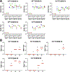
Figure 2.. Humoral immune responses in mpox challenged rhesus macaques.
Humoral immune responses were assessed at baseline, on day 14 and day 28 following challenge, and on day 42 following re-challenge by (A) binding antibody ELISA titers to A35, B6, and H3 and (B) neutralizing antibody PRNT titers to virus. Red horizontal bars reflect mean responses.
Figure 3.
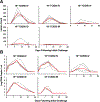
Figure 3.. Cellular immune responses in mpox challenged rhesus macaques.
(A) CD8+ and (B) CD4+ T cell responses to pooled A29, A35, B6, and M1 peptides by IFN-γ intracellular cytokine staining (ICS) assays on day 28 following challenge. Dotted lines reflect limit of quantitation. Red horizontal bars reflect mean responses. Responses depicted are % IFN-γ positive CD8+ or CD4+ T cells following peptide stimulation.
Figure 4.
Figure 4.. Poxvirus skin lesions and viral loads following mpox challenge.
(A) Poxvirus skin lesion count and (B) plasma log viral DNA copies/ml (limit 50 copies/ml) were assessed on days 0, 3, 7, 10, 14, 21, and 28 following challenge. Red lines reflect median values.
Figure 5.
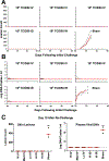
Figure 5.. Poxvirus skin lesions and viral loads following mpox re-challenge.
On day 28, the 18 convalescent animals and 3 concurrent naïve controls were re-challenged with 106 TCID50 by the i.v. route. (A) Poxvirus skin lesion count and (B) plasma log viral DNA copies/ml (limit 50 copies/ml) were assessed on study days 28, 31, 35, 38, 42, and 49, which reflect days 0, 3, 7, 10, 14, 21, and 28 following re-challenge. (C) Summary poxvirus skin lesion counts and plasma log viral DNA copes/ml on day 38, which reflects day 10 following re-challenge. **, two-sided Mann-Whitney tests. Dotted line reflects limit of quantitation. Red lines reflect median values.
Figure 6.
Figure 6.. Pathology of mpox skin lesions.
Three rhesus macaques were necropsied on day 10 following mpox challenge. (A) Hyperkeratotic epidermal hyperplasia with overlying and adjacent vesicle formation. (B) intracytoplasmic keratinocyte Guanieri-like body next to epidermal keratin pearl (insert, arrow). (C) Syncytial cells (arrows) in region of lymphohistiocytic dermatitis underlying epidermal vesicle. (D) ballooning degeneration and necrosis of sebaceous follicular epithelium (insert, arrow). (E) Poxvirus antigen by immunohistochemistry (brown) in epidemal vesicle, (F) Poxviral antigen by immunohistochemistry in regional of ballooning degeneration, proliferation, and epidermal edema in epidermal erosion. (G, H) Poxvirus RNA by RNAScope (red) in epidermal and follicular epithelium with ballooning degeneration, proliferation, and edema.
Figure 7.
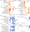
Figure 7.. Transcriptomic pathways increased or decreased following mpox challenge and re-challenge.
Gene set enrichment analysis GSEA was performed on differential expression genes (DEGs) on days 1, 3, 7, 14, 21, and 28 after challenge and on days 1, 3, 7, and 10 following re-challenge (study days 29, 31, 35, and 38). The GSEA normalized enrichment score (NES) is shown for each pathway across time. Circle size is proportional to the NES, and circle color is proportional to the -log10 FDR q value. Color gradient corresponds to the NES, where increased pathways (A–D) were shown in red and downregulated pathways (E, G) in blue. Insignificant pathways were shown in small open dark circles (FDR q value >0.05). F, H show the GSEA rank of the top downregulated genes associated with extracellular matrix (F) and metabolism (H).

References

    1. Basgoz N, Brown CM, Smole SC, Madoff LC, Biddinger PD, Baugh JJ, and Shenoy ES (2022). Case 24–2022: A 31-Year-Old Man with Perianal and Penile Ulcers, Rectal Pain, and Rash. N Engl J Med 387, 547–556. 10.1056/NEJMcpc2201244. - DOI - PubMed
    1. Thornhill JP, Barkati S, Walmsley S, Rockstroh J, Antinori A, Harrison LB, Palich R, Nori A, Reeves I, Habibi MS, et al. (2022). Monkeypox Virus Infection in Humans across 16 Countries - April-June 2022. N Engl J Med 387, 679–691. 10.1056/NEJMoa2207323. - DOI - PubMed
    1. Gessain A, Nakoune E, and Yazdanpanah Y (2022). Monkeypox. N Engl J Med 387, 1783–1793. 10.1056/NEJMra2208860. - DOI - PubMed
    1. Grandpre LE, Duke-Cohan JS, Ewald BA, Devoy C, Barouch DH, Letvin NL, Reinherz EL, Baden LR, Dolin R, and Seaman MS (2009). Immunogenicity of recombinant Modified Vaccinia Ankara following a single or multi-dose vaccine regimen in rhesus monkeys. Vaccine 27, 1549–1556. S0264–410X(09)00020–6 [pii] 10.1016/j.vaccine.2009.01.010. - DOI - PMC - PubMed
    1. Maksyutov RA, Gavrilova EV, and Shchelkunov SN (2016). Species-specific differentiation of variola, monkeypox, and varicella-zoster viruses by multiplex real-time PCR assay. Journal of virological methods 236, 215–220. 10.1016/j.jviromet.2016.07.024. - DOI - PMC - PubMed

Publication types