Review
. 2023 Nov;299(11):105263.
doi: 10.1016/j.jbc.2023.105263. Epub 2023 Sep 20.

Diversity in connexin biology

Affiliations
Review

Diversity in connexin biology

Sergiu A Lucaciu et al. J Biol Chem. 2023 Nov.

Abstract

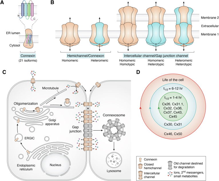
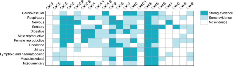
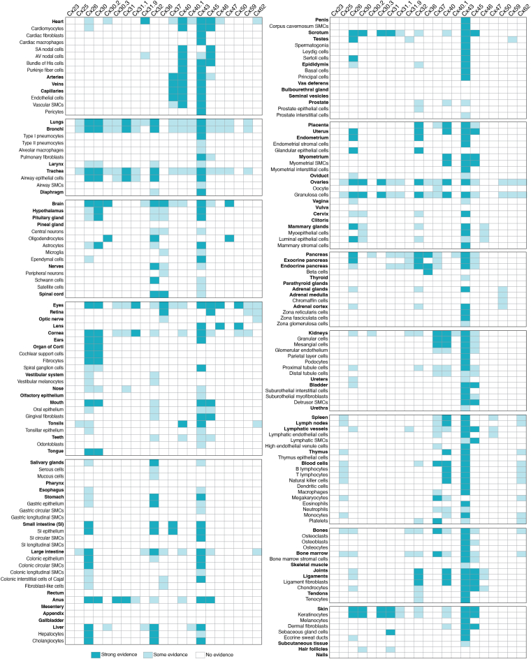
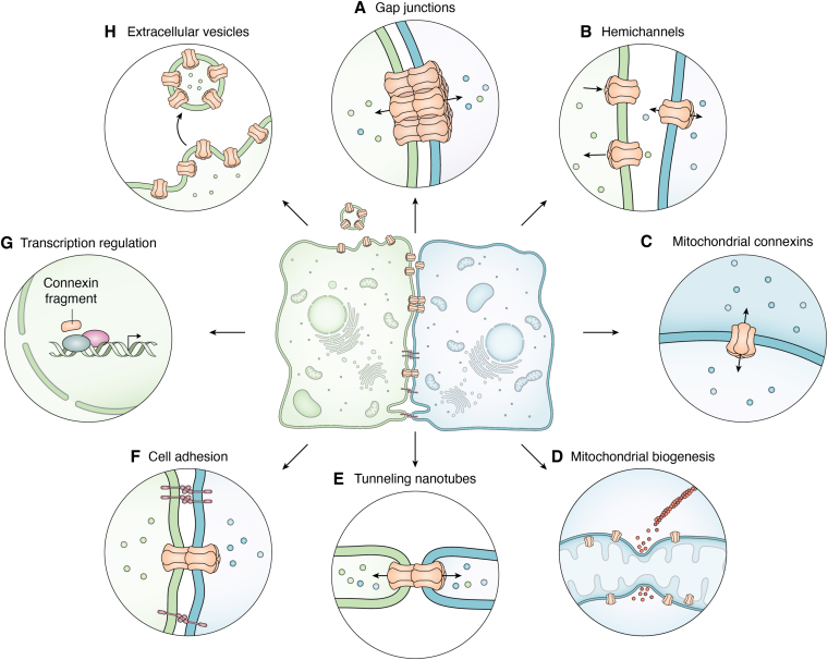
Over 35 years ago the cell biology community was introduced to connexins as the subunit employed to assemble semicrystalline clusters of intercellular channels that had been well described morphologically as gap junctions. The decade that followed would see knowledge of the unexpectedly large 21-member human connexin family grow to reflect unique and overlapping expression patterns in all organ systems. While connexin biology initially focused on their role in constructing highly regulated intercellular channels, this was destined to change as discoveries revealed that connexin hemichannels at the cell surface had novel roles in many cell types, especially when considering connexin pathologies. Acceptance of connexins as having bifunctional channel properties was initially met with some resistance, which has given way in recent years to the premise that connexins have multifunctional properties. Depending on the connexin isoform and cell of origin, connexins have wide-ranging half-lives that vary from a couple of hours to the life expectancy of the cell. Diversity in connexin channel characteristics and molecular properties were further revealed by X-ray crystallography and single-particle cryo-EM. New avenues have seen connexins or connexin fragments playing roles in cell adhesion, tunneling nanotubes, extracellular vesicles, mitochondrial membranes, transcription regulation, and in other emerging cellular functions. These discoveries were largely linked to Cx43, which is prominent in most human organs. Here, we will review the evolution of knowledge on connexin expression in human adults and more recent evidence linking connexins to a highly diverse array of cellular functions.

Keywords: canonical; connexin; expression; gap junctional intercellular communication; gap junctions; hemichannel; human; noncanonical.


Conflict of interest statement

Conflict of interest The authors declare that they have no conflicts of interest with the contents of this article.

Figures

Figure 1
Figure 1
Growth of gap junction research. Since the discovery of gap junctions in the 1960s, connexins in the 1980s, and hemichannels in the 1990s, there has been a rapid and steady increase in knowledge on these topics as evidenced by the explosion of articles published each year. It is notable that over half of published papers in the field have focused on Cx43. Since 2015 the connexin community has seen a slight decline in articles that feature gap junctions, connexins, and Cx43, while hemichannel papers remain steady.
Figure 2
Figure 2
Organization and life cycle of connexins.A, connexins are polytopic integral membrane proteins that pass through the lipid bilayer four times during their insertion into the endoplasmic reticulum (ER) membrane. B, properly folded connexins oligomerize into hexamers termed connexons that can function as hemichannels in single membranes. Connexons can be homomeric or engage two or more connexin isoforms and be heteromeric. At the cell surface connexons assemble into a wide combination of homomeric, heteromeric, homotypic, or heterotypic channel arrangements. C, the typical life cycle of connexins as elucidated largely on the interrogation of Cx43. Connexins undergo biosynthesis and cotranslational import into the endoplasmic reticulum prior to being transported through the ER-Golgi intermediate compartment (ERGIC) to the Golgi apparatus. After oligomerization, connexons are delivered to the cell surface, where they may function as hemichannels prior to docking with adjacent connexons to form gap junction (GJ) channels that are suitable for the direct intercellular exchange of metabolites. GJ channels cluster into gap junction plaques with newer channels being found at the edges of the plaques. GJs and GJ fragments may proceed to internalize into double-membrane structures called connexosomes that proceed to lysosomes for degradation. D, diversity in the half-life of connexins. The majority of interrogated connexins have been found to have a relatively short half-life of 1 to 4 h with others having longer relative half-lives depending on the connexin isoform and the cellular context in which they are found with some connexins (e.g., Cx46, Cx50) existing for the entire life of the cell as seen in anuclear lens fiber cells.
Figure 3
Figure 3
Complexity of GJIC. GJIC may involve the potential passage of literally thousands of members of the metabolome through homotypic or heterotypic channels. Selective passage of ions, signaling molecules, and metabolites is highly dependent on the connexin isoforms (depicted as red/blue subunits) used to build gap junction channels. Identification of transjunctional molecules is difficult, so not surprising, direct evidence for the passage of metabolites exists for only a small subset of molecules found within the metabolome. Part of the figure was generated using BioRender. For a movie animation of this figure see: https://www.schulich.uwo.ca/lairdlab/img/cell-animation-small.mp4. GJIC, gap junctional intercellular communication.
Figure 4
Figure 4
Expression of human connexin isoforms in all 12 adult body systems. Heatmap depiction of the expression of 21 human connexin isoforms in all adult human body systems as revealed by a systematic analysis of articles available on PubMed. Strong evidence for the expression of a connexin reflects situations where a connexin was observed by multiple laboratories using various detection approaches. Some evidence of connexin expression reflects situations where there is limited evidence of the connexin expression, and no evidence of connexin expression denotes situations where the connexin was either not investigated or negative data was obtained. Note that Cx43 is found in all systems, while other connexins are variably expressed across the adult human anatomy. This data compilation should be seen as a guide as additional insights into where connexins are expressed in adult human cells and tissues continue to emerge and be confirmed at the mRNA and protein levels.
Figure 5
Figure 5
Heatmap depiction of connexin isoform expression in adult human organs, tissues, and cells as revealed in situ or primary cell culture. Note the near ubiquitous distribution of Cx43, the broad distribution of Cx26 and Cx32, and the highly restricted distribution of Cx23, Cx25, Cx59, and Cx62. Given that high-avidity antibodies do not exist for all 21 connexin isoforms and the fact that not all isoforms have been intensely investigated, it is likely that connexin isoforms will be detected in more adult human organs, tissues, and cell types in the future. Heatmap was generated as described in Figure 4. Organs/tissues are noted in bold. Compartmentalization of the list represents the 12 body systems. SMC, smooth muscle cell.
Figure 6
Figure 6
Schematic depiction of the functional diversity of connexins. Once thought to be solely linked to GJIC (A) and later to hemichannel functions within the plasma membrane (B), evidence now supports connexins or connexin fragments as having roles in the mitochondria (C and D), tunneling nanotubules (E), cell adhesion (F), transcription (G), and within extracellular vesicle membranes (H). Other emerging cellular roles for Cx43 are not shown.

References

    1. Sohl G., Willecke K. An update on connexin genes and their nomenclature in mouse and man. Cell Commun. Adhes. 2003;10:173–180. - PubMed
    1. Beyer E.C., Paul D.L., Goodenough D.A. Connexin43: a protein from rat heart homologous to a gap junction protein from liver. J. Cell Biol. 1987;105:2621–2629. - PMC - PubMed
    1. Sohl G., Willecke K. Gap junctions and the connexin protein family. Cardiovasc Res. 2004;62:228–232. - PubMed
    1. Kirichenko E.Y., Skatchkov S.N., Ermakov A.M. Structure and functions of gap junctions and their constituent connexins in the mammalian CNS. Biochem. (Mosc) Suppl. Ser. A. Membr. Cell Biol. 2021;15:107–119. - PMC - PubMed
    1. Mikalsen S.O., S I.K., Tausen M. Connexins during 500 Million years-from Cyclostomes to Mammals. Int. J. Mol. Sci. 2021;22:1584. - PMC - PubMed

Publication types

Grants and funding

LinkOut - more resources