. 2023 Dec;193(12):2017-2030.
doi: 10.1016/j.ajpath.2023.08.014. Epub 2023 Sep 19.

Neuroinflammation in the Dorsal Root Ganglia and Dorsal Horn Contributes to Persistence of Nociceptor Sensitization in SIV-Infected Antiretroviral Therapy-Treated Macaques

Affiliations

Neuroinflammation in the Dorsal Root Ganglia and Dorsal Horn Contributes to Persistence of Nociceptor Sensitization in SIV-Infected Antiretroviral Therapy-Treated Macaques

Rebecca Warfield et al. Am J Pathol. 2023 Dec.

Abstract

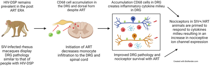
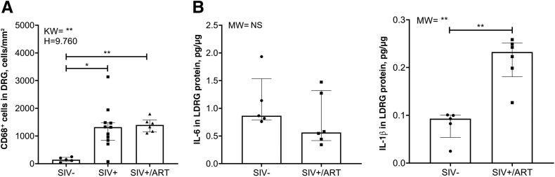
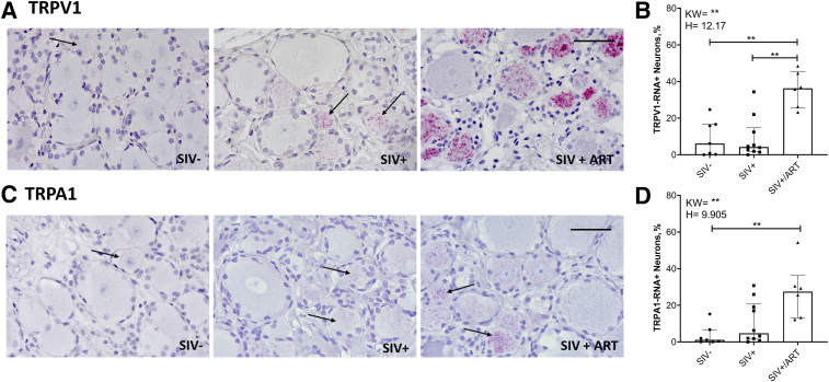
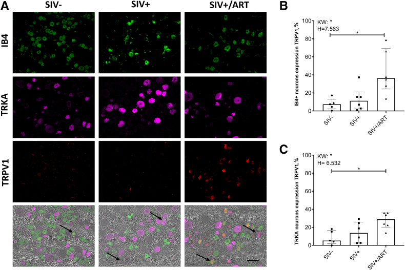
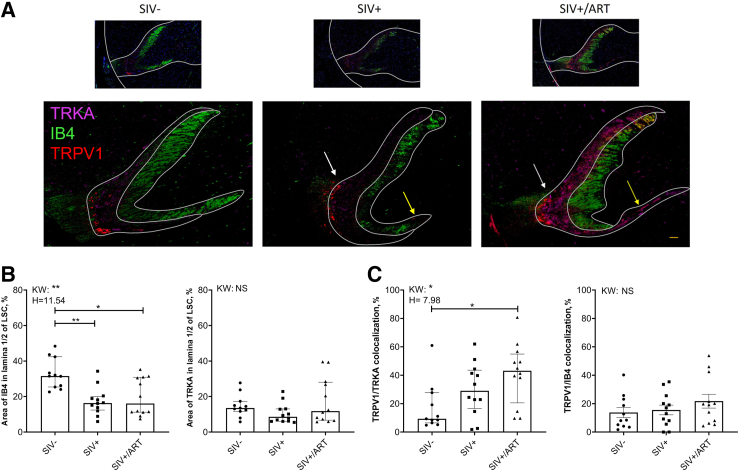
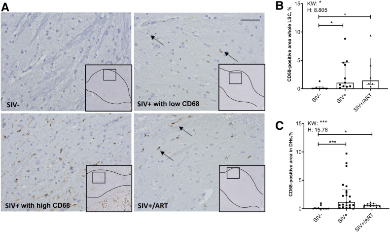
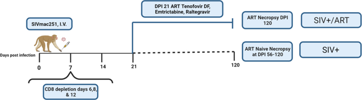
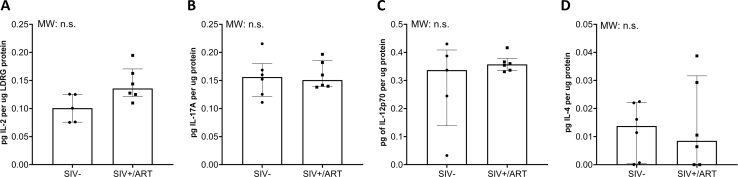
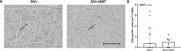
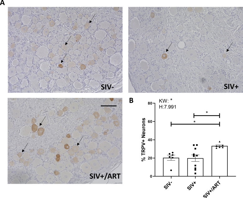
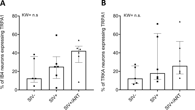
Despite the development of antiretroviral therapy (ART), HIV-associated distal sensory polyneuropathy remains prevalent. Using SIV-infected rhesus macaques, this study examined molecular mechanisms of peripheral and central sensitization to infer chronic pain from HIV infection. Previous studies identified atrophy in nociceptive neurons during SIV infection, which was associated with monocyte infiltration into the dorsal root ganglia (DRG). However, the sensory signaling mechanism connecting this pathology to symptoms remains unclear, especially because pain persists after resolution of high viremia and inflammation with ART. We hypothesized that residual DRG and dorsal horn neuroinflammation contributes to nociceptive sensitization. Using three cohorts of macaques [uninfected (SIV-), SIV-infected (SIV+), and SIV infected with ART (SIV+/ART)], this study showed an increase in the cellular and cytokine inflammatory profiles in the DRG of SIV+/ART macaques compared with uninfected animals. It found significant increase in the expression of nociceptive ion channels, TRPV1, and TRPA1 among DRG neurons in SIV+/ART compared with uninfected animals. SIV-infected and SIV+/ART animals showed reduced innervation of the nonpeptidergic nociceptors into the dorsal horn compared with uninfected animals. Finally, there were a significantly higher number of CD68+ cells in the dorsal horn of SIV+/ART macaques compared with uninfected animals. In summary, these data demonstrate that neuroinflammation, characteristics of nociceptor sensitization, and central terminal atrophy persists in SIV+/ART animals.


Figures

None
Graphical abstract
Figure 1
Figure 1
The number of CD68+ macrophages and IL-1β concentration in the dorsal root ganglion (DRG) remain elevated in simian immunodeficiency virus (SIV)–infected with antiretroviral therapy (SIV+ART) macaques. A: CD68+ cells were visualized through immunohistochemistry and calculated by division of image area for SIV uninfected (SIV–), SIV-infected (SIV+), and SIV-infected ART treated (SIV+/ART) macaques. B: Mesoscale Discovery S-Plex cytokine array determined the concentration of IL-6 and IL-1β per microgram of protein of DRG lysate (LDRG) from SIV—and SIV+/ART macaques. Statistical analysis was performed using a Kruskal-Wallis (KW) one-way analysis of variance and Dunn’s multiple comparison test or a U-test (UT). Data are expressed as medians ± interquartile ranges. SIV– (n = 5; A04-A05, A07-A09) (A), SIV+ (n = 10; A20-A30) (A), SIV+/ART (n = 6; A32-A37) (A); SIV– (n = 5; A04, A10-A13) (B), SIV+/ART (n = 6; A32-A37). ∗P < 0.05, ∗∗P < 0.01. NS, nonsignificant.
Figure 2
Figure 2
Expression of transient receptor potential vanilloid 1 (TRPV1) and transient receptor potential ankyrin 1 (TRPA1) is exclusively increased in the dorsal root ganglions (DRGs) simian immunodeficiency virus (SIV)–infected with antiretroviral therapy treated (SIV+/ART) macaques. A: Representative images from TRPV1 RNAscope DRG sections from SIV-uninfected (SIV–), SIV-infected (SIV+), and SIV+/ART macaques. Arrows indicate TRPV1 RNA-positive cells. B: The percentage of TRPV1-positive neurons in the DRG of SIV–, SIV+, and SIV+/ART macaques. C: Representative images from TRPA1 RNAscope DRG sections from SIV–, SIV+, and SIV+/ART macaques. Arrows indicate TRPA1 RNA-positive cells. D: The percentage of TRPA1-positive neurons in the DRG among groups. Statistical analysis was performed using a Kruskal-Wallis (KW) one-way analysis of variance and Dunn’s multiple comparison test. Data are expressed as medians ± interquartile ranges. SIV– (n = 7; A01-A03, A06-A09), SIV+ (n = 10; A20-A29), and SIV+/ART (n = 6; A32-A37) (B and D) ∗∗P < 0.01. Scale bars = 100 μm. Magnification, ×60.
Figure 3
Figure 3
Transient receptor potential vanilloid 1 (TRPV) expression is significantly increased in both tropomyosin receptor kinase A–expressing (TRKA+) and isolectin B4–expressing (IB4+) nociceptors of simian immunodeficiency virus (SIV)–infected with antiretroviral therapy treated (SIV+/ART) macaques. A: Representative images of Opal multiplex immunohistochemistry/RNAscope targeting TRPV1 RNA (red) within IB4+ (green) or TRKA+ (pink) nociceptors in the dorsal root ganglion of SIV-uninfected (SIV–), SIV-infected (SIV+), and SIV+/ART macaques. Arrows indicate TRPV1 RNA colocalized to either a IB4+ or TRKA+ nociceptor. B and C: Percentage of either TRPV1+/IB4+ (B) or TRPV1+/TRKA+ (C) reveals colocalization of TRPV1 in IB4+ and TRKA+ nociceptors. Statistical analysis was performed using a Kruskal-Wallis (KW) one-way analysis of variance and Dunn’s multiple comparison test. Data are expressed as medians ± interquartile ranges. n = 5 SIV– macaques (A02, A05-A08); n = 6 SIV+ macaques (A20-A21, A25-A27, A29); n = 6 SIV+/ART (A32-A37) macaques (A). ∗P < 0.05. Scale bar = 100 μm. Magnification, ×20.
Figure 4
Figure 4
Decreased reinnervation of nonpeptidergic central terminals in simian immunodeficiency virus (SIV)–infected with antiretroviral therapy treated (SIV+/ART) animals likely limits trafficking of transient receptor potential vanilloid 1 (TRPV1) to peptidergic nociceptors in the dorsal horn. A: Representative images from Opal multiplex immunohistochemical labeling dorsal horn innervating isolectin B4–expressing (IB4+) nonpeptidergic neurons (green) and tropomyosin receptor kinase A–expressing (TRKA+) peptidergic neurons (pink) to identify area of nociceptor innervation and direct colocalization of TRPV1 protein (red) within these nociceptor subtypes. White outline in the top images indicates the outline of white to gray matter in the dorsal horn, whereas the white outline in the lower images indicates the distinction between lamina 1 and lamina 2 of the dorsal horn. Representative image of IB4+ nonpeptidergic nociceptors loss and TRKA+ peptidergic neurons preservation in lamina 1 or 2 of the lumbar spinal cord (LSC) is indicated by the yellow arrow. Representative colocalization of TRPV1/TRKA or TRPV1/IB4 among groups is indicated by the white arrow. B and C: Individual dorsal horn values. Statistical analysis was performed using a Kruskal-Wallis (KW) one-way analysis of variance and Dunn’s multiple comparison test. Data are expressed as medians ± interquartile ranges. n = 11 SIV-uninfected (SIV–) macaques (A14-A19); n = 12 SIV-infected (SIV+) macaques; (A20, A22-A23, A25-A26, A29); n = 12 SIV+/ART macaques (A32-A37) (B and C). ∗P < 0.05, ∗∗P < 0.01. Scale bar = 100 μm. Original magnification: ×2 (A, top row); ×20 (A, bottom row). NS, nonsignificant.
Figure 5
Figure 5
CD68+ macrophages are increased in the dorsal horn on simian immunodeficiency virus (SIV) infection and SIV infection with antiretroviral therapy treated (SIV+/ART). A: Representative images from immunohistochemistry targeting CD68+ cells in the lumbar spinal cord. Images from both SIV-infected (SIV+) animals with low CD68 expression and SIV+ animals with high CD68 expression are included to represent animals with high levels of SIV myelitis. Arrows indicate CD68+ area. B: The percentage of CD68+ area in the whole lumbar spinal cord of SIV-uninfected (SIV–), SIV+, and SIV+/ART macaques. C: The percentage of the CD68+ area in the individual dorsal of SIV−, SIV+, and SIV+/ART macaques. Statistical analysis was performed using a Kruskal-Wallis (KW) one-way analysis of variance and Dunn’s multiple comparison test. Data are expressed as medians ± interquartile ranges. SIV– (n = 6; A14-A19), SIV+ (n = 11; A20-A26, A28-A31), and SIV+/ART (n = 6; A32-A37) macaques (B). SIV– (n = 12; A14-A19), SIV+ (n = 22; A20-A26, A28-A31), and SIV+/ART (n = 12; A32-A37) macaques. ∗P < 0.05, ∗∗∗P < 0.001. Scale bar = 100 μm. Original magnification: ×2 (A); ×40 (A, insets). DHs, dorsal horns.
Supplemental Figure S1
Timeline of simian immunodeficiency virus (SIV) infection, antiretroviral therapy (ART) intervention, and necropsy. Animals A01 to A19 serve as uninfected controls (referred to as SIV−). Animals A20 to A37 were infected with SIVmac251 viral swarm (5 ng of p27; Tulane National Primate Research Center’s Viral Core) followed by CD8 depletion (Nonhuman Primate Reagent Resource). Animals A20 to A31 did not receive ART and progressed to an SIV/AIDS animal (referred to as SIV+) as determined by simian AIDs criteria on necropsy. At postinfection day 21, animals A32 to A37 began receiving an ART regimen of raltegravir (22 mg/kg orally twice daily; Merck, Kenilworth, NJ), tenofovir disoproxil fumarate (30 mg/kg subcutaneously once daily; Gilead, Foster City, CA), and emtricitabine (10 mg/kg subcutaneously once daily; Gilead). SIV+/ART animals were time sacrificed at postinfection days 118 to 120. SIV+ animals were sacrificed based on humane end points consistent with the recommendations of the American Veterinary Medical Association Guidelines for the Euthanasia of Animals. Created with BioRender.com (Toronto, ON, Canada).
Supplemental Figure S2
Mesoscale Discovery S-Plex cytokine array determined the concentration of IL-2 (A), IL-17A (B), IL-12p70 (C), and IL-4 (D) per micrograms of protein of dorsal root ganglia lysate (LDRG) from simian immunodeficiency virus (SIV) uninfected (SIV−) and SIV-infected with antiretroviral therapy (SIV+/ART) macaques. Statistical analysis was performed using a U-test (MW). Concentrations of tumor necrosis factor-α, IL-10, and interferon-ᵧ fell below the lower limit of detection on most samples. Data are expressed as medians ± interquartile ranges. n = 5 SIV− macaques (A04, A10-A13); n = 6 SIV+/ART macaques (A32-A37). n.s., not significant.
Supplemental Figure S3
The number of simian immunodeficiency virus (SIV)mac239–positive cells in the dorsal root ganglia (DRG) area does not significantly differ among SIV-infected (SIV+) and SIV-infected with antiretroviral therapy (SIV+/ART) macaques. A: Representative images from RNAscope targeting SIVmac239 RNA in the DRG of SIV+ and SIV+/ART macaques. Arrows indicate SIVmac239 RNA+ cells. B: The number of SIVmac239+ cells/mm2 of DRG area in SIV+ compared with SIV+/ART macaques. Statistical analysis was performed using a U-test (MW). Data are expressed as means per animal (A) and medians ± interquartile range (B). n = 11 SIV+ macaques (A20-A30); n = 6 SIV+/ART macaques (A32-A37). Scale bar = 100 μm. n.s., not significant.
Supplemental Figure S4
A: Representative images of transient receptor potential vanilloid 1 (TRPV1) protein expression in the dorsal root ganglions (DRGs) of simian immunodeficiency virus (SIV)–uninfected (SIV−), SIV-infected (SIV+), and SIV-infected with antiretroviral therapy (SIV+/ART) macaques. Arrows indicate the range in intensity of diaminobenzidine staining indicative of a positive cell. B: The percentage of TRPV1-positive neurons in the DRG of SIV−, SIV+, and SIV+/ART macaques. Statistical analysis was performed using a Kruskal-Wallis (KW) one-way analysis of variance and Dunn’s multiple comparison test. Data are expressed as medians ± interquartile ranges (B). n = 6 SIV− macaques (A01, A03, A06-A09); n = 10 SIV+ macaques (A20-A29); n = 6 SIV+/ART macaques (A32-A37). ∗P < 0.05. Scale bar = 100 μm.
Supplemental Figure S5
A: Transient receptor potential vanilloid 1 (TRPA1) expression among isolectin B4–expressing (IB4+) and TRKA-expressing (TRKA+) nociceptors did not significantly differ among simian immunodeficiency virus (SIV)–uninfected (SIV−), SIV-infected (SIV+), and SIV-infected with antiretroviral therapy (SIV+/ART) animal groups. B: Percentage of IB4+ or TRKA+ nociceptors expressing TRPA1 RNA within the dorsal root ganglia (of SIV−, SIV+, and SIV+/ART macaques. Statistical analysis was performed using a Kruskal-Wallis (KW) one-way analysis of variance and Dunn’s multiple comparison test. Data are expressed as medians ± interquartile ranges (B). n = 5 SIV− macaques (A02, A05-A08), n = 6 SIV+ macaques (A20-A21, A25-A27, A29), and n = 6 SIV+/ART macaques (A32-A37). n.s., not significant.
Supplemental Figure S6
Single filter of representative images of dorsal horn immunofluorescence for tropomyosin receptor kinase A (TRKA) (pink), isolectin B4 (IB4) (green), and transient receptor potential vanilloid 1 (TRPV1) (red). Scale bar = 100 μm.

Similar articles

References

    1. Madden V.J., Parker R., Goodin B.R. Chronic pain in people with HIV: a common comorbidity and threat to quality of life. Pain Manag. 2020;10:253–260. - PMC - PubMed
    1. Aziz-Donnelly A., Harrison T.B. Update of HIV-associated sensory neuropathies. Curr Treat Options Neurol. 2017;19:36. - PubMed
    1. Mukoma J.N., Matheri J.M., Tawa N. Prevalence and clinical characteristics associated with peripheral neuropathy amongst persons on HAART in Busia County, Kenya. South Afr J Physiother. 2020;76:1430. - PMC - PubMed
    1. Ellis R.J., Rosario D., Clifford D.B., McArthur J.C., Simpson D., Alexander T., Gelman B.B., Vaida F., Collier A., Marra C.M., Ances B., Atkinson J.H., Dworkin R.H., Morgello S., Grant I. Continued high prevalence and adverse clinical impact of human immunodeficiency virus–associated sensory neuropathy in the era of combination antiretroviral therapy. Arch Neurol. 2010;67:552–558. - PMC - PubMed
    1. Anastasi J.K., Pakhomova A.M. Assessment and management of HIV distal sensory peripheral neuropathy: understanding the symptoms. J Nurse Pract. 2020;16:276–280. - PMC - PubMed

Publication types

MeSH terms