

The beta cell-immune cell interface in type 1 diabetes (T1D)
- PMID: 37734713
- PMCID: PMC10622886
- DOI: 10.1016/j.molmet.2023.101809
The beta cell-immune cell interface in type 1 diabetes (T1D)
Abstract
Background: T1D is an autoimmune disease in which pancreatic islets of Langerhans are infiltrated by immune cells resulting in the specific destruction of insulin-producing islet beta cells. Our understanding of the factors leading to islet infiltration and the interplay of the immune cells with target beta cells is incomplete, especially in human disease. While murine models of T1D have provided crucial information for both beta cell and autoimmune cell function, the translation of successful therapies in the murine model to human disease has been a challenge.
Scope of review: Here, we discuss current state of the art and consider knowledge gaps concerning the interface of the islet beta cell with immune infiltrates, with a focus on T cells. We discuss pancreatic and immune cell phenotypes and their impact on cell function in health and disease, which we deem important to investigate further to attain a more comprehensive understanding of human T1D disease etiology.
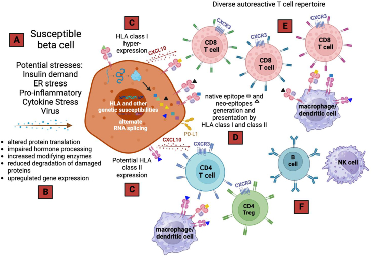
Major conclusions: The last years have seen accelerated development of approaches that allow comprehensive study of human T1D. Critically, recent studies have contributed to our revised understanding that the pancreatic beta cell assumes an active role, rather than a passive position, during autoimmune disease progression. The T cell-beta cell interface is a critical axis that dictates beta cell fate and shapes autoimmune responses. This includes the state of the beta cell after processing internal and external cues (e.g., stress, inflammation, genetic risk) that that contributes to the breaking of tolerance by hyperexpression of human leukocyte antigen (HLA) class I with presentation of native and neoepitopes and secretion of chemotactic factors to attract immune cells. We anticipate that emerging insights about the molecular and cellular aspects of disease initiation and progression processes will catalyze the development of novel and innovative intervention points to provide additional therapies to individuals affected by T1D.
Keywords: Beta cells; Islets; Neoepitopes; T cell infiltrates; Tolerance; Type 1 diabetes.
Copyright © 2023 The Authors. Published by Elsevier GmbH.. All rights reserved.
Conflict of interest statement
Declaration of competing interest AKL has been on a scientific advisory panel for Janssen Research & Development, LLC. AVJ has been a consultant for Pfizer Inc. and has received research funding from Mitsubishi-Tanabe Pharma. EAJ has been a consultant for ProventionBio and has research projects sponsored by Bristol-Myers Squibb and Novartis. HAR has been a SAB member for Prellis Biologics and Sigilon Therapeutics and consultant to Eli Lilly, Minutia, Guidepoint, and Axon. SCK is an SAB member for 4Immune Therapeutics.
Figures

References
-
- Atkinson M.A., Leiter E.H. The NOD mouse model of type 1 diabetes: as good as it gets? Nat Med. 1999;5:601–604. - PubMed
Publication types
MeSH terms
Grants and funding
LinkOut - more resources
Full Text Sources
Medical
Research Materials
