. 2023 Sep 1;37(17-18):801-817.
doi: 10.1101/gad.350814.123. Epub 2023 Sep 21.

Distinct accessory roles of Arabidopsis VEL proteins in Polycomb silencing

Affiliations

Distinct accessory roles of Arabidopsis VEL proteins in Polycomb silencing

Elsa Franco-Echevarría et al. Genes Dev. .

Abstract

Polycomb repressive complex 2 (PRC2) mediates epigenetic silencing of target genes in animals and plants. In Arabidopsis, PRC2 is required for the cold-induced epigenetic silencing of the FLC floral repressor locus to align flowering with spring. During this process, PRC2 relies on VEL accessory factors, including the constitutively expressed VRN5 and the cold-induced VIN3. The VEL proteins are physically associated with PRC2, but their individual functions remain unclear. Here, we show an intimate association between recombinant VRN5 and multiple components within a reconstituted PRC2, dependent on a compact conformation of VRN5 central domains. Key residues mediating this compact conformation are conserved among VRN5 orthologs across the plant kingdom. In contrast, VIN3 interacts with VAL1, a transcriptional repressor that binds directly to FLC These associations differentially affect their role in H3K27me deposition: Both proteins are required for H3K27me3, but only VRN5 is necessary for H3K27me2. Although originally defined as vernalization regulators, VIN3 and VRN5 coassociate with many targets in the Arabidopsis genome that are modified with H3K27me3. Our work therefore reveals the distinct accessory roles for VEL proteins in conferring cold-induced silencing on FLC, with broad relevance for PRC2 targets generally.

Keywords: PRC2; Polycomb silencing; VAL1; VIN3; VRN5; vernalization.


Figures

Figure 1.
Figure 1.
H3K27me2 enrichment at FLC in wt and mutant Arabidopsis. (A) Domain organization of VEL proteins. (B) Enrichment of H3K27me2 levels at FLC in ColFRI and different mutant plants vernalized for 6 wk (6WT0). Data are shown as the percentage input relative to H3. Nontransgenic ColFRI plants were used as a control sample. Error bars are means ± SEM from two independent experiments.
Figure 2.
Figure 2.
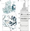
Analysis of the VRN5–PRC2 interaction. (A) Co-IP of HA-tagged PRC2 core components with GFP-tagged VEL proteins following coexpression in HEK293T cells, as indicated. (B) Western blots of α-GFP immunoprecipitates from extracts of vernalized vrn5 mutant plants bearing a VIN3-GFP transgene, probed with α-FIE antibody. (C) Co-IP of HA-tagged PRC2 core components with wt or mutant GFP-VRN5 bearing internal domain deletions (ΔN, ΔPHDs, ΔFN3, Δlinker, and ΔVEL correspond to the deletions of the N-terminal region, PHDsuper, FN3 domain, flexible linker, and polymerizing VEL domain, respectively, as depicted in Fig. 1A) in HEK293T cells. (D) Co-IP of HA-tagged PRC2 core components with wt or GFP-VRN5 mutants in HEK293T cells as described in G. (E) Molecular surface representation of PHDVRN5 predicted by AlphaFold2, colored according to electrostatic potential ([red] negative [blue] positive), showing conserved positively charged residues forming clusters 1 and 2 (front surface) and cluster 3 (rear surface). (F) Sequence logo conservation analysis of VRN5 linker connecting the FN3 and VEL domains in Viridiplantae, with residue numbers shown. (Dashed squares) Three conserved regions (residues 371–391, 420–445, and 493–518 in Arabidopsis) are shown. (G) Schematic representation of different VRN5 linker constructs used in D.
Figure 3.
Figure 3.
Structural differences between VRN5 and VIN3/VEL1 paralogs. (A) Sequence alignment between Arabidopsis thaliana (At) VRN541–339 and At VIN3126–412, with predicted secondary structure indicated for VRN5 (above) and VIN3 (below). Highlighted are the Zn2+-ligating residues of ZnF and PHD finger. (B) PAE plot obtained for PHDsuper and FN3 domains of At VRN541–339, At VIN3126–412, and At VEL1143–462. (Blue) Low error, (red) high error. See also Supplemental Fig. S4. (C) Orthogonal views of superpositions of At VRN541–339 (blue), At VIN3126–412 (yellow), and At VEL1143–462 (brown) in ribbon representation, as predicted by AlphaFold2. The zinc ions are from Pd PHDVIN3 (PDB: 7QCE). (D) Superpositions of PHD superdomains of At VRN541–240 (light blue) and VIN3123–307 (light yellow), as in C, with secondary structure elements indicated (Gray balls) Zinc ions (RMSD 1.70). Close-up view shows superimposed proximal PHD fingers as in D. (E) Co-IP of HA-tagged PRC2 core components with wt or mutant GFP-VRN5 or GFP-VIN3 bearing internal deletions or domain swaps (indicated at the top), as in Figure 2A.
Figure 4.
Figure 4.
Analysis of intramolecular interdomain interactions within VRN5. (A) Structural prediction of At PHDsuper–FN3VRN5 (in ribbon and surface representations), with key structural elements mediating interdomain interactions highlighted. (Light blue) PHDsuper, (teal) FN3 domain. Close-up view of PHDsuper–FN3 interactions, with key interacting residues in stick representation. (Dashed lines) Hydrogen bonds or salt bridges. (B) Co-IP of HA-tagged PRC2 core components with wt or selected interdomain mutants of GFP-VRN5 (indicated at the top), as in Figure 2A.
Figure 5.
Figure 5.
Interaction between VRN5 and the regulatory module of PRC2. (A) Co-IP of different combinations of HA-tagged PRC2 with GFP-VRN5, as in Figure 2A. (B) Co-IP of HA-tagged catalytic or regulatory modules of PRC2 with GFP-VRN5, as in Figure 2A. (N) VRN2 N-lobe, (C) VRN2 C-lobe. (C) Model of the interaction between structure prediction of VRN5 and the At PRC2 complex, with a catalytic module (purple background) comprising SWN (purple), FIE (light purple), and VRN2 C-lobe (light orange) and a regulatory module (light pink) comprising MSI1 (pink) and VRN2 N-lobe (orange).
Figure 6.
Figure 6.
Association between VIN3 and VAL1. (A) Domain architecture of VAL1. (Dark gray) VIN3-interacting CW finger. (BF) Co-IP of GFP-tagged wt or internal deletions of VAL1, with positive controls TPL and BMI1B tagged with FLAG-dsRed (star indicates short exposure) and wt or mutant FLAG-dsRed-tagged VEL proteins, revealing the critical role of VIN3KRFK (Δ506–509) in E; note also that KRFK suffices to confer some VAL1 interaction on VEL1 in F (also see the text). ΔN, ΔPHDs, ΔFN3, Δlinker, and ΔVEL correspond to deletions of the N-terminal region, PHDsuper, FN3, flexible linker, and polymerizing VEL domain, respectively, as depicted in Figure 1A. RR>AD is a polymerization mutant (R554A R556D). (G) VIN3 enrichment at FLC in wt and val1-2 mutant Arabidopsis vernalized for 6 wk (6WT0), with nontransgenic ColFRI as a negative control. Data are shown as percentage input; error bars are means ± SEM from two independent experiments. (H) Model based on AlphaFold2 predictions of the VRN5–PRC2 complex and its interaction with VIN3–VAL1 mediated by a heterotypic VEL–VEL interaction (star), with VAL1 being bound to the nucleation region of FLC (for simplicity, some flexible regions are not shown).
Figure 7.
Figure 7.
Genome-wide occupancy of VEL1, VIN3, and VRN5. (A) Table showing the number of VEL1, VIN3, and VRN5 target genes at NVs and 6WT0. Target genes present in at least two replicates were counted. We called 47,547 and 30,492 VEL1 peaks and assigned them to 11,591 and 8291 genes at NVs and 6WT0, respectively. We identified 175 peaks/137 genes (NVs) and 5672 peaks/2089 genes (6WT0) as potential targets for VIN3, and 6185 peaks/2159 genes (NVs) and 12,133 peaks/3372 genes (6WT0) as potential targets for VRN5. (B) Heat map showing the multiple overlaps among the VEL1, VIN3, and VRN5 at NVs and 6WT0 peaks (left) and the percentage overlap between peaks identified for VEL1, VIN3, and VRN5 at NVs and 6WT0 (right). (C) IGV screenshots showing the colocalization of the VEL proteins at the TSS of RSH3. (D) Metagene plots of VEL distribution over transcription units and flanking regions. (TSS) Transcription start site, (TTS) transcription termination site, (RPGC) reads per genomic content. Inputs from the respective VEL IPs were used as controls. (E) IGV screenshots showing the colocalization of the VEL proteins at the TTS of DOG1. (F) ChIP-qPCR showing enrichment of VRN2 after 6 wk of cold treatment (6WT0) at 10 potential target genes. Data are shown as the percentage input. Nontransgenic ColFRI plants were used as a negative control sample. FLC was used as a positive control locus, and SN1 was used as a negative control locus. Error bars are means ± SEM from two independent experiments. (G) Expression of several potential target genes in the mutant vin3-1 FRI relative to the wt ColFRI at 6WT0. Data are shown normalized to the expression level of the respective gene in ColFRI. Error bars are means ± SEM from at least three biological replicates. (*) P < 0.05, (**) P < 0.01 for statistical tests between samples by Student's t-test.

Similar articles

Cited by

  • Epigenetics in the modern era of crop improvements.
    Xue Y, Cao X, Chen X, Deng X, Deng XW, Ding Y, Dong A, Duan CG, Fang X, Gong L, Gong Z, Gu X, He C, He H, He S, He XJ, He Y, He Y, Jia G, Jiang D, Jiang J, Lai J, Lang Z, Li C, Li Q, Li X, Liu B, Liu B, Luo X, Qi Y, Qian W, Ren G, Song Q, Song X, Tian Z, Wang JW, Wang Y, Wu L, Wu Z, Xia R, Xiao J, Xu L, Xu ZY, Yan W, Yang H, Zhai J, Zhang Y, Zhao Y, Zhong X, Zhou DX, Zhou M, Zhou Y, Zhu B, Zhu JK, Liu Q. Xue Y, et al. Sci China Life Sci. 2025 Jun;68(6):1570-1609. doi: 10.1007/s11427-024-2784-3. Epub 2025 Jan 8. Sci China Life Sci. 2025. PMID: 39808224 Review.
  • Flowering time: From physiology, through genetics to mechanism.
    Maple R, Zhu P, Hepworth J, Wang JW, Dean C. Maple R, et al. Plant Physiol. 2024 Apr 30;195(1):190-212. doi: 10.1093/plphys/kiae109. Plant Physiol. 2024. PMID: 38417841 Free PMC article. Review.
  • Cold-induced nucleosome dynamics linked to silencing of Arabidopsis FLC.
    Montez M, Zhu D, Huertas J, Maristany MJ, Rutjens B, Nielsen M, Collepardo-Guevara R, Dean C. Montez M, et al. Nat Commun. 2025 Jul 1;16(1):5550. doi: 10.1038/s41467-025-60735-z. Nat Commun. 2025. PMID: 40593581 Free PMC article.
  • Targets and mechanisms of epigenetic regulation in the temperate cereal vernalisation process.
    Daneels T, Martinez-Barrales G, Bosmans C, Geuten K. Daneels T, et al. Front Plant Sci. 2025 Aug 5;16:1520593. doi: 10.3389/fpls.2025.1520593. eCollection 2025. Front Plant Sci. 2025. PMID: 40838075 Free PMC article. Review.
  • COOLAIR and PRC2 function in parallel to silence FLC during vernalization.
    Nielsen M, Menon G, Zhao Y, Mateo-Bonmati E, Wolff P, Zhou S, Howard M, Dean C. Nielsen M, et al. Proc Natl Acad Sci U S A. 2024 Jan 23;121(4):e2311474121. doi: 10.1073/pnas.2311474121. Epub 2024 Jan 18. Proc Natl Acad Sci U S A. 2024. PMID: 38236739 Free PMC article.

References

    1. Alabert C, Barth TK, Reverón-Gómez N, Sidoli S, Schmidt A, Jensen ON, Imhof A, Groth A. 2015. Two distinct modes for propagation of histone PTMs across the cell cycle. Genes Dev 29: 585–590. 10.1101/gad.256354.114 - DOI - PMC - PubMed
    1. Baile F, Merini W, Hidalgo I, Calonje M. 2021. EAR domain-containing transcription factors trigger PRC2-mediated chromatin marking in Arabidopsis. Plant Cell 33: 2701–2715. 10.1093/plcell/koab139 - DOI - PMC - PubMed
    1. Batth TS, Tollenaere MAX, Rüther P, Gonzalez-Franquesa A, Prabhakar BS, Bekker-Jensen S, Deshmukh AS, Olsen JV. 2019. Protein aggregation capture on microparticles enables multipurpose proteomics sample preparation. Mol Cell Proteomics 18: 1027–1035. 10.1074/mcp.TIR118.001270 - DOI - PMC - PubMed
    1. Berry S, Dean C, Howard M. 2017. Slow chromatin dynamics allow Polycomb target genes to filter fluctuations in transcription factor activity. Cell Syst 4: 445–457.e8. 10.1016/j.cels.2017.02.013 - DOI - PMC - PubMed
    1. Bienz M. 2014. Signalosome assembly by domains undergoing dynamic head-to-tail polymerization. Trends Biochem Sci 39: 487–495. 10.1016/j.tibs.2014.08.006 - DOI - PubMed

Publication types

MeSH terms

LinkOut - more resources