. 2023 Dec;64(12):3377-3388.
doi: 10.1111/epi.17776. Epub 2023 Oct 17.

GRIN1 variants associated with neurodevelopmental disorders reveal channel gating pathomechanisms

Affiliations

GRIN1 variants associated with neurodevelopmental disorders reveal channel gating pathomechanisms

Lotten Ragnarsson et al. Epilepsia. 2023 Dec.

Abstract

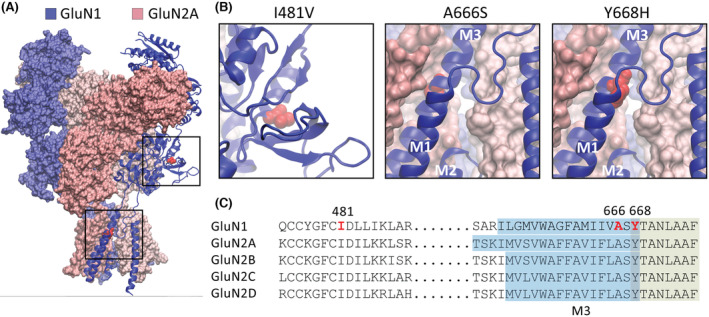
Objective: N-methyl-d-aspartate (NMDA) receptors are expressed at synaptic sites, where they mediate fast excitatory neurotransmission. NMDA receptors are critical to brain development and cognitive function. Natural variants to the GRIN1 gene, which encodes the obligatory GluN1 subunit of the NMDA receptor, are associated with severe neurological disorders that include epilepsy, intellectual disability, and developmental delay. Here, we investigated the pathogenicity of three missense variants to the GRIN1 gene, p. Ile148Val (GluN1-3b[I481V]), p.Ala666Ser (GluN1-3b[A666S]), and p.Tyr668His (GluN1-3b[Y668H]).

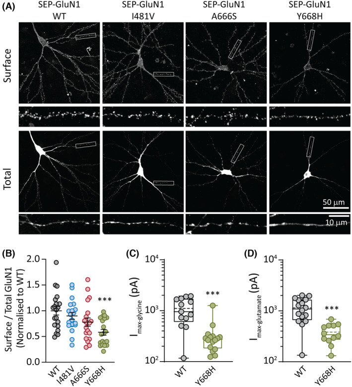
Methods: Wild-type and variant-containing NMDA receptors were expressed in HEK293 cells and primary hippocampal neurons. Patch-clamp electrophysiology and pharmacology were used to profile the functional properties of the receptors. Receptor surface expression was evaluated using fluorescently tagged receptors and microscopy.

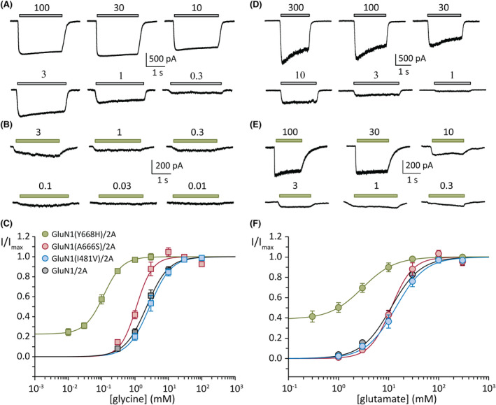
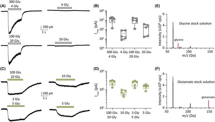
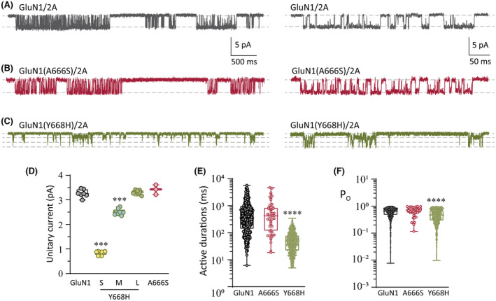
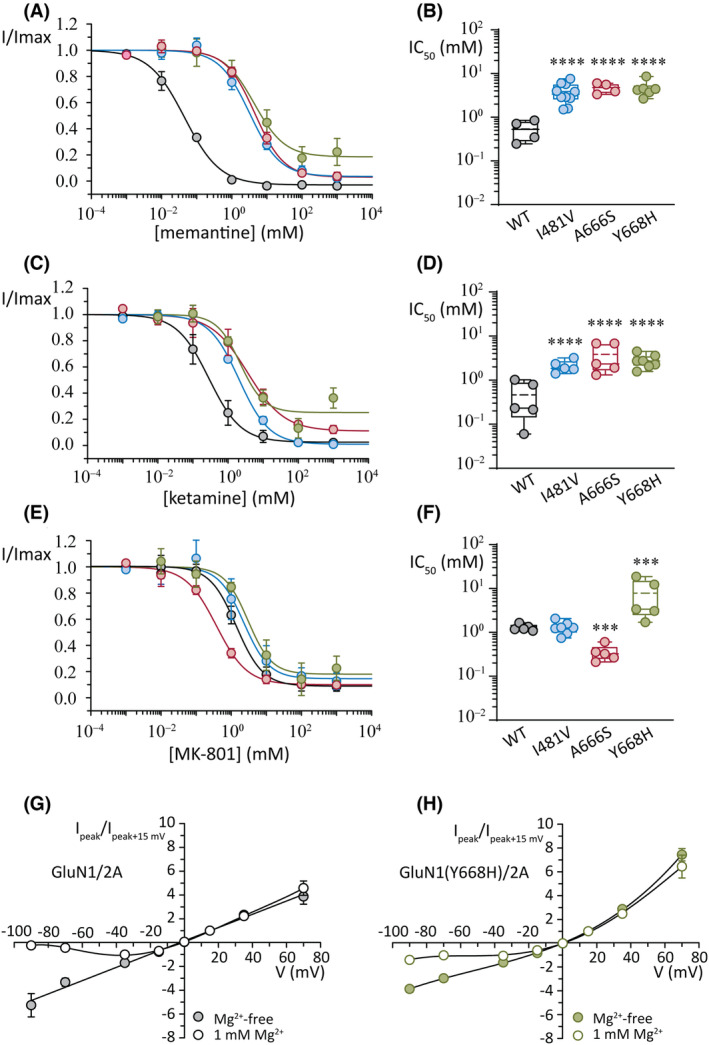
Results: Our data demonstrate that the GluN1(I481V) variant is inhibited by the open pore blockers ketamine and memantine with reduce potency but otherwise has little effect on receptor function. By contrast, the other two variants exhibit gain-of-function molecular phenotypes. Glycine sensitivity was enhanced in receptors containing the GluN1(A666S) variant and the potency of pore block by memantine and ketamine was reduced, whereas that for MK-801 was increased. The most pronounced functional deficits, however, were found in receptors containing the GluN1(Y668H) variant. GluN1(Y668H)/2A receptors showed impaired surface expression, were more sensitive to glycine and glutamate by an order of magnitude, and exhibited impaired block by extracellular magnesium ions, memantine, ketamine, and MK-801. These variant receptors were also activated by either glutamate or glycine alone. Single-receptor recordings revealed that this receptor variant opened to several conductance levels and activated more frequently than wild-type GluN1/2A receptors.

Significance: Our study reveals a critical functional locus of the receptor (GluN1[Y668]) that couples receptor gating to ion channel conductance, which when mutated may be associated with neurological disorder.

Keywords: NMDA receptors; developmental delay; epilepsy.


Conflict of interest statement

The authors report no competing interests. We confirm that we have read the Journal's position on issues involved in ethical publication and affirm that this report is consistent with those guidelines.

Figures

FIGURE 1
FIGURE 1
GluN1 missense variants. (A) Schematic representation of the GluN1/2A N‐methyl‐d‐aspartate receptor in the agonist‐bound state showing the overall subunit arrangement and the position of the three GluN1 variants. (B) Expanded views of the boxed areas in A showing the amino acid substitutions as red spheres. The schematics were made in Visual Molecular Dynamics software using the 7EOS PDB file. (C) Segments of amino acid sequences that encompass the three variants showing the sequence identity between the GluN1 and GluN2 subunits. The substituted positions in the GluN1‐3b subunit are shown in red. The M3 transmembrane domain is highlighted in blue, and the contiguous segment that is a key signal transduction element is highlighted in green.
FIGURE 2
FIGURE 2
GluN1 variant surface expression. (A) Rat hippocampal neurons transfected with plasmids encoding superecliptic pHluorin (SEP)‐GluN1 (wild‐type [WT]) and the three variant subunits SEP‐GluN1(I481V), SEP‐GluN1(A666S), and SEP‐GluN1(Y668H). Representative images are shown of the surface and total SEP‐GluN1 in a neuron from each group, together with expanded views of the boxed regions, shown below. (B) Quantification of surface expression of the surface/total GluN1 ratio normalized to the value of control neurons expressing SEP‐GluN1 WT. Data are presented as mean ± SEM (WT, n = 20 neurons; I481V, n = 20; A666S, n = 20; Y668H, n = 20; from three independent cultures). (C) Quantification of peak whole‐cell currents mediated by the indicated receptors in response to saturating concentrations of glycine (and half‐maximal effective concentration [EC50] of glutamate). (D) Quantification of peak whole‐cell currents in response to saturating concentrations of glutamate (and an EC50 concentration of glycine). ***p < .001.
FIGURE 3
FIGURE 3
Concentration–response plots of glycine and glutamate. (A, B) Representative whole‐cell currents in response to the indicated micromolar concentrations of glycine (and half‐maximal effective concentration [EC50] of glutamate) from cells expressing the wild‐type (WT) GluN1/2A (A) and GluN1(Y668H)/2A variant (B) receptors. (C) Normalized glycine concentration–response plots of group data for the indicated WT and variant receptors. (D, E) Representative whole‐cell currents in response to the indicated micromolar concentrations of glutamate (and an EC50 concentration of glycine) from cells expressing the WT GluN1/2A (D) and GluN1(Y668H)/2A variant (E) receptors. (C) Normalized glutamate concentration–response plots of group data for the indicated WT and variant receptors. Currents were obtained at a holding potential of −70 mV.
FIGURE 4
FIGURE 4
Activation of GluN1(Y668H)/2A by single neurotransmitter. (A) Representative whole‐cell currents in response to the indicated micromolar concentrations of agonists (left) and single agonist (right) for wild‐type (WT) GluN1/2A receptors. (B) Quantification of peak currents from group data showing peak currents from agonist combinations compared to single agonists for WT receptors. (C) Representative whole‐cell currents in response to the indicated micromolar concentrations of agonists (left) and a single agonist (right) for GluN1(Y668H)/2A variant receptors. (D) Quantification of peak currents from group data showing peak currents from agonist combinations compared to single agonists for GluN1(Y668H)/2A variant receptors. (E, F). Mass spectra of the agonist stock solutions used in this study. The glycine stock (E) shows a clear peak at the glycine molecular mass of 75.07 g mol−1 (red peak), whereas the glutamate stock (F) shows a peak at the molecular mass of glutamate of 147.13 g mol−1 (red peak). Note that the plots in E and F show additional peaks that correspond to other masses in the extracellular solution.
FIGURE 5
FIGURE 5
Single‐receptor currents. (A–C) Representative single‐receptor currents recorded from excised outside‐out membrane patches expressing the WT GluN1/2A (A), GluN1(A666S)/2A (B), and GluN1(Y668H)/2A (C). Expanded views of single‐receptor activity are shown on the left. (D) Quantification of unitary current for the indicated receptors. Note that the GluN1(Y668H)/2A variant receptors have a wild‐type‐like amplitude (large [L]) and two smaller amplitudes (small [S] and medium [M]). (E) Plots of discrete activations (active periods) of single‐receptor current for the indicated receptors. (F) Plots of the open state occupancy (PO) within active periods for the indicated receptors. ***p < .001, ****p < .0001.
FIGURE 6
FIGURE 6
Effects of open channel blockers. (A, B) Concentration–response plots for peak current (A) and summary of the half‐maximal inhibitory concentration (IC50) values (B) for the inhibition by memantine. (C, D) Concentration–response plots for peak current (C) and summary of the IC50 values (D) for the inhibition by ketamine. (E, F) Concentration–response plots for peak current (E) and summary of the IC50 values (F) for the inhibition by MK‐801. Currents were obtained at a holding potential of −100 mV. (G) Normalized whole‐cell current–voltage plots for wild‐type (WT) GluN1/2A receptors in the absence (filled circles) and presence (open circles) of extracellular Mg2+ ions (1 mmol·L−1). Note the block of current at negative voltages in 1 mmol·L−1 Mg2+. (H) Normalized whole‐cell current–voltage plots for GluN1(Y668H)/2A variant receptors in the absence (filled circles) and presence (open circles) of extracellular Mg2+ ions (1 mmol·L−1). Note the impaired block at negative voltages in 1 mmol·L−1 Mg2+ and the greater relative outward current compared to WT receptors. Currents were normalized to +15 mV. ***p < .001, ****p < .0001.

References

    1. Wang H, Lv S, Stroebel D, Zhang J, Pan Y, Huang X, et al. Gating mechanism and a modulatory niche of human GluN1–GluN2A NMDA receptors. Neuron. 2021;109:2443–2456.e2445. 10.1016/j.neuron.2021.05.031 - DOI - PubMed
    1. Paoletti P, Bellone C, Zhou Q. NMDA receptor subunit diversity: impact on receptor properties, synaptic plasticity and disease. Nat Rev Neurosci. 2013;14:383–400. 10.1038/nrn3504 - DOI - PubMed
    1. Traynelis SF, Wollmuth LP, McBain CJ, Menniti FS, Vance KM, Ogden KK, et al. Glutamate receptor ion channels: structure, regulation, and function. Pharmacol Rev. 2010;62:405–496. 10.1124/pr.109.002451 - DOI - PMC - PubMed
    1. Ascher P, Nowak L. The role of divalent cations in the N‐methyl‐d‐aspartate responses of mouse central neurones in culture. J Physiol. 1988;399:247–266. - PMC - PubMed
    1. Burnashev N, Schoepfer R, Monyer H, Ruppersberg JP, Günther W, Seeburg PH, et al. Control by asparagine residues of calcium permeability and magnesium blockade in the NMDA receptor. Science. 1992;257:1415–1419. - PubMed