. 2023 Sep 21;21(1):340.
doi: 10.1186/s12951-023-02098-5.

Hypoxic mesenchymal stem cell-derived exosomes promote the survival of skin flaps after ischaemia-reperfusion injury via mTOR/ULK1/FUNDC1 pathways

Affiliations

Hypoxic mesenchymal stem cell-derived exosomes promote the survival of skin flaps after ischaemia-reperfusion injury via mTOR/ULK1/FUNDC1 pathways

Chao Deng et al. J Nanobiotechnology. .

Abstract

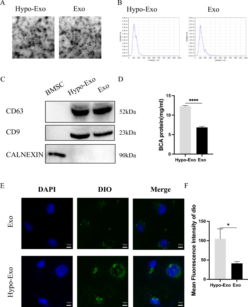
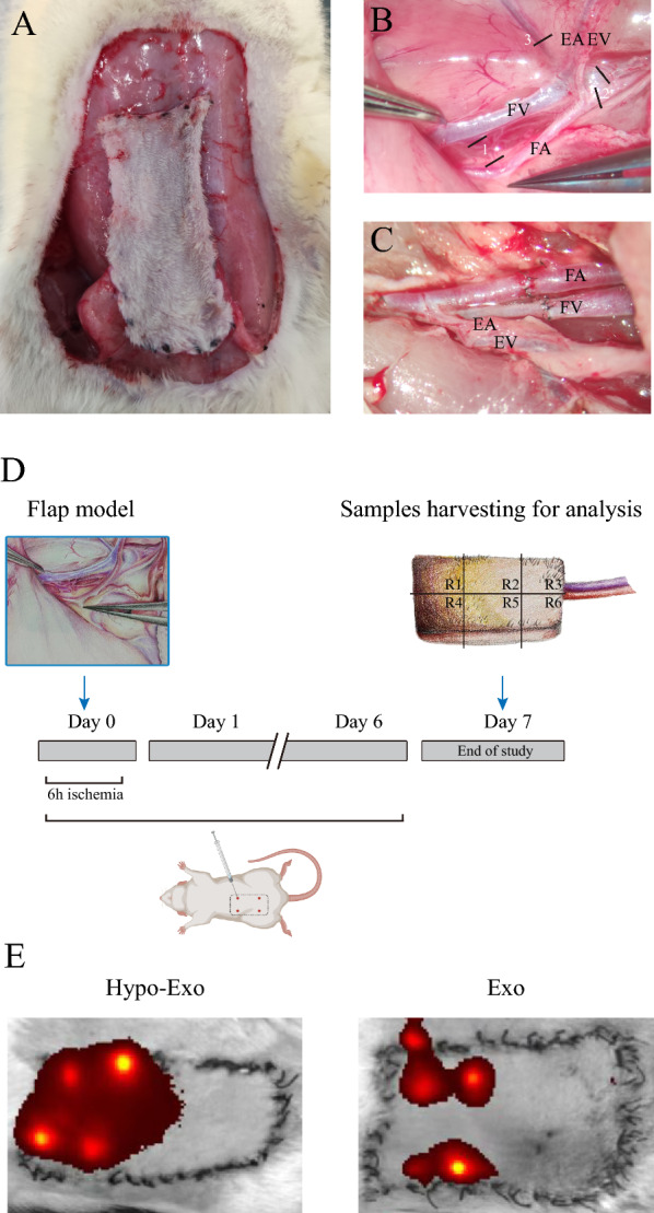
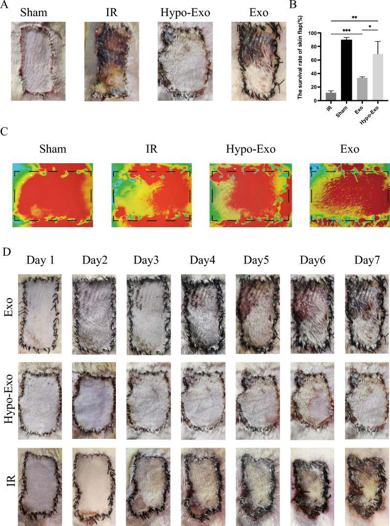
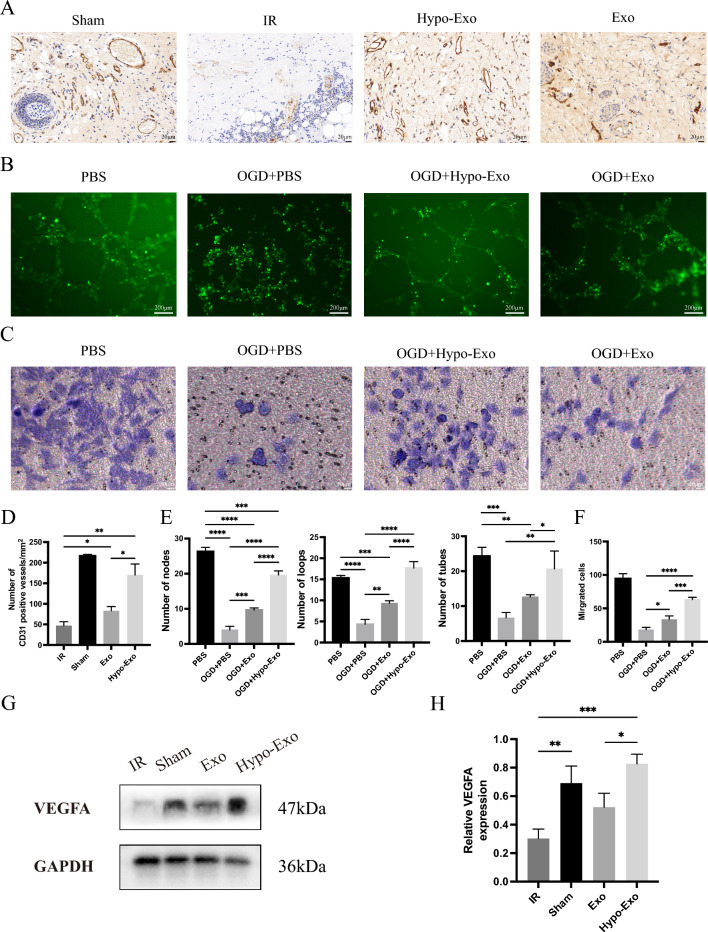
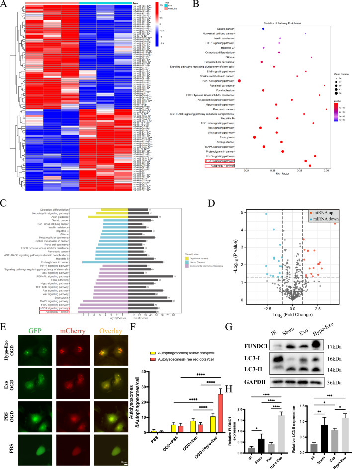
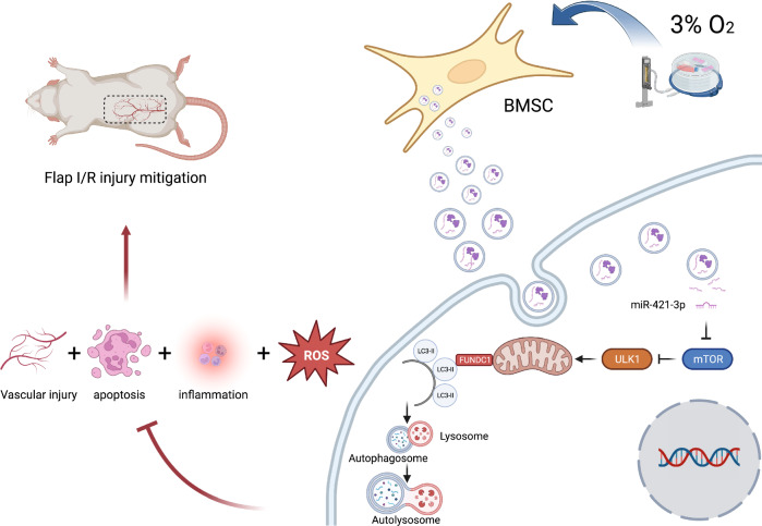
Flap necrosis, the most prevalent postoperative complication of reconstructive surgery, is significantly associated with ischaemia-reperfusion injury. Recent research indicates that exosomes derived from bone marrow mesenchymal stem cells (BMSCs) hold potential therapeutic applications in several diseases. Traditionally, BMSCs are cultured under normoxic conditions, a setting that diverges from their physiological hypoxic environment in vivo. Consequently, we propose a method involving the hypoxic preconditioning of BMSCs, aimed at exploring the function and the specific mechanisms of their exosomes in ischaemia-reperfusion skin flaps. This study constructed a 3 × 6 cm2 caudal superficial epigastric skin flap model and subjected it to ischaemic conditions for 6 h. Our findings reveal that exosomes from hypoxia-pretreated BMSCs significantly promoted flap survival, decrease MCP-1, IL-1β, and IL-6 levels in ischaemia-reperfusion injured flap, and reduce oxidative stress injury and apoptosis. Moreover, results indicated that Hypo-Exo provides protection to vascular endothelial cells from ischaemia-reperfusion injury both in vivo and in vitro. Through high-throughput sequencing and bioinformatics analysis, we further compared the differential miRNA expression profiles between Hypo-Exo and normoxic exosomes. Results display the enrichment of several pathways, including autophagy and mTOR. We have also elucidated a mechanism wherein Hypo-Exo promotes the survival of ischaemia-reperfusion injured flaps. This mechanism involves carrying large amounts of miR-421-3p, which target and regulate mTOR, thereby upregulating the expression of phosphorylated ULK1 and FUNDC1, and subsequently further activating autophagy. In summary, hypoxic preconditioning constitutes an effective and promising method for optimizing the therapeutic effects of BMSC-derived exosomes in the treatment of flap ischaemia-reperfusion injury.

Keywords: 3-MA; Autophagy; Bone marrow mesenchymal stem cells; Exosomes; Hypoxia; Ischaemia–reperfusion; Skin flap; miRNA.


Conflict of interest statement

The authors declare that they have no competing interests.

Figures

Fig. 1
Fig. 1
Characterization of Exosomes. A Morphology of Hypo-Exo and Exo under TEM. B NTA particle analysis of Hypo-Exo and Exo, two groups share similar size ranges (40–150 nm). C Western blotting analysis showing the the expression of CD63, CD9 and CALENXIN. D The BCA assay was used to measure the exosome protein concentration in the two groups (****P < 0.0001). E Uptake of the green fluorescence dye Dio labelled Hypo-Exo and Exo into HUVECs (bar size 10 μm). F Statistical evaluation of Mean fluorescence intensities in the two groups (*P < 0.05). Bars indicated means ± SD
Fig. 2
Fig. 2
Flap model, Schematic diagram of surgery and experimental design. A The caudal superficial epigastric skin free flap (3 × 6 cm2). B Intraoperative Vascular Anatomy: EA epigastric artery, EV epigastric vein, FA femoral artery, FV femoral vein, 1 (ligatures of lateral circumflex FA and FV), 2 (ligatures of saphenous artery and vein), 3 (ligatures of proximal caudal FA and FV). C Anastomose the femoral artery and femoral vein with 10–0 nylon suture. D Experimental process design. E In vivo tracking of DiR-labeled exosomes
Fig. 3
Fig. 3
Flap appearance and survival rate. A The skin flap appearance of each group on the 7th day after operation. B The flap survival rate of each group on the 7th day after operation (*P < 0.05, **P < 0.01, ***P < 0.001). C Angiography images of each group on the 7th day after operation. D Changes of skin flap appearance in each group from the 1st day to the 7th day after operation. Bars indicated means ± SD
Fig. 4
Fig. 4
Hypo-Exo improved the pathological state of flaps after I/R injury, inhibited apoptosis, reduced the release of ROS in flap and reduced the production of inflammatory factors. A Representative histology images (Original magnification 40X, bar size 20 μm). B Detection of apoptosis by TUNEL assay (bar size 100 μm). C Double immunofluorescence staining of Dihydroethidium (DHE)-ROS (red) and DAPI (bule) in flap.D Relative quantitative data of apoptotic cells and TUNEL cells (**P < 0.01, ****P < 0.0001). E Statistical evaluation of ROS mean fluorescence intensities in each group (*P < 0.05, **P < 0.01). F Protein expression levels of Bax, Bcl-2 and GAPDH. G Quantitative analysis of the protein expression levels of Bax/Bcl-2 (*P < 0.05). H Relative MCP-1, IL-6, IL-1β mRNA expression level in skin flap was measured by qPCR in Sham, IR, Hypo-Exo and Exo groups (*P < 0.05, ***P < 0.001, ****P < 0.0001), Bars indicated means ± SD
Fig. 5
Fig. 5
Hypo-Exo protected vascular endothelial cells from I/R damage in vivo and vitro. A CD31 immunohistochemical detection in each group (Original magnification 40X, bar size 20 μm). B Representative images showing tube formation in HUVECs treated with PBS, OGD + PBS, OGD + Hypo-Exo or OGD + Exo (bar size 200 μm). C Representative images showing migrated HUVECs by transwell assay treated with PBS, OGD + PBS, OGD + Hypo-Exo or OGD + Exo (bar size 50 μm). D The number of CD31 positive blood vessels in each group (**P < 0.01, *P < 0.05). E Quantitative data of tube formation using Image J (*P < 0.05, **P < 0.01, ***P < 0.001, ****P < 0.0001). F Quantitative data of the migrated cells (*P < 0.05, ***P < 0.001, ****P < 0.0001) G, H Western blot shows the expression of VEGF-A in flap treated with each group, and quantitative data of western blot (*P < 0.05, **P < 0.01, ***P < 0.001), Bars indicated means ± SD
Fig. 6
Fig. 6
High-throughput sequencing analysis of miRNAs in Hypo-Exo and Exo. A Clustered heat map of differentially expressed miRNAs depicting up and down regulated miRNAs. B, C GO and KEGG enrichment of differentially expressed miRNAs. D Volcano plot of differentially expressed miRNAs depicting up and down regulated miRNAs. E Autophagosomes and autolysosomes in each group (bar size 10 μm). F Quantitative analysis of autophagosomes and autolysosomes in each group (****P < 0.0001). G, H Western blot shows the expression of FUNDC1, LC3-I, LC3-II in flap treated with each group, and quantitative data of western blot analysis (*P < 0.05, **P < 0.01, ***P < 0.001, ****P < 0.0001), Bars indicated means ± SD
Fig. 7
Fig. 7
3-MA inhibit autophagy reversed the biological effects promoted by Hypo-Exo in vivo and vitro. A Representative skin flap appearance of each group on the7th day after operation. B Representative histology images of each group on the 7th day after operation C CD31 immunohistochemical detection in each group (Original magnification 40X, bar size 20 μm) D Quantitative data of CD31 positive blood vessels in each group (**P < 0.01) E Double immunofluorescence staining of Dihydroethidium (DHE)-ROS (red) and DAPI (bule) in each group. F Statistical evaluation of Mean fluorescence intensities in each group (**P < 0.01). G Double immunofluorescence staining of TUNEL (green) and DAPI (bule) in each group. H Quantitative data of apoptotic cells and TUNEL cells (**P < 0.01). I, J Western blot analysis of the expression of proteins in flap, and quantitative data of western blot (*P < 0.05). K Representative images showing tube formation in HUVECs treated with OGD + Hypo-Exo, OGD + Hypo-Exo + 3-MA (bar size 200 μm). L Representative images showing migrated HUVECs by transwell assay treated with OGD + Hypo-Exo, OGD + Hypo-Exo + 3-MA (bar size 50 μm). M Quantitative data of tube formation using ImageJ (*P < 0.05, ** P < 0.01). N Quantitative data of the migrated cells (**P < 0.01), Bars indicated means ± SD
Fig. 8
Fig. 8
Hypo-Exo enhances autophagy by reducing mTOR protein expression by delivering miR-421-3p. A Network diagram for predicting miRNA target genes. B Bioinformatics analysis predicted miRNAs that were potentially capable of binding to mTOR. C qPCR analysis revealed the expression levels of miR-421-3p, miR-199a-3p, miR-505-3p, and miR-195-5p in Hypo-Exo and Exo (*P < 0.05). D schematic diagram illustrating the predicted binding sites of miR-421-3p in the 3'UTR of mTOR mRNA. E Dual-Luciferase Reporter System was used to detect luciferase activity. (***P < 0.001). F qPCR analysis revealed the expression level of miR-421-3p in each group (****P < 0.0001). G qPCR analysis revealed miR-421-3p expression levels in HUVECs after transfer with miR-421-3p inhibitors and NC (*P < 0.05). H, I Western blot shows the expression of mTOR, p-mTOR, ULK1, p-ULK1, P62, LC3-I, LC3-II, FUNDC1, p-FUNDC1 in each group, and quantitative data of western blot (*P < 0.05, **P < 0.01, ***P < 0.001), Bars indicated means ± SD
Fig. 9
Fig. 9
Antagomir-421-3p abolishes the therapeutic effect of Hypo-Exo. A Representative skin flap appearance of each group on the7th day after operation. B The flap survival rate of each group on the 7th day after operation (*P < 0.05, **P < 0.01). C Representative histology images (Original magnification 40X, bar size 20 μm). D CD31 immunohistochemical detection in each group (Original magnification 40X, bar size 20 μm). E Double immunofluorescence staining of mTOR(red) and DAPI (bule) in each group (bar size 50 μm). F Quantitative data of CD31 positive blood vessels in each group (***P < 0.001, ****P < 0.0001). G Statistical evaluation of Mean fluorescence intensities in each group (*P < 0.05). Bars indicated means ± SD
Fig. 10
Fig. 10
Schematic Diagram: Hypo-Exo, carrying miR-421-3p, activates autophagy by targeting mTOR and upregulating the expression of phosphorylated ULK1 and FUNDC1. This in turn mitigates the release of inflammatory factors in the skin flap, reduces ROS generation, counteracts cell apoptosis, and protects blood vessels, ultimately promoting the survival of ischemia–reperfusion flaps

References

    1. Varghese BT, Vijayakumar P, Ani Raj R, Thomas S. Salvaging skin loss of free fibular osteo-cutaneous flaps in oral oncological reconstruction. Oral Oncol. 2019;97:131–132. - PubMed
    1. Celik N, Wei F-C, Lin C-H, Cheng M-H, Chen H-C, Jeng S-F, Kuo Y-R. Technique and strategy in anterolateral thigh perforator flap surgery, based on an analysis of 15 complete and partial failures in 439 cases. Plast Reconstr Surg. 2002;109:2211–2216. - PubMed
    1. Geng L, Zhang G, Yao M, Fang Y. Rip 1-dependent endothelial necroptosis participates in ischemia-reperfusion injury of mouse flap. J Dermatol Sci. 2020;97:30–40. - PubMed
    1. Luo X, Zhao B, Chu T, Chen H, Li B, Li Z, Yan H. Improvement of multiterritory perforator flap survival supported by a hybrid perfusion mode: a novel strategy and literature review. J Tissue Viability. 2021;30:276–281. - PubMed
    1. Harder Y, Amon M, Laschke MW, et al. An old dream revitalised: preconditioning strategies to protect surgical flaps from critical ischaemia and ischaemia-reperfusion injury. J Plast Reconstr Aesthet Surg. 2008;61:503–511. - PubMed

LinkOut - more resources