Review
. 2023 Sep 21;18(1):64.
doi: 10.1186/s13024-023-00655-y.

Retinal ganglion cell repopulation for vision restoration in optic neuropathy: a roadmap from the RReSTORe Consortium

Collaborators, Affiliations
Review

Retinal ganglion cell repopulation for vision restoration in optic neuropathy: a roadmap from the RReSTORe Consortium

Jonathan R Soucy et al. Mol Neurodegener. .

Abstract

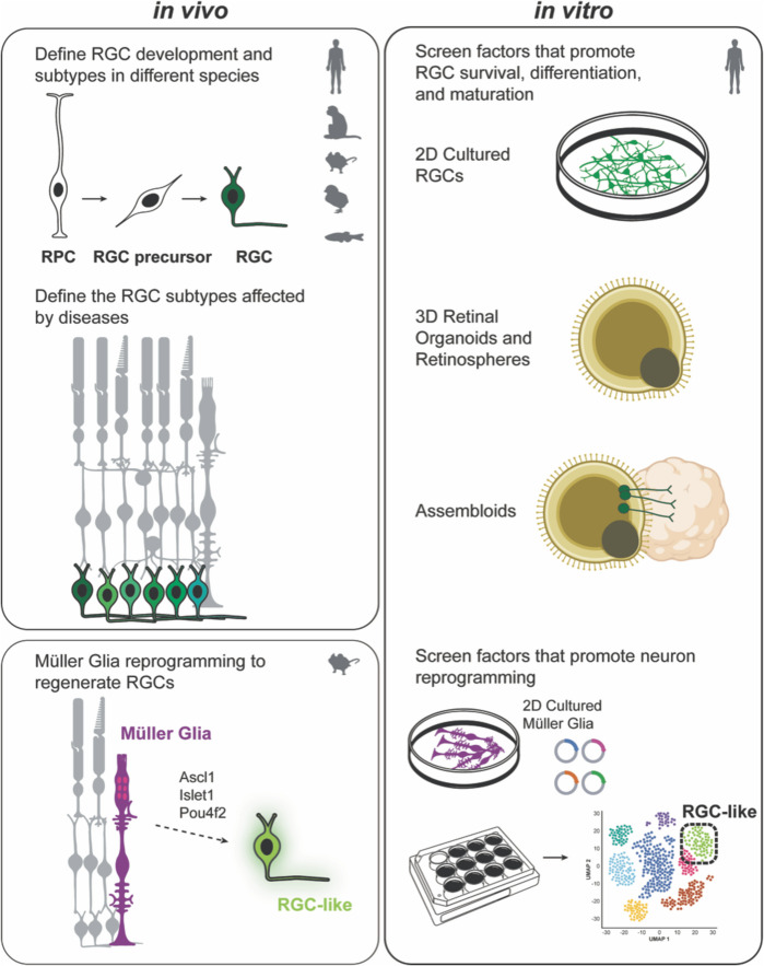
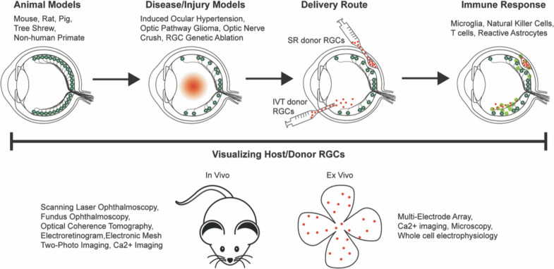
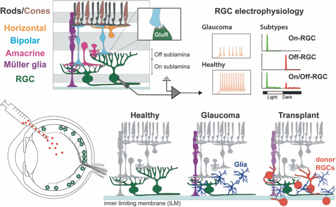
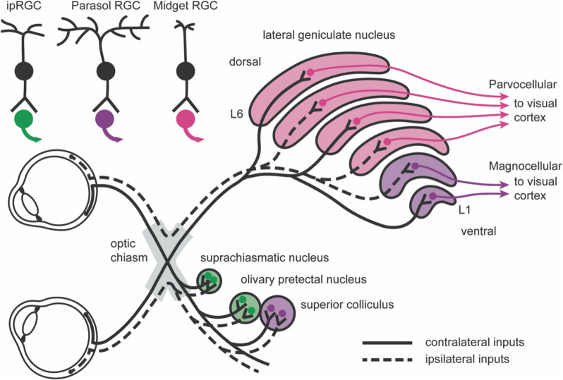
Retinal ganglion cell (RGC) death in glaucoma and other optic neuropathies results in irreversible vision loss due to the mammalian central nervous system's limited regenerative capacity. RGC repopulation is a promising therapeutic approach to reverse vision loss from optic neuropathies if the newly introduced neurons can reestablish functional retinal and thalamic circuits. In theory, RGCs might be repopulated through the transplantation of stem cell-derived neurons or via the induction of endogenous transdifferentiation. The RGC Repopulation, Stem Cell Transplantation, and Optic Nerve Regeneration (RReSTORe) Consortium was established to address the challenges associated with the therapeutic repair of the visual pathway in optic neuropathy. In 2022, the RReSTORe Consortium initiated ongoing international collaborative discussions to advance the RGC repopulation field and has identified five critical areas of focus: (1) RGC development and differentiation, (2) Transplantation methods and models, (3) RGC survival, maturation, and host interactions, (4) Inner retinal wiring, and (5) Eye-to-brain connectivity. Here, we discuss the most pertinent questions and challenges that exist on the path to clinical translation and suggest experimental directions to propel this work going forward. Using these five subtopic discussion groups (SDGs) as a framework, we suggest multidisciplinary approaches to restore the diseased visual pathway by leveraging groundbreaking insights from developmental neuroscience, stem cell biology, molecular biology, optical imaging, animal models of optic neuropathy, immunology & immunotolerance, neuropathology & neuroprotection, materials science & biomedical engineering, and regenerative neuroscience. While significant hurdles remain, the RReSTORe Consortium's efforts provide a comprehensive roadmap for advancing the RGC repopulation field and hold potential for transformative progress in restoring vision in patients suffering from optic neuropathies.

Keywords: Glaucoma; Neuroprotection; Ophthalmology; Optic neuropathy; Organoids; Regenerative medicine; Retinal ganglion cells; Stem cells; Transplantation.


Conflict of interest statement

The authors declare that they have no competing interests.

Figures

Fig. 1
Fig. 1
RGC development, subtype specification, differentiation, and regeneration. Retinal progenitor cells (RPC), RGC precursors, and mature RGCs can be defined and isolated from various species. Neuronal replacement therapies will be advanced by defining the RGC subtypes affected by various optic neuropathies and developing methods to target donor RGC maturation into specific subtypes. In vitro systems serve as a source of RGCs for cell replacement therapies and are useful for screening factors that promote RGC differentiation, maturation, and survival. RGCs can be cultured in monolayers, 3D retinal organoids, retinospheres, and assembloids. Through direct reprogramming in vivo and in vitro, Müller glia can also be a source for newborn mammalian RGCs, and factors that promote neuron reprogramming can be identified. While RGCs can be isolated from various species, RGC repopulation through reprogramming is currently only studied in mice, but in vitro studies can be performed using human samples
Fig. 2
Fig. 2
RGC transplantation models, methods, and assessment. Each animal and disease/injury model possesses advantages and disadvantages for studying essential aspects of RGC replacement and mimicking different characteristics of optic neuropathies. Donor RGCs can be delivered to the intravitreal (IVT) or subretinal (SR) space, but each route has unique barriers to overcome to achieve structural integration. In addition to integrating within the host retina, donor RGCs must avoid being targeted by the adaptive and innate immune systems. Visualizing donor and host RGCs is essential to translate cell replacement therapies to the clinic, and a combination of techniques is required to properly assess the structural and functional integration of the transplanted cells
Fig. 3
Fig. 3
RGC neurocircuitries in healthy, diseased, and transplanted retinas. Bipolar and amacrine cells establish direct contact with RGCs to relay visual information. Different RGC subtypes extend their dendrites into ON and OFF sublamina in the inner plexiform layer and exhibit different electrophysiological responses. Glaucoma causes dendrite retraction and eventual death of RGCs and the activation of astrocytes, microglia, and Müller glia, while photoreceptor, bipolar, amacrine, and horizontal cells are relatively unaffected. RGC transplantation must replace lost RGCs, return the diseased retina to a homeostatic state, and establish neurocircuitry between host and donor cells. While donor RGCs have been shown to survive in the retina, few are currently able to migrate into the ganglion cell layer, with the inner limiting membrane (ILM) serving as a major barrier for intravitreal (IVT) delivery, and even fewer form de novo neurocircuits in the retina
Fig. 4
Fig. 4
Retinal ganglion cell (RGC) pathways in the human brain. Visual information travels from each retina through the optic nerve and converges at the optic chiasm. Uncrossed ipsilateral inputs connect to L2, L3, and L5 in the lateral geniculate nucleus, whereas crossed contralateral inputs connect to L1, L4, and L6 in the lateral geniculate nucleus. Both ipsilateral and contralateral inputs connect to the suprachiasmatic nucleus, olivary pretectal nucleus, and superior colliculus. Intrinsically photosensitive RGCs (ipRGCs), among others, connect to the neurons in the suprachiasmatic nucleus and olivary pretectal nucleus (green) to regulate circadian rhythms and the pupillary light reflex, respectively. Parasol RGCs, among others, connect to the neurons in the superior colliculus (purple) to orient head and eye movements and to L1 and L2 in the lateral geniculate nucleus as a part of the magnocellular pathway (purple). Midget RGCs, among others, connect to the neurons in L3, L4, L5, and L6 of the lateral geniculate nucleus as a part of the parvocellular pathway (pink). The magnocellular and parvocellular pathways connect to the visual cortex to primarily process motion and high-contrast information, respectively

References

    1. Goldberg JL, Espinosa JS, Xu Y, Davidson N, Kovacs GTA, Barres BA. Retinal ganglion cells do not extend axons by default promotion by neurotrophic signaling and electrical activity. Neuron. 2002;33:689–702. - PubMed
    1. Carelli V, Morgia CL, Ross-Cisneros FN, Sadun AA. Optic neuropathies: the tip of the neurodegeneration iceberg. Hum Mol Genet. 2017;26:R139–50. - PMC - PubMed
    1. Gokoffski KK, Peng M, Alas B, Lam P. Neuro-protection and neuro-regeneration of the optic nerve: recent advances and future directions. Curr Opin Neurol. 2020;33:93–105. - PMC - PubMed
    1. Sharma R, Bose D, Maminishkis A, Bharti K. Retinal pigment epithelium replacement therapy for age-related macular degeneration: are we there yet? Annu Rev Pharmacol. 2020;60:553–72.
    1. Patterson SS, Bembry BN, Mazzaferri MA, Neitz M, Rieke F, Soetedjo R, et al. Conserved circuits for direction selectivity in the primate retina. Curr Biol. 2022;32:2529-2538.e4. - PMC - PubMed

Publication types