. 2023 Sep 21;14(1):14.
doi: 10.1186/s13227-023-00218-8.

Nematostella vectensis exemplifies the exceptional expansion and diversity of opsins in the eyeless Hexacorallia

Affiliations

Nematostella vectensis exemplifies the exceptional expansion and diversity of opsins in the eyeless Hexacorallia

Kyle J McCulloch et al. Evodevo. .

Abstract

Background: Opsins are the primary proteins responsible for light detection in animals. Cnidarians (jellyfish, sea anemones, corals) have diverse visual systems that have evolved in parallel with bilaterians (squid, flies, fish) for hundreds of millions of years. Medusozoans (e.g., jellyfish, hydroids) have evolved eyes multiple times, each time independently incorporating distinct opsin orthologs. Anthozoans (e.g., corals, sea anemones,) have diverse light-mediated behaviors and, despite being eyeless, exhibit more extensive opsin duplications than medusozoans. To better understand the evolution of photosensitivity in animals without eyes, we increased anthozoan representation in the phylogeny of animal opsins and investigated the large but poorly characterized opsin family in the sea anemone Nematostella vectensis.

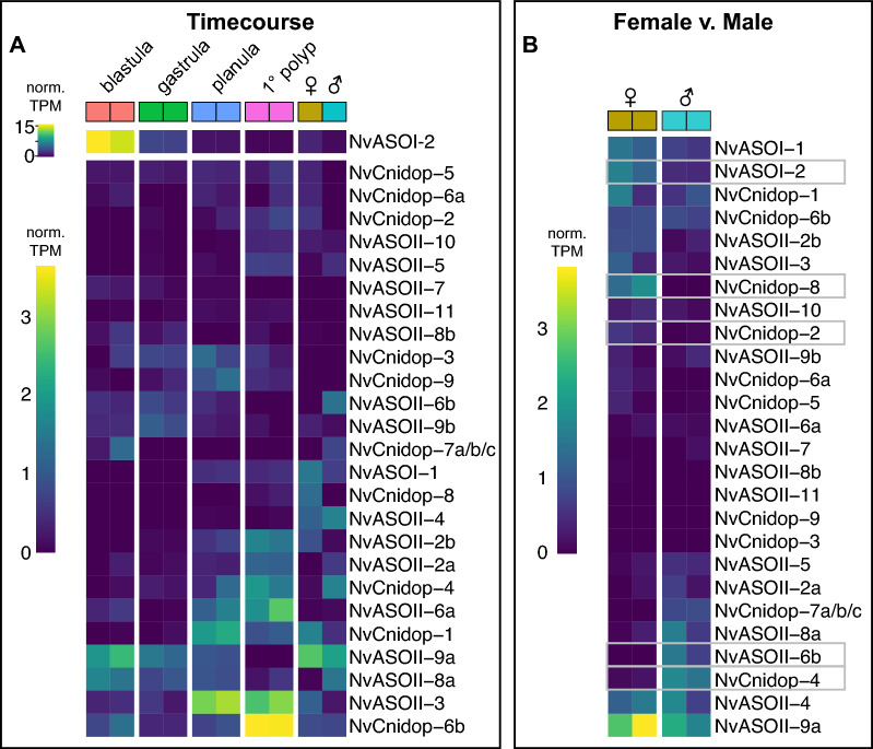
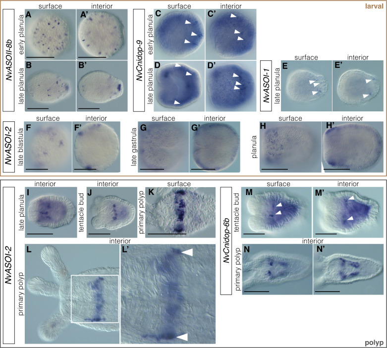
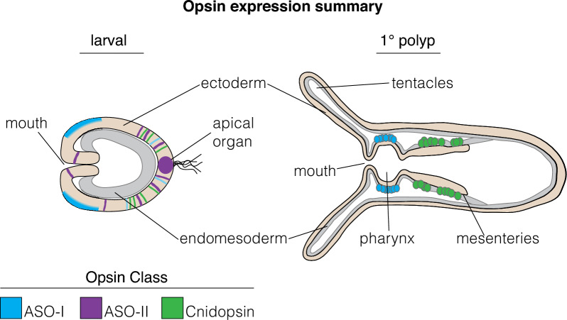
Results: We analyzed genomic and transcriptomic data from 16 species of cnidarians to generate a large opsin phylogeny (708 sequences) with the largest sampling of anthozoan sequences to date. We identified 29 opsins from N. vectensis (NvOpsins) with high confidence, using transcriptomic and genomic datasets. We found that lineage-specific opsin duplications are common across Cnidaria, with anthozoan lineages exhibiting among the highest numbers of opsins in animals. To establish putative photosensory function of NvOpsins, we identified canonically conserved protein domains and amino acid sequences essential for opsin function in other animal species. We show high sequence diversity among NvOpsins at sites important for photoreception and transduction, suggesting potentially diverse functions. We further examined the spatiotemporal expression of NvOpsins and found both dynamic expression of opsins during embryonic development and sexually dimorphic opsin expression in adults.

Conclusions: These data show that lineage-specific duplication and divergence has led to expansive diversity of opsins in eyeless cnidarians, suggesting opsins from these animals may exhibit novel biochemical functions. The variable expression patterns of opsins in N. vectensis suggest opsin gene duplications allowed for a radiation of unique sensory cell types with tissue- and stage-specific functions. This diffuse network of distinct sensory cell types could be an adaptive solution for varied sensory tasks experienced in distinct life history stages in Anthozoans.

Keywords: Anthozoa; Cnidaria; Hexacorallia; Nematostella; Opsin; Photoreceptor; Rhodopsin; Sea anemone.


Conflict of interest statement

The authors declare that they have no competing interests.

Figures

Fig. 1
Fig. 1
Phylogenetic placement and genomic architecture of the 29 N. vectensis opsins. A Simplified cnidarian phylogeny adapted from [91]. The range of known opsins found in a single species in each group is indicated on the phylogeny (from literature review). N. vectensis is in Actiniaria, Hexacorallia, in the Anthozoa (in blue) and has 29 opsins, the most so far identified of any anthozoan. B, C Light blue corresponds to ASO-I, purple to ASO-II, and green to cnidopsins. B N. vectensis chromosomes with opsin loci are shown. Nearly all N. vectensis opsins segregate on chromosomes by clade. Numbers below each chromosome are length in megabases, ( +) indicates recent tandem duplicates with highly similar sequences. Arrowheads indicate direction the gene is found in the genome. C Maximum likelihood tree of 708 opsins, with major animal opsin clades labeled (Tetraopsins include RGR/Go opsins/Group 4 opsins). Cnidarian-specific clades are colored and in bold. IQtree branch support is defined by ultrafast bootstraps (Ufboot) and likelihood ratio test (SH-aLRT). The number of N. vectensis opsins in each clade is listed (blue numbers). D Conservation of intron structure in the cnidops/xenops clade. A representative xenopsin from the oyster Crassostrea gigas shares an intron/exon boundary with all other xenopsins investigated [29] and several N. vectensis cnidopsins. Red box shows intron/exon boundary mapped on to amino acid alignment. Gray bars represent aligned sequence, black lines are gaps in the alignment
Fig. 2
Fig. 2
Current and ancestral anthozoan-specific opsin duplications. A Anthozoan species with opsin data are listed. Each column is the subclade number within the three major anthozoan opsin clades. “Un” is ASO-II opsins that are unspecified, or not found in previously identified clades. The number of opsins for each species and opsin subclade is listed in the boxes. Gray boxes with 0 signify genomic evidence of no opsins, while white boxes signify no opsin identified from transcriptomic evidence. Species names in bold have genomic evidence available, asterisks are for species with newly reported opsins in this study. B Anthozoan lineage tree, with numbers of opsins represented by line thickness and estimated by parsimony based on opsin numbers and phylogenetic positions (Fig. 3). Numbers in boxes represent the estimated number of opsins present in the last common ancestor of each anthozoan lineage. An increase in thickness of lines signifies an opsin duplication while an x signifies loss of the gene. The dashed line signifies uncertainty due to limited sampling in Octocorallia
Fig. 3
Fig. 3
Anthozoan-specific opsin evolutionary patterns of duplication and loss. Trees are zoomed subsets of the maximum-likelihood tree (Fig. 1C) for each anthozoan opsin group. Species names and branches are color coded according to lineage. Support for branches is denoted with a black circle or a white circle. A ASO-I group opsins are split into two main subclades, with most anthozoans having an opsin duplicate in each subclade. B The ASO-II group is sister to c-opsins and comprised exclusively of opsins from hexacorals. Previously identified ASO-II Groups 2.1 and 2.2 [6] are well-supported but ASO-II Group 1 is not. C Anthozoan cnidopsins form a single well-supported clade within the larger cnidopsin/xenopsin clade. Within Anthozoan cnidopsins there are two sister clades, one of which is well-supported while the other is not. For clarity, branch lengths are transformed, and branch support is not shown for branches leading to the two shallow-most nodes on these trees (For full tree topology and support values see Fig. 1C, Additional File 2)
Fig. 4
Fig. 4
N. vectensis opsins have high levels of sequence diversity at canonically conserved functional sites. A Left, cartoon bovine rhodopsin structure showing 7 transmembrane domains surrounding the vitamin A derived chromophore and the highly conserved lysine at position 296 (black) required for chromophore binding. The glutamic acid counterion (orange) is conserved among vertebrate c-opsins and important for chromophore binding. Right, cartoon diagram of bovine rhodopsin sequence with select functional sites color coded by functional category, matching the numbered sites in B. Cartoon G protein subunits are shown bound to rhodopsin. The specific G protein alpha subunit can vary (letters in parentheses) depending on opsin sequence, with functional implications for type of signaling cascade activated. B Left, maximum-likelihood phylogeny of N. vectensis opsins, with IQtree2 support. Right, select N. vectensis opsin amino acids are aligned with bovine rhodopsin (c-opsin), D. melanogaster r-opsin, and T. cystophora cnidopsin. Canonically conserved functional residues and positions follow bovine rhodopsin numbering and correspond to A. From left to right, the first box contains structural features minimally required for function in all opsins. 7TM indicates protein sequence has seven transmembrane domains. Black is the conserved Lys(K)296; yellow shows conserved cysteine residues that form a stabilizing disulfide bridge. Second box lists known counterions from bilaterian and box jelly opsins (orange). Third box shows a conserved spectral tuning site across opsin clades (blue). A second spectral tuning site is also found at counterion site Asp(D)83. Substitutions known to cause a blue shift in both sites are shaded blue. Fourth box contains conserved sites known to be important for G-protein signaling (light gray). NvOpsin residues that are conserved at the known functional sites are shaded in dark gray. Rightmost, NvOpsins are labeled by subclade within each major anthozoan opsin clade
Fig. 5
Fig. 5
NvOpsins are expressed dynamically throughout development and between sexes. A Expression levels from time course analysis are plotted for opsins across all stages and both sexes. Each box is from a single replicate library. For visual clarity, highly expressing NvASOI-2 is shown using a separate scale. Heatmap illustrates variable opsin expression across development in all opsin transcripts. NvCnidop7a/b/c loci are highly similar in coding sequence, such that transcripts are not distinguished between the three sequences. NvASOII-1 had no high-identity transcript in our transcriptome or in NvERTx. B A separate analysis comparing male and female adult opsin expression is visualized in a heatmap. The gray boxes indicate opsins that are qualitatively distinct between the sexes
Fig. 6
Fig. 6
NvOpsin spatial expression patterns suggest a diversity of functions throughout development. AB′ NvASOII-8b is only expressed at the swimming planula stage. Expression is ectodermal, in individual cells scattered throughout the ectoderm in early planula. In later stage planula (B′) the expression is also concentrated at the aboral end in the sensory apical organ. CD′ NvCnidop-8 is expressed in a subset of aboral ectodermal cells at planula stage (arrowheads). EE′ NvASOI-1 is also expressed in a subset of cells in the aboral ectoderm. FH′ NvASOI-2 is expressed in the ectoderm at early stages. Starting at blastula stage (F) through gastrula (G), expression is patchy in clusters of cells which tend to be concentrated more orally later in development (H). By late planula/tentacle bud stage expression has concentrated in an endomesodermal ring at the aboral end of the pharynx, where it can be seen into the primary polyp stage (IL′). MN′ NvCnidop-6b begins to express at late planula/tentacle bud stage in the pharyngeal endomesoderm, and by polyp stage is expressed in specific cells throughout the mesenteries. Scale bar 100 um; in all images oral is left, aboral is right
Fig. 7
Fig. 7
Summary of opsin expression patterns in development. Two summary stages are shown for the primarily early larval expression, and the primarily later primary polyp expression patterns of N. vectensis opsins. Opsin expression is color-coded by clade, ectoderm is beige, endomesoderm is gray. Opsins are expressed throughout developmental stages and tissue types

Similar articles

Cited by

References

    1. Feuda R, Hamilton SC, McInerney JO, Pisani D. Metazoan opsin evolution reveals a simple route to animal vision. Proc Natl Acad Sci. 2012;109:18868–18872. doi: 10.1073/pnas.1204609109. - DOI - PMC - PubMed
    1. Terakita A. The opsins. Genome Biol. 2005;6:1–9. doi: 10.1186/gb-2005-6-3-213. - DOI - PMC - PubMed
    1. Kozmik Z, Ruzickova J, Jonasova K, Matsumoto Y, Vopalensky P, Kozmikova I, et al. Assembly of the cnidarian camera-type eye from vertebrate-like components. Proc Natl Acad Sci. 2008;105:8989–8993. doi: 10.1073/pnas.0800388105. - DOI - PMC - PubMed
    1. Ramirez MD, Pairett AN, Pankey MS, Serb JM, Speiser DI, Swafford AJ, et al. The last common ancestor of most bilaterian animals possessed at least nine opsins. Genome Biol Evol. 2016;8:3640–3652. - PMC - PubMed
    1. Picciani N, Kerlin JR, Sierra N, Swafford AJM, Ramirez MD, Roberts NG, et al. Prolific origination of eyes in cnidaria with co-option of non-visual opsins. Curr Biol. 2018;28:2413–2419.e4. doi: 10.1016/j.cub.2018.05.055. - DOI - PubMed