

Wnt activation as a potential therapeutic approach to treat partial limbal stem cell deficiency
- PMID: 37735479
- PMCID: PMC10514048
- DOI: 10.1038/s41598-023-42794-8
Wnt activation as a potential therapeutic approach to treat partial limbal stem cell deficiency
Abstract
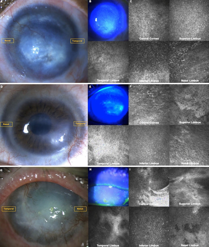
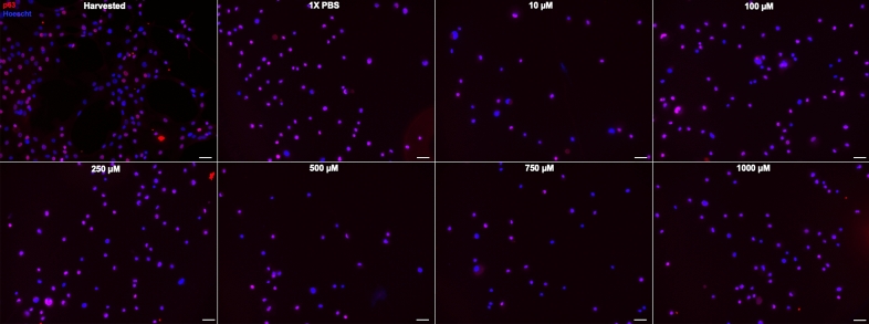
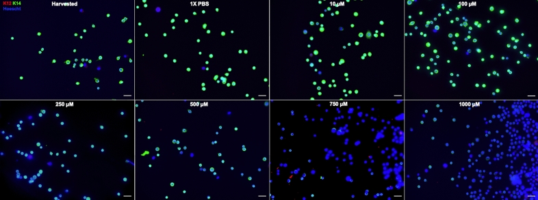
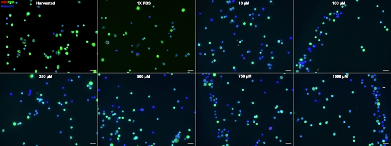
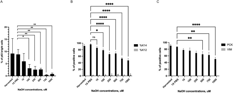
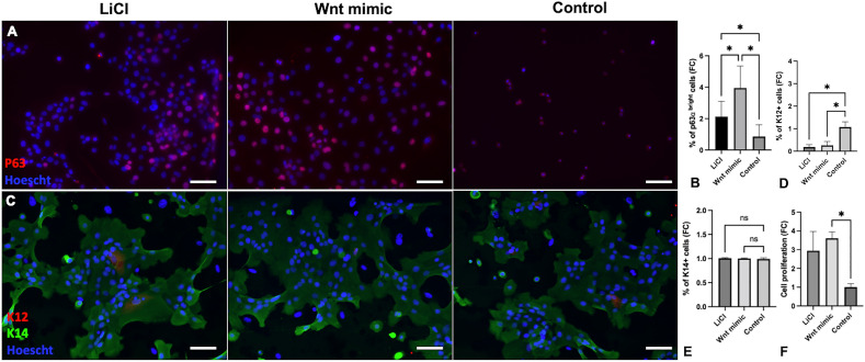
Limbal epithelial stem/progenitor cells (LSCs) are adult stem cells located at the limbus, tightly regulated by their niche involving numerous signaling pathways, such as Wnt. Wnt proteins are secreted morphogens that play critical roles in embryonic development, stem cell proliferation, self-renewal, tissue regeneration, and remodeling in adults. It has been shown that a small molecule Wnt mimic could improve LSCs expansion ex vivo. Damage to the LSCs and/or their niche can lead to limbal stem cell deficiency (LSCD), a condition that can cause corneal blindness and is difficult to treat. This study explored if repopulating residual LSCs in partial LSCD through Wnt activation could be a novel therapeutic approach. To mimic LSCD due to a chemical injury, single cultured LSCs were exposed to various concentrations of sodium hydroxide. A progressive loss of the LSCs phenotype was observed: the percentage of p63bright cells and cytokeratin (K)14+ cells decreased while the percentage of K12+ increased. Wnt activation was attained by treating the LSCs with lithium chloride (LiCl) and a small-molecule Wnt mimic, respectively. After 18 h of treatment, LSCs proliferation was increased, and the LSCs phenotype was recovered, while the untreated cells did not proliferate and lost their phenotype. The percentage of p63bright cells was significantly higher in the Wnt mimic-treated cells compared with untreated cells, while the percentage of K12+ cells was significantly lower. These findings suggest that local Wnt activation may rescue LSCs upon alkaline injury.
© 2023. Springer Nature Limited.
Conflict of interest statement
The authors declare no competing interests.
Figures
Similar articles
-
Ocular Surface Regeneration by Limbal Stem Cells Therapies: State of the Art, Challenges, and Perspectives.Stem Cells Transl Med. 2023 Nov 3;12(11):714-719. doi: 10.1093/stcltm/szad058. Stem Cells Transl Med. 2023. PMID: 37715946 Free PMC article.
-
Loss of corneal epithelial stem cell properties in outgrowths from human limbal explants cultured on intact amniotic membrane.Regen Med. 2008 May;3(3):329-42. doi: 10.2217/17460751.3.3.329. Regen Med. 2008. PMID: 18462056
-
New characterization and safety evaluation of human limbal stem cells used in clinical application: fidelity of mitotic process and mitotic spindle morphologies.Stem Cell Res Ther. 2023 Dec 13;14(1):368. doi: 10.1186/s13287-023-03586-z. Stem Cell Res Ther. 2023. PMID: 38093301 Free PMC article.
-
Regulation of Limbal Epithelial Stem Cells: Importance of the Niche.Int J Mol Sci. 2021 Nov 5;22(21):11975. doi: 10.3390/ijms222111975. Int J Mol Sci. 2021. PMID: 34769405 Free PMC article. Review.
-
Pathogenesis of Alkali Injury-Induced Limbal Stem Cell Deficiency: A Literature Survey of Animal Models.Cells. 2023 May 1;12(9):1294. doi: 10.3390/cells12091294. Cells. 2023. PMID: 37174694 Free PMC article. Review.
Cited by
-
Advances in the Diagnosis and Management of Limbal Stem Cell Deficiency.Cornea. 2025 Apr 1;44(4):405-411. doi: 10.1097/ICO.0000000000003775. Epub 2024 Dec 19. Cornea. 2025. PMID: 39729420 Review.
-
PAX6-WNK2 Axis Governs Corneal Epithelial Homeostasis.Invest Ophthalmol Vis Sci. 2024 Oct 1;65(12):40. doi: 10.1167/iovs.65.12.40. Invest Ophthalmol Vis Sci. 2024. PMID: 39453672 Free PMC article.
