

Imidazo[1,2-c]quinazolines as a novel and potent scaffold of α-glucosidase inhibitors: design, synthesis, biological evaluations, and in silico studies
- PMID: 37735489
- PMCID: PMC10514295
- DOI: 10.1038/s41598-023-42549-5
Imidazo[1,2-c]quinazolines as a novel and potent scaffold of α-glucosidase inhibitors: design, synthesis, biological evaluations, and in silico studies
Abstract
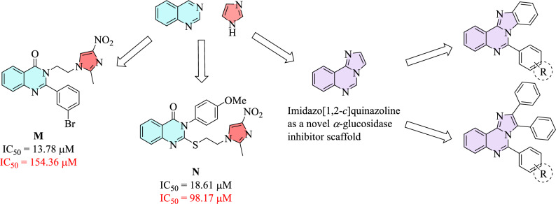
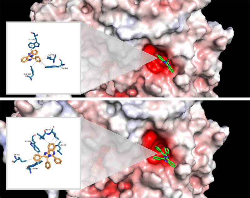
α-Glucosidase inhibition is an approved treatment for type 2 diabetes mellitus (T2DM). In an attempt to develop novel anti-α-glucosidase agents, two series of substituted imidazo[1,2-c]quinazolines, namely 6a-c and 11a-o, were synthesized using a simple, straightforward synthetic routes. These compounds were thoroughly characterized by IR, 1H and 13C NMR spectroscopy, as well as mass spectrometry and elemental analysis. Subsequently, the inhibitory activities of these compounds were evaluated against Saccharomyces cerevisiae α-glucosidase. In present study, acarbose was utilized as a positive control. These imidazoquinazolines exhibited excellent to great inhibitory potencies with IC50 values ranging from 12.44 ± 0.38 μM to 308.33 ± 0.06 μM, which were several times more potent than standard drug with IC50 value of 750.0 ± 1.5 μM. Representatively, compound 11j showed remarkable anti-α-glucosidase potency with IC50 = 12.44 ± 0.38 μM, which was 60.3 times more potent than positive control acarbose. To explore the potential inhibition mechanism, further evaluations including kinetic analysis, circular dichroism, fluorescence spectroscopy, and thermodynamic profile were carried out for the most potent compound 11j. Moreover, molecular docking studies and in silico ADME prediction for all imidazoquinazolines 6a-c and 11a-o were performed to reveal their important binding interactions, as well as their physicochemical and drug-likeness properties, respectively.
© 2023. Springer Nature Limited.
Conflict of interest statement
The authors declare no competing interests.
Figures


References
-
- Sun H, Saeedi P, Karuranga S, Pinkepank M, Ogurtsova K, Duncan BB, Stein C, Basit A, Chan JC, Mbanya JC. IDF Diabetes Atlas: Global, regional and country-level diabetes prevalence estimates for 2021 and projections for 2045. Diabetes Res. Clin. Pract. 2022;183:109119. doi: 10.1016/j.diabres.2021.109119. - DOI - PMC - PubMed
-
- Ojebiyi AO. The impacts of pharmacological and other interventions for preventing the onset of diabetes. Int. J. Diabet. Metab. Disord. 2023;8:268–276.
Publication types
MeSH terms
Substances
LinkOut - more resources
Full Text Sources
Medical
Molecular Biology Databases
