. 2023 Sep 21;14(1):5883.
doi: 10.1038/s41467-023-41577-z.

Structural basis of the activation of TRPV5 channels by long-chain acyl-Coenzyme-A

Affiliations

Structural basis of the activation of TRPV5 channels by long-chain acyl-Coenzyme-A

Bo-Hyun Lee et al. Nat Commun. .

Abstract

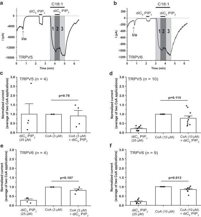
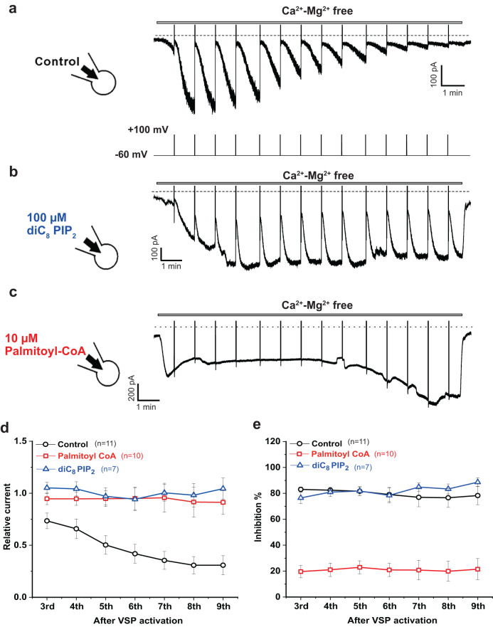
Long-chain acyl-coenzyme A (LC-CoA) is a crucial metabolic intermediate that plays important cellular regulatory roles, including activation and inhibition of ion channels. The structural basis of ion channel regulation by LC-CoA is not known. Transient receptor potential vanilloid 5 and 6 (TRPV5 and TRPV6) are epithelial calcium-selective ion channels. Here, we demonstrate that LC-CoA activates TRPV5 and TRPV6 in inside-out patches, and both exogenously supplied and endogenously produced LC-CoA can substitute for the natural ligand phosphatidylinositol 4,5-bisphosphate (PI(4,5)P2) in maintaining channel activity in intact cells. Utilizing cryo-electron microscopy, we determined the structure of LC-CoA-bound TRPV5, revealing an open configuration with LC-CoA occupying the same binding site as PI(4,5)P2 in previous studies. This is consistent with our finding that PI(4,5)P2 could not further activate the channels in the presence of LC-CoA. Our data provide molecular insights into ion channel regulation by a metabolic signaling molecule.


Conflict of interest statement

The authors declare no competing interests

Figures

Fig. 1
Fig. 1. Long chain acyl-CoA activates TRPV5 and TRPV6 channels.
Chemical structures of oleoyl-CoA (C18:1) (a) and dioctanoyl (diC8) PI(4,5)P2 (b). c–h Excised inside-out patch clamp recordings on TRPV5- and TRPV6-expressing Xenopus laevis oocytes were performed as described in the Methods. Traces show currents at +100 (upper) and −100 (lower) mV. Dashed lines show zero current. The establishment of the inside-out (i/o) configuration is indicated by the arrows. The applications of 25 µM diC8 PI(4,5)P2 (PIP2) and different concentrations of octanoyl (C8:0) CoA, and C18:1 CoA are shown by the horizontal lines. Representative trace for the effects of C8:0 CoA and C18:1 CoA on TRPV5 (c) and TRPCV6 (e). Summary of the effects of C8:0 CoA and C18:1 CoA on TRPV5 (d) and TRPV6 (f). Summary of the effects of the different fatty acyl-CoAs (10 µM) on TRPV5 (g) and TRPV6 (h). Bar graphs show mean ± SEM and scatter plots. The symbols represent individual oocytes.
Fig. 2
Fig. 2. The 3′-phosphate is required for LC-CoA to activate TRPV5 and TRPV6 channels.
a Chemical structure of 3’-dephospho-palmitoyl CoA. b-e Excised inside-out patch clamp recordings on Xenopus laevis oocytes expressing TRPV5 and TRPV6 were performed as described in Methods. Traces show currents at −100 mV, dashed lines show zero current. The establishment of the inside-out configuration (i/o) is indicated by the arrows. The application of 25 µM diC8 PI(4,5)P2, 10 µM 3′-dephospho-palmitoyl-CoA, and 10 µM palmitoyl (C16:0) CoA are shown by the horizontal lines. Representative traces for the effect of diC8 PI(4,5)P2, 3′-dephospho-palmitoyl-CoA, and C16:0 CoA on TRPV5 (b) and TRPV6 (d). Summary of the effects of diC8 PI(4,5)P2, 3′-dephospho-palmitoyl-CoA, and C16:0 CoA on TRPV5 (c) and TRPV6 (e). Bar graphs show mean ± SEM and scatter plots. The symbols represent experiments from individual oocytes.
Fig. 3
Fig. 3. DGS-NTA does not activate TRPV5 and TRPV6 channels.
a Chemical structure of DGS-NTA. b-e Excised inside-out patch clamp recordings on Xenopus laevis oocytes expressing TRPV5 and TRPV6 were performed as described in Methods. The establishment of the inside-out configuration is indicated by the arrows. In panel b an air bubble was applied to break the membrane vesicle. Traces show currents at −100 mV. Dashed lines show zero current. The applications of 25 µM diC8 PI(4,5)P2, and 10 and 100 μM DGS-NTA, and 10 μM oleoyl-CoA (C18:1) are shown by the horizontal lines. b, d Representative traces for the effect of diC8 PI(4,5)P2, DGS-NTA, and C18:1 CoA on TRPV5 (b) and TRPV6 (d). c, e Summary of the effects of diC8 PI(4,5)P2, DGS-NTA, and C18:1 CoA on TRPV5 (c) and TRPV6 (e). Bar graphs show mean ± SEM and scatter plots. The symbols represent experiments from individual oocytes.
Fig. 4
Fig. 4. Oleoyl-CoA binding site and conformational changes of TRPV5.
The TRPV5 CryoEM map in presence of oleoyl-CoA in open (a) and closed state (c) at a resolution of 3.25 Å and 3.09 Å, respectively. The CoA density is highlighted in pink and lipids in yellow. Coordinate model of CoA binding site in open (TRPV5CoA Open) (b) and closed (TRPV5CoA Closed) (d) state with overlaid density contoured at σ=3.5. Pore profile of TRPV5 in presence of PI(4,5)P2 (PDB 8FFO, TRPV5PIP2) (e), TRPV5CoA Open (f), and TRPV5CoA Closed (g). h Pore radius along the conducting pathway of TRPV5Apo (blue), TRPV5PIP2 (orange), TRPV5CoA Open (green), and TRPV5CoA Closed (purple). Dotted gray line at 1.1 Å is the dehydrated calcium radius. Conservation of the PI(4,5)P2 (i) and CoA (j) binding site. Distance between activator and the residues forming the binding site (R302, K484, R492, R584) are shown in blue. Comparison of pore domain movements (S5, S6 helixes) of TRPV5CoA Open versus TRPV5PIP2 (k) and TRPV5CoA Open versus TRPV5CoA Closed (l) from the side (left) and the bottom (right) view.
Fig. 5
Fig. 5. The effect of co-application of LC-CoA and PI(4,5)P2 on TRPV5 and TRPV6 channels.
Excised inside-out patch clamp recordings on Xenopus oocytes expressing TRPV5 (a, c, d) and TRPV6 (b, e, f) were performed as described in Methods. Traces show currents at −100 mV. Dashed lines show zero current. The establishment of the inside-out configuration is indicated by the arrows. The application of 25 µM diC8 PI(4,5)P2 and C18:1 CoA are shown by the horizontal lines. Representative trace shows the effect of co-application of 10 μM C18:1 CoA and 25 μM diC8 PI(4,5)P2 (PIP2) on TRPV5 (a) and TRPV6 (b). Each number in the gray area indicates a different application solution; 1 and 3 are applications of C18:1 CoA and 2 is a co-application of C18:1 CoA and diC8 PI(4,5)P2. Summary of the data from 3 µM C18:1 CoA (c, e) and 10 µM C18:1 CoA (d, f) in the presence or in the absence of 25 µM diC8 PI(4,5)P2. In each panel, the middle bar indicates the average current at gray areas 1 and 3 (current from the application of C18:1 CoA). P < 0.05 (Paired Sample t-test, two-tailed). Bar graphs show mean ± SEM and scatter plots. The symbols represent experiments from individual oocytes.
Fig. 6
Fig. 6. Palmitoyl-CoA activates TRPV6 channels after PI(4,5)P2 depletion in intact cells.
Whole-cell voltage-clamp recordings in HEK293 cells transiently cotransfected with TRPV6 and ci-VSP were performed as described in Methods. In these measurements, constant holding at −60 mV for 59 s, then a depolarizing step to +100 mV for 1 s was applied to activate ci-VSP and induce PI(4,5)P2 hydrolysis. This was repeated up to 14 times continuously (a, bottom). The intracellular pipette solution did not contain ATP. a monovalent TRPV6 currents were measured by application of Mg2+- and Ca2+-free solution, as described in the methods section. Standard intracellular solution was supplemented with 100 µM diC8 PI(4,5)P2 (b) or 10 µM palmitoyl-CoA (c). d Summary of relative recovered current after +100 mV pulse compared to peak current. e Summary of the ratio between the steady-state current before the +100 mV pulse and inhibited current right after the +100 mV pulse. Line graphs in d and e show mean ± SEM from individual cells.
Fig. 7
Fig. 7. Endogenous LC-CoA maintains TRPV6 activity.
a-d Representative traces for whole-cell patch-clamp recordings in HEK293T cells transiently cotransfected with TRPV6 and ci-VSP, performed as described in the Methods section. The membrane potential was clamped at −60 mV for 59 s, then a depolarizing step to +100 mV for 1 s was applied (arrows) to activate ci-VSP, after which the holding potential returned to −160 mV. Monovalent currents were measured in a Ca2+- Mg2+-free solution, and at the end of the experiment a solution containing 1 mM Mg2+ was applied to inhibit monovalent currents, see methods for details. The transfected cells were pretreated with (b) or without (a) 164 µM oleic acid overnight in DMEM containing high glucose (25 mM). For the low glucose condition, transfected cells were pretreated with 164 µM oleic acid (c) or 164 µM oleic acid with 100 µM etomoxir (d) overnight in the DMEM containing normal glucose (5.5 mM). e Summary of the inhibited currents right after the +100 mV pulse. Statistical significance was calculated with one way analysis of variance, overall ANOVA F = 16.53, p = 3.96 ×10⁻⁶. f Cartoon explaining the various treatments in these experiments. Long chain fatty acids (oleic acid) are taken up by the cell and converted to LC-CoA, which is transported from the cytoplasm to the mitochondria by CPT-1. High glucose, and etomoxir inhibits CPT-1, leading to increased cytoplasmic LC-CoA levels (created with Biorender). Bar graph shows mean ± SEM and scatter plots. The symbols represent experiments from individual cells.

Similar articles

Cited by

References

    1. Shrago E. Long-chain acyl-CoA as a multi-effector ligand in cellular metabolism. J. Nutr. 2000;130:290S–293S. - PubMed
    1. Larsson O, Deeney JT, Branstrom R, Berggren PO, Corkey BE. Activation of the ATP-sensitive K+ channel by long chain acyl-CoA. A role in modulation of pancreatic beta-cell glucose sensitivity. J. Biol. Chem. 1996;271:10623–10626. - PubMed
    1. Rohacs T, et al. Specificity of activation by phosphoinositides determines lipid regulation of Kir channels. Proc. Natl Acad. Sci. USA. 2003;100:745–750. - PMC - PubMed
    1. Schulze D, Rapedius M, Krauter T, Baukrowitz T. Long-chain acyl-CoA esters and phosphatidylinositol phosphates modulate ATP inhibition of KATP channels by the same mechanism. J. Physiol. 2003;552:357–367. - PMC - PubMed
    1. Liu GX, Hanley PJ, Ray J, Daut J. Long-chain acyl-coenzyme A esters and fatty acids directly link metabolism to KATP channels in the heart. Circ. Res. 2001;88:918–924. - PubMed

Publication types