. 2023 Oct;25(10):1535-1545.
doi: 10.1038/s41556-023-01229-2. Epub 2023 Sep 21.

Defining E3 ligase-substrate relationships through multiplex CRISPR screening

Affiliations

Defining E3 ligase-substrate relationships through multiplex CRISPR screening

Richard T Timms et al. Nat Cell Biol. 2023 Oct.

Erratum in

Abstract

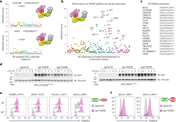
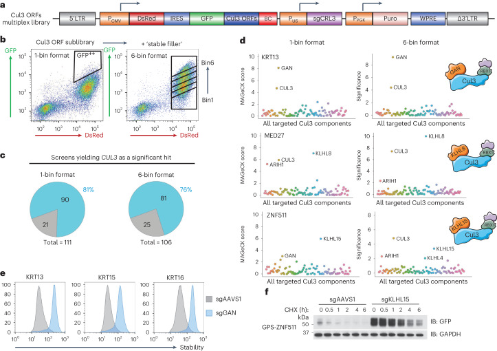
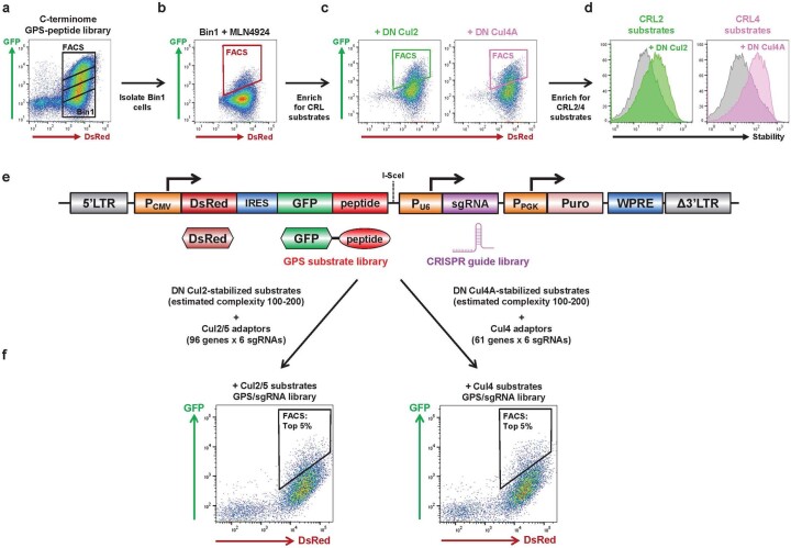
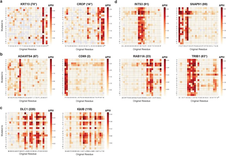
Specificity within the ubiquitin-proteasome system is primarily achieved through E3 ubiquitin ligases, but for many E3s their substrates-and in particular the molecular features (degrons) that they recognize-remain largely unknown. Current approaches for assigning E3s to their cognate substrates are tedious and low throughput. Here we developed a multiplex CRISPR screening platform to assign E3 ligases to their cognate substrates at scale. A proof-of-principle multiplex screen successfully performed ~100 CRISPR screens in a single experiment, refining known C-degron pathways and identifying an additional pathway through which Cul2FEM1B targets C-terminal proline. Further, by identifying substrates for Cul1FBXO38, Cul2APPBP2, Cul3GAN, Cul3KLHL8, Cul3KLHL9/13 and Cul3KLHL15, we demonstrate that the approach is compatible with pools of full-length protein substrates of varying stabilities and, when combined with site-saturation mutagenesis, can assign E3 ligases to their cognate degron motifs. Thus, multiplex CRISPR screening will accelerate our understanding of how specificity is achieved within the ubiquitin-proteasome system.


Conflict of interest statement

S.J.E. is a founder of MAZE Therapeutics, Mirimus, TSCAN Therapeutics and ImmuneID, and serves on the scientific advisory board of Homology Medicines, TSCAN Therapeutics and MAZE Therapeutics; none of these associations impacts this work. All other authors declare no competing interests.

Figures

Fig. 1
Fig. 1. Design of the multiplex CRISPR screening platform.
a, Individual FACS-based CRISPR screens are highly effective at identifying the cognate E3 ligase for unstable substrates tagged with a fluorescent protein such as GFP, but suffer from limited throughput as they are only capable of analysing a single substrate per screen. b, In contrast, multiplex CRISPR screening aims to identify the cognate E3 ligases for tens or hundreds of substrates in a single experiment. By encoding a library of GFP-tagged substrates and CRISPR sgRNAs targeting E3 ligases on the same lentiviral vector, cells expressing stabilized substrates paired with an sgRNA targeting the cognate E3 ligase can be enriched by FACS and the combination identified by paired-end sequencing. LTR, long terminal repeat; PCMV, human cytomegalovirus promoter; IRES, internal ribosome entry site; PPGK, phosphoglycerate kinase promoter; WPRE, Woodchuck Hepatitis Virus post-transcriptional regulatory element.
Fig. 2
Fig. 2. A proof-of-principle multiplex CRISPR screen recapitulates known C-degron pathways.
a, Schematic representation of the dual GPS/CRISPR multiplex screening library, in which the GFP-fusion substrates were a pool of peptides enriched for C-terminal degrons targeted by Cul2 or Cul4 E3 ligase complexes, and the CRISPR sgRNA library targeted either Cul2/5 or Cul4 adaptors. b,c, Identification of KLHDC2 substrates bearing C-terminal di-glycine motifs: the multiplex screen results for six example substrates, all of which terminate with two glycine residues (b); the performance of sgRNAs targeting KLHDC2 across all substrates (c). d,e, Cullin adaptors are correctly assigned to their cognate C-terminal degrons. A range of peptide substrates bearing canonical C-degron motifs targeted by Cul2 (d) and Cul4 (e) adaptors were successfully identified. All source numerical data are available in Supplementary Tables 1–6.
Fig. 3
Fig. 3. Cul2FEM1B regulates a C-degron pathway specific for proline.
ac, FEM1B substrates are highly enriched for C-terminal proline: screen results for two example substrates (a), the performance of sgRNAs targeting FEM1B across all substrates (b) and a tabulation of the sequences of all substrates for which FEM1B was a significant hit (c), with terminal proline residues indicated in red. d, Cycloheximide chase assays to monitor the degradation of the indicated GPS substrates in control (sgAAVS1) or FEM1B knockout (sgFEM1B) cells by immunoblot (IB). e,f, FEM1B targets C-terminal proline: C-terminal 23-mer peptides derived from the indicated genes, either with (wild type, WT) or without (ΔP) their terminal proline residue, were expressed in control (sgAAVS1) and FEM1B knockout (sgFEM1B) cells in the context of the GPS system and their stability measured by flow cytometry (e); full-length ORFs of the BEX family terminating in proline were more stable in FEM1B knockout cells (f). Immunoblot and flow cytometry experiments were performed twice with similar results. All source numerical data are available in Supplementary Tables 1–6; unprocessed blots are available in source data. Source data
Fig. 4
Fig. 4. FEM1B uses multiple pockets to bind diverse degrons.
ac, Structural analysis of FEM1B–degron interactions: overview of existing structures of FEM1B (purple) bound to the cysteine-rich substrate FNIP1 (yellow) or the Arg-ended CDK5R1 C-terminus (blue), compared with AlphaFold predictions of FEM1B bound to a representative Pro-end degron (POLD2, orange) (a); the Arg/Pro -1 pocket of FEM1B (purple) is shown bound to the CDK5R1 Arg-end substrate (blue) and the POLD2 Pro-end substrate (orange) (b); the aromatic-binding pocket of FEM1B (purple) is shown bound to three substrates (orange) that each requires a Phe, Trp or His to be recognized by FEM1B (c). df, Pro-ended FEM1B substrates require a hydrophobic residue ~15–20 residues from the C-terminus for efficient degradation. Saturation mutagenesis results for three representative Pro-ended substrates are shown, the C-terminus of PSMB5 (d), the C-terminus of SNF8 (e) and the C-terminus of CCDC89 (f); the darker the red colour, the greater the stabilizing effect of the mutation. The Add column indicates the effect of appending each individual amino acid at the extreme C-terminus of the peptide substrate. g, Evolutionary conservation of FEM1B binding pockets. The surface of FEM1B (residues 86–400) is coloured by conservation on the basis of an alignment of sequences from 12 diverse animal species. The sites for binding the Pro (orange) and Arg (blue) C-termini, FNIP1 (yellow) and zinc ions (cyan), and aromatic residues (orange) are shown for reference. Source numerical data are available in Supplementary Table 7.
Fig. 5
Fig. 5. Stability profiling of the human ORFeome identifies substrates of Cullin-RING E3 ubiquitin ligases.
ad, Identifying substrates of Cullin-RING E3 ligases: schematic representation of the GPS ORFeome library, comprising approximately 14,000 full-length, sequence-verified barcoded human ORFs (a); schematic representation of the comparative stability profiling screen using the pan-Cullin inhibitor MLN4924, where the human ORFeome library was expressed in HEK-293T cells and partitioned into six equal bins by FACS, and using the same settings and gates, the process was repeated for cells treated with MLN4924 (b); overall distribution of stability scores, comparing untreated (grey) and MLN4924-treated (red) cells (c); and 1,554 ORFs exhibited stabilization >0.5 PSI units following MLN4924 treatment (d). e,f, Assigning substrates to individual Cullin complexes: schematic representation of the barcoded sublibrary comprising the top 540 ORFs exhibiting the greatest stabilization from d (e); comparative stability profiling was performed as depicted in b to assess the stability of the library in cells expressing either an empty vector (grey) versus C-terminally truncated DN versions of Cul1 (yellow), Cul2 (light green), Cul3 (light blue), Cul4A (pink), Cul4B (purple) or Cul5 (dark green) (f). Screen profiles for four example substrates are shown. Source numerical data are available in Supplementary Tables 8–12.
Fig. 6
Fig. 6. A multiplex CRISPR screen to identify the cognate adaptors required for full-length protein substrates targeted by Cul3 complexes.
a, Schematic representation of the multiplex CRISPR screening vector, wherein ~100 full-length ORFs targeted by Cul3 complexes were fused to the C-terminus of GFP, and the CRISPR sgRNA library targeted known BTB adaptors. b, The multiplex CRISPR screen was performed in two ways: in the 1-bin format (left), the top ~5% of the population was sorted into a single bin, while in the 6-bin format (right), a pool of cells expressing stable substrates was spiked-in to broaden the stability distribution of the library, followed by partitioning into six equal bins by FACS to enable measurement of the stability of each ORF–sgRNA pair. c,d, Summary of the screen results: the majority of screens identified CUL3 as a significant hit (c); example results from successful screens, where both the 1-bin and 6-bin approaches concordantly identified the same BTB adaptor (d). e,f, Validation of the screen results: GAN was correctly identified as the BTB adaptor targeting keratins that we validated in a panel of individual experiments by flow cytometry (e), and KLHL15 targets ZNF511 as assayed by cycloheximide chase assays in control (sgAAVS1) versus KLHL15 knockout (sgKLHL15) cells (f). Immunoblot (IB) and flow cytometry experiments were performed twice with similar results. Source numerical data are available in Supplementary Tables 13–17; unprocessed blots are available in source data. Source data
Fig. 7
Fig. 7. Systematic identification of linear motifs targeted by Cullin-RING E3 ubiquitin ligases.
ac, Schematic representation of the experimental strategy: a lentiviral GPS library of peptides substrates was generated through microarray oligonucleotide synthesis, wherein the same 540 ORFs exhibiting the greatest degree of stabilization upon MLN4924 treatment were expressed as a series of overlapping 24-mer tiles (a); comparative stability profiling in the presence and absence of MLN4924 then identified GFP–peptide fusions which were targeted by CRLs (b); and for the peptide substrates which exhibited the largest degree of stabilization, saturation mutagenesis was performed to quantify the stability of a panel of mutants in which each residue was mutated to all other possible residues, thereby defining degron motifs at amino acid resolution (c). dg, Example degron motifs targeted by Cullin-RING E3 ligases. Saturation mutagenesis results for substrates derived from the C-terminus of ALKBH7 (d) and internal peptides derived from ESRRA (e), MATN2 (f) and TOR1AIP2 (g) are shown. Source numerical data are available in Supplementary Tables 18 and 19.
Fig. 8
Fig. 8. A multiplex CRISPR screen assigns Cullin-RING E3 ligases to their cognate degrons.
a, Schematic representation of the multiplex CRISPR screening vector, wherein peptides with mapped degrons were fused to the C-terminus of GFP and the CRISPR sgRNA library targeted all known Cullin substrate adaptors. be, Assigning Cullin-RING E3 ligases to their cognate linear degrons. Data are shown for substrates derived from the C-terminus of CCDC89 (b) and internal peptides derived from NECAB1 (c), EPB41L3 (d) and GMCL1 (e): in each case, the saturation mutagenesis results mapping the degron motif are shown (left), alongside the multiplex CRISPR screen results after both one sort and two sorts (right). Source numerical data are available in Supplementary Tables 20–38.
Extended Data Fig. 1
Extended Data Fig. 1. Generation of multiplex CRISPR screen libraries to interrogate C-terminal degrons targeted by Cullin-RING E3 ligases.
a-d Derivation of cells expressing C-terminal peptides targeted by Cul2 complex or Cul4 complexes. Starting from HEK-293T cells expressing the C-terminome GPS-peptide library, we isolated the most unstable GFP-peptide fusions (Bin1) (a), treated them with MLN4924, and then isolated cells in which the GFP-peptides fusions were stabilized (b). After recovery in the absence of MLN4924, to further purify substrates of specific Cullin complexes we expressed dominant-negative (DN) versions of either Cul2 (green) or Cul4A (pink) and again isolated the cells in which the GFP-peptides fusions were stabilized by FACS (c). After recovery, we re-challenged the sorted cells with the DN Cullins to verify that the final populations of cells were highly enriched for CRL substrates (d). e,f, Generation of the multiplex CRISPR screening vector to examine C-terminal degrons targeted by Cullins. e, Schematic representation of the multiplex CRISPR screening vector. GFP-fusion peptides were amplified from the genomic DNA of the cells in (d) by PCR and cloned into the lentiviral vector downstream of GFP; the CRISPR sgRNA library targeting either Cul2 or Cul4 adaptors was then cloned into the resulting substrate library using the I-SceI site to generate the dual GPS-sgRNA multiplex CRISPR screening library. f, The library was then introduced into HEK-293T stably expressing Cas9, and the top ~5% of cells expressing the most stable substrates were isolated by FACS. Genomic DNA was then extracted from both the sorted cells and the unsorted libraries, and substrate-sgRNA pairs enriched in the sorted cells quantified by paired-end Illumina sequencing.
Extended Data Fig. 2
Extended Data Fig. 2. Novel aspects of C-degron pathways revealed by the multiplex CRISPR screen.
a-d, DCAF12 recognizes a wider variety of motifs characterized by a glutamic acid at the penultimate position (a,b), while TRPC4AP can recognize substrates with arginine residues at the -4 and -5 position as well as those with arginine at -3 (c,d). e, Peptide substrates of FEM1B are enriched for C-terminal proline. Screen results for six substrates in which FEM1B was identified as a significant hit are shown; inspection of the sequences of the peptides revealed that they all terminate with a C-terminal proline residue (highlighted in red). f, Cycloheximide chase assays to monitor the degradation of the indicated GPS substrates in control (sgAAVS1) or FEM1B knockout (sgFEM1B) cells by immunoblot. Immunoblot experiments were performed twice with similar results. All source numerical data are available in Supplementary Tables 1-6; unprocessed blots are available in source data. Source data
Extended Data Fig. 3
Extended Data Fig. 3. Analysis of FEM1B degrons.
a, Additional AlphaFold structural predictions for the interactions between FEM1B and Pro-ended peptide substrates. b, The Arg/Pro -1 pocket of FEM1B is shown bound to four different Pro-ended degrons predicted to make similar interactions with FEM1B. c, Many of the Pro-end (orange) or Arg-end (blue) substrates use a leucine at the -3 position to interact with a surface on FEM1B (purple). d-f, Saturation mutagenesis results for additional Pro-ended substrates. Source numerical data are available in Supplementary Table 7.
Extended Data Fig. 4
Extended Data Fig. 4. Genetic complementation of FEM1B knockout cells.
a-b, Assessing the role of the predicted FEM1B binding pockets in the recognition of three peptide substrates terminating with proline. Three example GFP-tagged peptide substrates were selected: the C-termini of PSMB5 and SNF8, predicted by both AlphaFold and the saturation mutagenesis data to make essential contacts with both the Arg/Pro -1 pocket and the aromatic-binding pocket (Extended Data Fig. 3a,d,e), and the C-terminus of BEX2, which AlphaFold suggested also binds both pockets but for which the saturation mutagenesis suggested only the contacts with the Arg/Pro -1 pocket were essential for efficient degradation (Extended Data Fig. 3a,f). FEM1B knockout cells were first transduced with lentiviral vectors encoding FLAG-tagged wild-type FEM1B or FEM1B variants harboring the indicated mutations. Subsequently the cells were transduced with GPS vectors encoding the indicated peptide substrates of FEM1B terminating with C-terminal proline and their stability measured by flow cytometry (a). Relative expression levels of the exogenous FEM1B constructs were assayed by immunoblot (b). We attempted to validate that the -1 binding pocket mutants were competent for the degradation of GFP fused to a peptide degron from FNIP1 (ref. ), but we found that the FNIP1degron peptide was only minimally stabilized in FEM1B knockout cells when expressed in the context of the GPS system (c). However, overexpression of FEM1B harboring the R126A mutation did result in substantial destabilization of the FNIP1 construct in wild-type cells (d). Immunoblot and flow cytometry experiments were performed twice with similar results. Unprocessed blots are available in source data. Source data
Extended Data Fig. 5
Extended Data Fig. 5. Proteome-wide stability profiling to identify substrates of Cullin-RING E3 ligases.
a, The GPS-ORFeome screen exhibited high reproducibility. Example screen profiles for the indicated ORFs are shown, reflecting the distribution of sequencing reads across the 6 stability bins. Colored lines (grays, control; reds, MLN4924) represent different barcodes attached to the same ORF. b, The GPS-ORFeome screen identified known CRL substrates. Example screen profiles for a range of positive control substrates are shown. c, Generation of barcoded sub-library of ORFs stabilized by MLN4924. Bacteria harboring individual ORF constructs in Gateway entry vectors were grown up, pooled, and a Gateway LR reaction performed to shuttle the ORFs into a barcoded GPS lentiviral destination vector. Barcode-ORF pairs were subsequently assigned by paired-end Illumina sequencing.
Extended Data Fig. 6
Extended Data Fig. 6. A multiplex CRISPR screen with full-length proteins targeted by Cul3 complexes.
a, Validation of the Cul3 ORF sub-library. A sub-library of 116 ORFs was generated as in Extended Data Fig. 3c; the barcoded pool exhibited robust stabilization upon expression of dominant-negative Cul3. b, Broadening the stability distribution of the library for the screen in the ‘6-bin’ format. To enable the population to be sorted into 6 equal bins across the full stability spectrum, cells expressing orthogonal GPS ORF-sgRNA dual expression vectors in which the ORF substrates were stable were spiked-in at the appropriate ratio to yield an approximately even stability distribution. c,d, Example screen results for three substrates in which KLHL9 and/or KLHL13 (two paralogous BTB adaptors) were identified, together with a list of all putative KLHL9/13 substrates. Source numerical data are available in Supplementary Tables 13-17.
Extended Data Fig. 7
Extended Data Fig. 7. Linear degrons targeted by Cullin-RING E3 ligases.
a-d, Example degron motifs delineated through site-saturation mutagenesis: (a) C-terminal degrons, (b) ‘narrow hydrophobic’ degrons, (c) ‘broad hydrophobic’ degrons, and (d) more complex degrons. Source numerical data are available in Supplementary Tables 18-19.
Extended Data Fig. 8
Extended Data Fig. 8. Assigning linear degron motifs to their cognate Cullin-RING E3 ligase.
a, Sorting plots for the multiplex CRISPR screen. Three multiplex CRISPR screening libraries were generated in which substrates were divided into three groups based on their expected stability; in each case, the top ~5% of cells based on the stability of the GFP-peptide fusion were isolated by FACS. b-d, Assigning Cullin-RING substrate adaptors to cognate degron motifs: a C-terminal degron harboring an E-2 motif correctly assigned to DCAF12 (b), a ‘broad hydrophobic’ degron assigned to FBXO38 (c), and a complex degron assigned to KLHL15 (d). e,f, Individual validation of the multiplex CRISPR screening results. e, The indicated peptide substrates expressed in the context of the GPS system were introduced into either control (sgAAVS1, gray) or knockout (yellow, green and blue) HEK-293T cells and their stability measured by flow cytometry. f, A cycloheximide chase assay was also used to monitor the degradation of the ZNF511 (4) peptide substrates in control (sgAAVS1) or KLHL15 knockout (sgKLHL15) cells by immunoblot. Immunoblot and flow cytometry experiments were performed twice with similar results. Source numerical data are available in Supplementary Tables 20-38; unprocessed blots are available in source data. Source data
Extended Data Fig. 9
Extended Data Fig. 9. Comparison of multiplex CRISPR screen hits with physical interactions in BioGRID.
a, Across all multiplex screens there were 1013 unique E3-substrate hits, of which 31 were present in the BioGRID interaction database. Datasets were simulated with 1013 hits chosen at random and we most frequently found that 7 of these hits were present in the BioGRID database. The 31 hits that we observe is therefore highly significant (p < 0.0001). b, The same analysis was performed as in (a) except that all E3-substrate hits that included a Cullin were excluded.
Extended Data Fig. 10
Representative FACS gating strategy for all GPS experiments.

References

    1. Rape M. Ubiquitylation at the crossroads of development and disease. Nat. Rev. Mol. Cell Biol. 2018;19:59–70. doi: 10.1038/nrm.2017.83. - DOI - PubMed
    1. Mészáros B, Kumar M, Gibson TJ, Uyar B, Dosztányi Z. Degrons in cancer. Sci. Signal. 2017;10:eaak9982. doi: 10.1126/scisignal.aak9982. - DOI - PubMed
    1. Lee JM, Hammarén HM, Savitski MM, Baek SH. Control of protein stability by post-translational modifications. Nat. Commun. 2023;14:201. doi: 10.1038/s41467-023-35795-8. - DOI - PMC - PubMed
    1. Clague MJ, Heride C, Urbé S. The demographics of the ubiquitin system. Trends Cell Biol. 2015;25:417–426. doi: 10.1016/j.tcb.2015.03.002. - DOI - PubMed
    1. Iconomou, M. & Saunders, D. N. Systematic approaches to identify E3 ligase substrates. Biochem. J.473, 4083–4101 (2016). - PMC - PubMed

Publication types

MeSH terms