

Validation of the diagnostic efficacy of O-RADS in adnexal masses
- PMID: 37735610
- PMCID: PMC10514283
- DOI: 10.1038/s41598-023-42836-1
Validation of the diagnostic efficacy of O-RADS in adnexal masses
Abstract
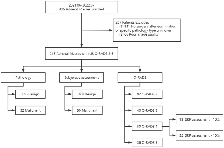
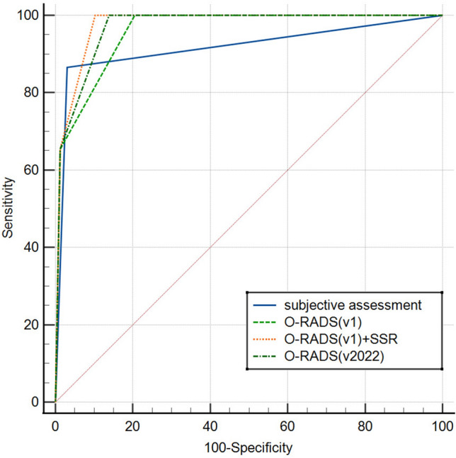
The aim of this study was to validate the performance of the Ovarian-Adnexal Reporting and Data Systems (O-RADS) series models proposed by the American College of Radiology (ACR) in the preoperative diagnosis of adnexal masses (AMs). Two experienced sonologists examined 218 patients with AMs and gave the assessment results after the examination. Pathological findings were used as a reference standard. Of the 218 lesions, 166 were benign and 52 were malignant. Based on the receiver operating characteristic (ROC) curve, we defined a malignant lesion as O-RADS > 3 (i.e., lesions in O-RADS categories 4 and 5 were malignant). The area under the curve (AUC) of O-RADS (v2022) was 0.970 (95% CI 0.938-0.988), which wasn't statistically significantly different from the O-RADS (v1) combined Simple Rules Risk (SRR) assessment model with the largest AUC of 0.976 (95% CI 0.946-0.992) (p = 0.1534), but was significantly higher than the O-RADS (v1) (AUC = 0.959, p = 0.0133) and subjective assessment (AUC = 0.918, p = 0.0255). The O-RADS series models have good diagnostic performance for AMs. Where, O-RADS (v2022) has higher accuracy and specificity than O-RADS (v1). The accuracy and specificity of O-RADS (v1), however, can be further improved when combined with SRR assessment.
© 2023. Springer Nature Limited.
Conflict of interest statement
The authors declare no competing interests.
Figures

Similar articles
-
Ovarian-Adnexal Imaging-Reporting and Data System (O-RADS) ultrasound version 2019: a prospective validation and comparison to updated version (v2022) in pathologically confirmed adnexal masses.Eur Radiol. 2025 Jun;35(6):3080-3095. doi: 10.1007/s00330-024-11235-z. Epub 2024 Nov 28. Eur Radiol. 2025. PMID: 39604652
-
Performance of IOTA Simple Rules, Simple Rules risk assessment, ADNEX model and O-RADS in differentiating between benign and malignant adnexal lesions in North American women.Ultrasound Obstet Gynecol. 2022 May;59(5):668-676. doi: 10.1002/uog.24777. Epub 2022 Apr 8. Ultrasound Obstet Gynecol. 2022. PMID: 34533862
-
Diagnostic Performance of O-RADS US (Version 2019 and Version 2022) Incorporating Acoustic Shadowing by Junior Radiologists: Analyzing 1061 Adnexal Masses.J Ultrasound Med. 2025 May;44(5):845-855. doi: 10.1002/jum.16644. Epub 2025 Jan 10. J Ultrasound Med. 2025. PMID: 39791550
-
O-RADS US v2022: An Update from the American College of Radiology's Ovarian-Adnexal Reporting and Data System US Committee.Radiology. 2023 Sep;308(3):e230685. doi: 10.1148/radiol.230685. Radiology. 2023. PMID: 37698472 Review.
-
O-RADS Ultrasound Version 1: A Scenario-Based Review of Implementation Challenges.AJR Am J Roentgenol. 2022 Dec;219(6):916-927. doi: 10.2214/AJR.22.28061. Epub 2022 Jul 20. AJR Am J Roentgenol. 2022. PMID: 35856453 Review.
Cited by
-
Accurate prediction of benign and malignant adnexal tumors in surgical resection and conservative treatment: construction and external validation of a diagnostic model based on CEUS, HE4, and O-RADS US v2022 evaluation.J Ovarian Res. 2025 Jun 6;18(1):123. doi: 10.1186/s13048-025-01707-1. J Ovarian Res. 2025. PMID: 40481532 Free PMC article.
-
Integrating Contrast-enhanced US to O-RADS US for Classification of Adnexal Lesions with Solid Components: Time-intensity Curve Analysis versus Visual Assessment.Radiol Imaging Cancer. 2024 Nov;6(6):e240024. doi: 10.1148/rycan.240024. Radiol Imaging Cancer. 2024. PMID: 39392388 Free PMC article.
-
Ovarian-Adnexal Imaging-Reporting and Data System (O-RADS) ultrasound version 2019: a prospective validation and comparison to updated version (v2022) in pathologically confirmed adnexal masses.Eur Radiol. 2025 Jun;35(6):3080-3095. doi: 10.1007/s00330-024-11235-z. Epub 2024 Nov 28. Eur Radiol. 2025. PMID: 39604652
-
Clinical value of ACR O-RADS combined with CA125 in the risk stratification of adnexal masses.Front Oncol. 2024 Aug 30;14:1369900. doi: 10.3389/fonc.2024.1369900. eCollection 2024. Front Oncol. 2024. PMID: 39281376 Free PMC article.
-
Can transabdominal shear wave elastography play a role in solving the dilemma of complex cystic and solid ovarian tumors by ultrasound?J Ultrasound. 2025 May 31. doi: 10.1007/s40477-025-01027-6. Online ahead of print. J Ultrasound. 2025. PMID: 40448793
References
-
- Higgins, R. V., van Nagell, J. R. Jr, Woods, C. H., Thompson, E. A. & Kryscio, R. J. Interobserver variation in ovarian measurements using transvaginal sonography. Gynecol. Oncol.39(1), 69–71 (1990). - PubMed
Publication types
MeSH terms
LinkOut - more resources
Full Text Sources
