. 2023 Nov;30(11):1640-1652.
doi: 10.1038/s41594-023-01096-3. Epub 2023 Sep 21.

Aberrant gene activation in synovial sarcoma relies on SSX specificity and increased PRC1.1 stability

Affiliations

Aberrant gene activation in synovial sarcoma relies on SSX specificity and increased PRC1.1 stability

Nezha S Benabdallah et al. Nat Struct Mol Biol. 2023 Nov.

Abstract

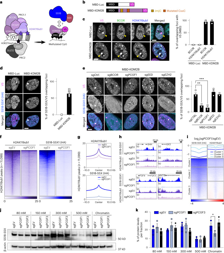
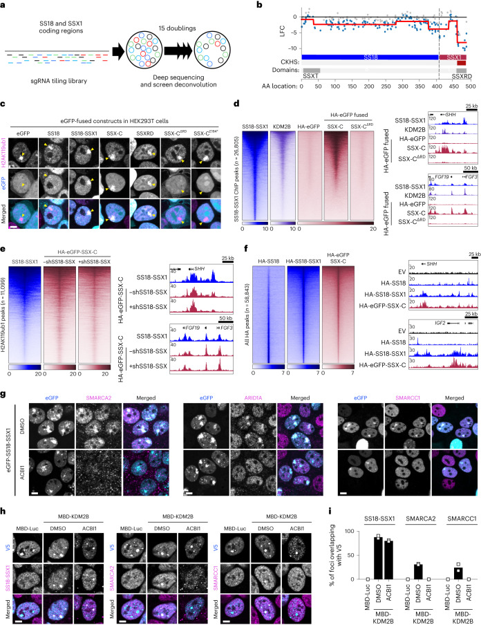
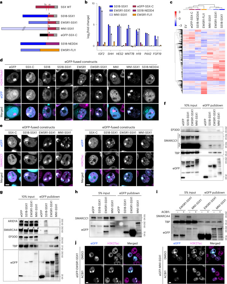
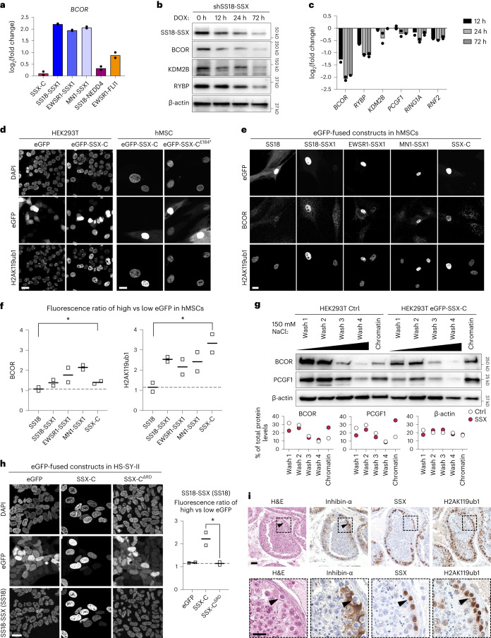
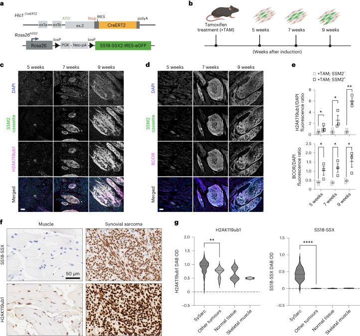
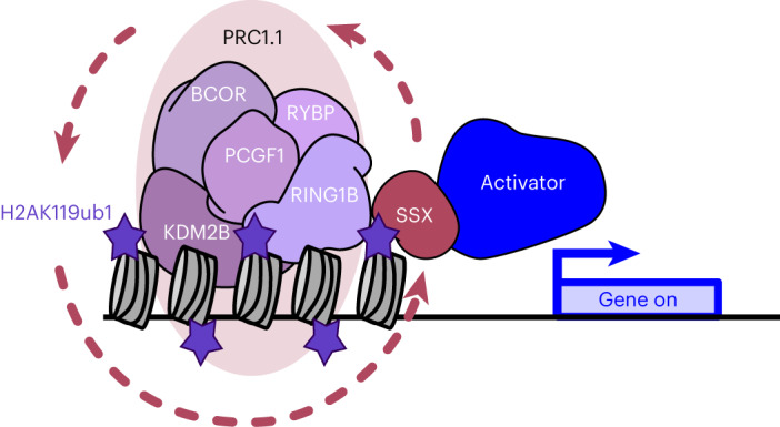
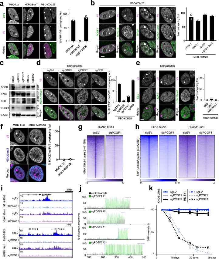
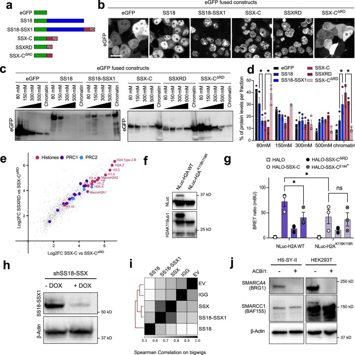
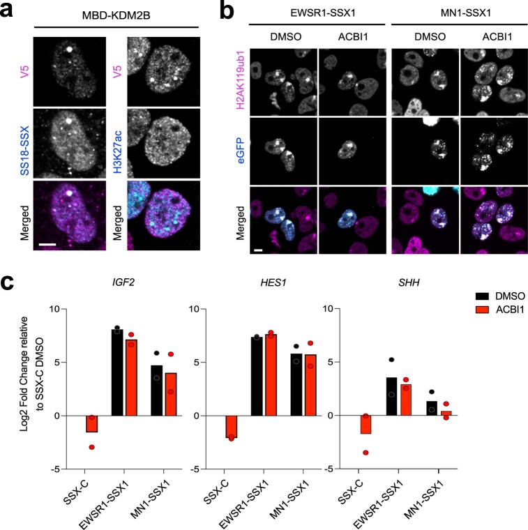
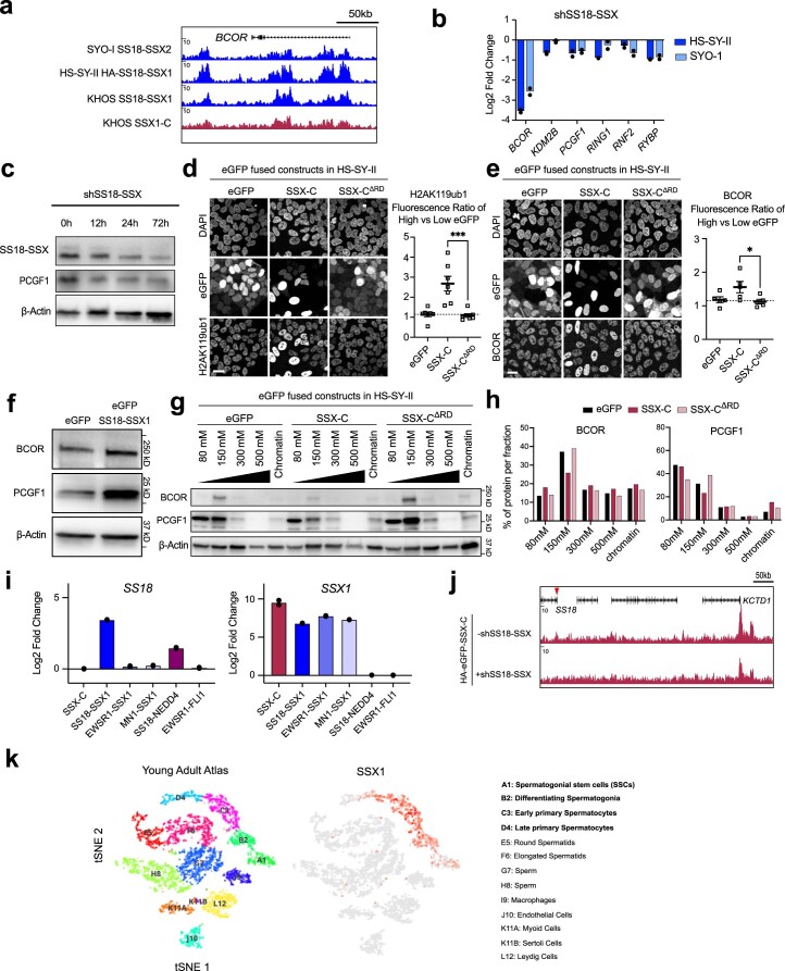
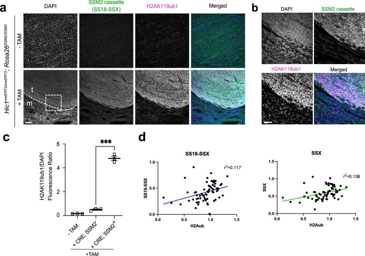
The SS18-SSX fusion drives oncogenic transformation in synovial sarcoma by bridging SS18, a member of the mSWI/SNF (BAF) complex, to Polycomb repressive complex 1 (PRC1) target genes. Here we show that the ability of SS18-SSX to occupy H2AK119ub1-rich regions is an intrinsic property of its SSX C terminus, which can be exploited by fusion to transcriptional regulators beyond SS18. Accordingly, SS18-SSX recruitment occurs in a manner that is independent of the core components and catalytic activity of BAF. Alternative SSX fusions are also recruited to H2AK119ub1-rich chromatin and reproduce the expression signatures of SS18-SSX by engaging with transcriptional activators. Variant Polycomb repressive complex 1.1 (PRC1.1) acts as the main depositor of H2AK119ub1 and is therefore required for SS18-SSX occupancy. Importantly, the SSX C terminus not only depends on H2AK119ub1 for localization, but also further increases it by promoting PRC1.1 complex stability. Consequently, high H2AK119ub1 levels are a feature of murine and human synovial sarcomas. These results uncover a critical role for SSX-C in mediating gene deregulation in synovial sarcoma by providing specificity to chromatin and further enabling oncofusion binding by enhancing PRC1.1 stability and H2AK119ub1 deposition.


Conflict of interest statement

The authors declare no competing interests.

Figures

Fig. 1
Fig. 1. PRC1.1 regulates SS18-SSX recruitment independently of PRC2.
a, MBD-mediated targeting to methylated CpG. Here, KDM2B is redirected to methylated CpG via the MBD. b, Schematic of MBD-Luc and MBD-KDM2B fusions containing a V5 tag. MBD-KDM2B contains the histone demethylase domain (JmjC) and a mutated CxxC. c, Left: immunofluorescence in HS-SY-II cells of the MBD constructs (V5, magenta) with BCOR (green) and H2AK119ub1 (cyan). Arrowheads point to the MBD foci. Scale bars, 5 μm. Right: percentage of BCOR or H2AK119ub1 foci overlapping a V5 focus in n = 3 (MBD-Luc) or n = 4 (MBD-KDM2B) biological replicates. Data represent the mean and the s.e.m. d, Left: immunofluorescence for V5 (magenta) and SS18-SSX1 (HA, cyan). Right: percentage of HA (SS18-SSX1) foci overlapping a V5 focus. Data represent the mean ± s.e.m. in n = 5 biological replicates. e, Left: immunofluorescence of MBD-KDM2B (V5, magenta) in the presence of different sgRNAs (eGFP background fluorescence) with SS18-SSX1 (HA, cyan) in HS-SY-II-Cas9 cells. Right: percentage of HA (SS18-SSX1) foci overlapping a V5 focus in n = 2 (sgBCOR, sgEZH2), n = 3 (sgPCGF1, sgEED) or n = 4 (sgCTRL) biological replicates. Data represent the mean ± s.e.m. P values determined by unpaired one-tailed t-test between groups (**P = 0.003; ***P = 0.0005). f, Heatmaps of H2AK119ub1 calibrated ChIP (purple) and SS18-SSX scaled HA CUT&RUN signals (blue) in HS-SY-II-Cas9 cells expressing empty sgRNA as control (sgEV) or targeting PCGF1 (sgPCGF1). Both heatmaps represent signals over H2AK119ub1 peaks (n = 11,099) called using H2AK119ub1 CUT&RUN in HS-SY-II (Extended Data Fig. 1h). Rows correspond to ±10-kb regions across the midpoint of each enriched region, ranked by increasing signal. g, H2AK119ub1 and SS18-SSX corresponding score distributions. h, Gene tracks for H2AK119ub1 and SS18-SSX at the SHH, IGF2, FGF4-FGF3 and WNT7B loci. i, k-means clustering of H2AK119ub1 and SS18-SSX log2 ratio of sgPCGF1 over sgEV. j, Salt extraction assay displaying SS18-SSX1 levels by western blot in HS-SY-II-Cas9 cells expressing an empty vector (EV) or sgRNAs against PCGF1 or PCGF3. k, Percentage of total SS18-SSX per salt extraction fractions. Data represent the mean ± s.e.m. of n = 3 biological replicates. P values determined by paired one-tailed t-test between groups (*P = 0.03). Source data
Fig. 2
Fig. 2. SSX C terminus directs SS18-SSX chromatin binding independently of BAF.
a, Layout of CRISPR–Cas9 knockout tiling screen. b, Mapping of CKHS regions in SS18-SSX1 using ProTiler based on log2(fold change) (LFC) in representation of sgRNAs targeting SS18-SSX1 in HS-SY-II. The CKHS region is highlighted in dark red and corresponds to the SSXRD Pfam sequence (PF09514). c, Immunofluorescence of HEK293T cells expressing eGFP constructs (cyan) stained for H2AK119ub1 (magenta). Images are representative of three biological replicates. Yellow arrowheads indicate the Barr body. Scale bar, 5 μm. d, Left: heatmaps for SS18-SSX1 (endogenously HA tagged) and KDM2B ChIP–seq from ref. and HA ChIP in HS-SY-II cells expressing HA-eGFP fused to SSX-C or SSX-CΔRD over SS18-SSX1 peaks (n = 26,805). Rows correspond to ±10-kb regions across the midpoint of each HA-enriched region, ranked by increasing signal. Right: gene tracks for SS18-SSX1, KDM2B and HA ChIP–seq at the SHH and FGF4-FGF3 loci. e, Left: CUT&RUN heatmaps in HS-SY-II cells for SS18-SSX1 (endogenously HA tagged) and HA-eGFP fused to SSX-C without and with SS18-SSX depletion mediated by shRNA. Heatmaps represent CUT&RUN signals over H2AK119ub1 peaks (n = 11,099). Rows correspond to ±10-kb regions across the midpoint of each HA-enriched region, ranked by increasing signal. Right: CUT&RUN gene tracks at the SHH and FGF4-FGF3 loci. f, Left: heatmaps for HA CUT&RUN in KHOS-240S cells expressing HA-SS18, HA-SS18-SSX1 or HA-eGFP-SSX-C. Heatmaps represent CUT&RUN signals over all HA peaks (n = 58,843). Rows correspond to ±10-kb regions across the midpoint of each HA-enriched region, ranked by increasing signal. Right: CUT&RUN gene tracks at the SHH and FGF4-FGF3 loci. g, Immunofluorescence of HEK293T cells expressing eGFP-SS18-SSX1 (cyan) treated with DMSO (top) or 500 nM ACBI1 (bottom) stained for BAF subunits (magenta) SMARCA2 (BRM, left), ARID1A (middle) or SMARCC1 (right). Images are representative of two biological replicates. h, Immunofluorescence of MBD constructs (V5, cyan) and SS18-SSX1 (HA), SMARCA2 or SMARCC1 (magenta) in HS-SY-II cells not treated (MBD-Luc) or treated with DMSO or 500 nM ACBI1 (MBD-KDM2B). i, Percentage of SS18-SSX1 (HA), SMARCA2 or SMARCC1 foci overlapping a V5 focus. Data represent the mean of two biological replicates. Source data
Fig. 3
Fig. 3. New SSX fusions drive similar gene signature via alternative activators.
a, Schematic representing cloned constructs. SS18-SSX1 and SSX-C contain the canonical breakpoint ‘a’, whereas EWSR1-SSX1 and MN1-SSX1 exhibit an alternative breakpoint ‘b’. WT, wild type. b, The log2-transformed fold change of FPKM values in hMSCs expressing the new fusion constructs and controls at synovial sarcoma signature genes. Data represent the mean of two biological replicates. c, RNA-seq heatmap showing the 1,000 most variable genes with a cutoff z-score of 4. d, Immunofluorescence of HEK293T cells expressing eGFP constructs (cyan) stained for H2AK119ub1 (magenta). Images are representative of three biological replicates. Scale bar, 5 μm. e, SMARCC1 (left) or H3K27ac (right) immunofluorescence of HEK293T cells expressing the indicated eGFP constructs. Bottom panels display merge channels with eGFP (cyan) and SMARCC1 or H3K27ac (magenta). Images are representative of three biological replicates. f, Co-immunoprecipitation pulling down on eGFP in HEK293T cells expressing eGFP constructs representing one replicate. gi, Co-immunoprecipitation pulling down on eGFP in HEK293T cells expressing eGFP constructs with harsher chromatin shearing conditions. All co-immunoprecipitations were repeated in two independent replicates. j, H3K27ac immunofluorescence of HEK293T cells expressing eGFP-EWSR1-SSX1 (left) or eGFP-MN1-SSX1 (right) treated with DMSO (top) or 500 nM ACBI1 (bottom) for SMARCA2 (BRM, left), ARID1A (middle) or SMARCC1 (right). Merge channels display eGFP (cyan) and BAF H3K27ac (magenta) overlays. Images are representative of three biological replicates. Source data
Fig. 4
Fig. 4. SSX-C increases PRC1.1 stability, thus reinforcing H2AK119ub1 levels and SS18-SSX occupancy.
a, The log2-transformed fold change of FPKM values in hMSCs expressing the new fusion constructs and controls for BCOR mRNA levels. Data represent the mean of two biological replicates. b, Western blot of whole cell extracts of HS-SY-II cells expressing shRNA against SS18-SSX over a time course of 0–72 h of doxycycline (DOX) induction. Blot is representative of four biological replicates. c, Quantitative PCR (qPCR) displaying log2-transformed fold change of mRNA levels normalized by GAPDH HS-SY-II cells expressing shRNA against SS18-SSX over a time course of 0–72 h of doxycycline induction. Data are relative to time 0 and represent the mean of two biological replicates. d, Immunofluorescence in HEK293T cells (left) or hMSCs (right) expressing the indicated eGFP-fused constructs with nuclei stained with DAPI, eGFP signals and H2AK119ub1 stainings. Scale bars, 20 μm. Images are representative of two independent replicates throughout the figure. e, Immunofluorescence against BCOR and H2AK119ub1 in hMSCs expressing eGFP-fused constructs. f, Quantification of BCOR and H2AK119ub1 fluorescence ratio in high versus low eGFP in hMSCs. Data represent the mean of two biological replicates. P values determined by ratio of paired one-tailed t-test between groups (*P = 0.047 for BCOR and *P = 0.02 for H2AK119ub1). g, Top: sequential chromatin washes assay using 150 mM salt buffer in uninduced control (Ctrl) or eGFP-SSX-C-expressing HEK293T cells. BCOR, PCGF1 or β-actin as loading control was detected by western blot. Bottom: quantification of the protein distribution for BCOR, PCGF1 or β-actin in the various washes. Data represent the percentage of total protein levels of one replicate. h, Left: immunofluorescence against SS18 in HS-SY-II cells expressing the indicated eGFP-fused constructs. Right: quantification of the SS18 fluorescence ratio in high versus low eGFP cells. Data represent the mean of two biological replicates. P values determined by ratio of paired one-tailed t-test between groups (P = 0.03). i, Hematoxylin and eosin (H&E) and immunohistochemical staining for inhibin-α, SSX and H2AK119ub1 in human testis. Scale bar (top), 40 μm. Bottom panel displays insets of the areas marked by dashed lines in the top panel. Scale bar (bottom), 20 μm. Source data
Fig. 5
Fig. 5. High levels of H2AK119ub1 are acquired during synovial sarcoma development.
a, Overview of the Hic1CreERT2 knock-in allele and of the Rosa26-hSSM2 allele (Rosa26hSS2) for conditional induction of SS18-SSX2 in Hic1-expressing mesenchymal progenitors. Upon tamoxifen treatment, CreERT2 mediates recombination between the two LoxP sites in SSM2 mice, thereby removing the transcriptional stop signal and allowing transcription of SS18-SSX2IRES-EGFP from the endogenous ROSA26 promoter. b, Illustration of the timeline for the tissue sample collection of samples analyzed in c and d. Eight-week-old mice were treated with tamoxifen, and tongue muscle tissues were collected at 5, 7 and 9 weeks after induction. c,d, Immunofluorescence of Hic1creERT2/creERT2; Rosa26SSM2/SSM2, Cre-positive mouse tongue tissue at 5, 7 or 9 weeks after induction. The cells are stained for DAPI, SSM2 (eGFP) and H2AK119ub1 (c) or BCOR (d). Scale bars, 100 μm. e, Quantification of H2AK119ub1 (top) and BCOR (bottom) signal intensity normalized to DAPI signal intensity in three biological replicates (three different mice) in tamoxifen treated mice (+TAM) expressing or not expressing the SSM2 cassette (human SS18-SSX2) and showing normal tongue muscle (+TAM; SSM2) adjacent to synovial sarcoma tumors (+TAM; SSM2+). P values determined by paired one-tailed t-test between groups (from left to right, *P = 0.04, *P = 0.04, **P = 0.002 (H2AK119ub1); *P = 0.03, *P = 0.04, *P = 0.04 (BCOR)). f, Immunohistochemical staining for H2AK119ub1 on a tissue microarray of human surgical excised tissue specimens (left, skeletal muscle; right, synovial sarcoma). Scale bar, 50 μm. g, Quantification of H2AK119ub DAB signal intensity across 37 synovial sarcomas (sample cores in duplicate), other sarcomas (one case each of epithelioid sarcoma, sarcomatoid mesothelioma, Ewing sarcoma, sarcomatoid renal cell carcinoma, clear cell sarcoma, dedifferentiated liposarcoma and myxoid liposarcoma) and normal tissues (normal skeletal muscle, ovarian stroma, breast glandular tissue and testis controls). Quantification for the two skeletal muscle samples is also shown separately in the graph. All samples were stained in parallel on the same formalin-fixed, paraffin-embedded tissue microarray slide. P values determined by Mann–Whitney U-test between groups (**P = 0.001 (H2AK119ub1), ****P < 0.0001 (SS18-SSX)). Source data
Fig. 6
Fig. 6. Model.
Model depicting the strong interplay between SSX and H2AK119ub1. SSX-C interacts with regions rich in ubiquitinated H2A on lysine K119 and therefore determines oncofusion chromatin occupancy. SSX-C binding further enhances H2AK119ub1 levels, reinforcing the presence of the SSX fusion on chromatin. Aberrant activation of Polycomb target genes is mediated by the recruitment of different transcriptional activators via their SSX-C domain.
Extended Data Fig. 1
Extended Data Fig. 1. PRC1.1 is suficient to initiate SS18-SSX recruitment.
a) Left, Immunofluorescence for the MBD-Luc, MBD-KDM2B or for KDM2B-WT fused to a V5 tag (V5, magenta) and HP1 (green). Right, percentage of V5 foci overlapping HP1 foci. Data represents the mean of 2 biological replicates. Scale bars 5um throughout the figure. b) Left, Immunofluorescence of MBD-KDM2B (V5, magenta) with PCGF1, RING1B, RYBP and H2AK119ub1 (green). Right, percentage of foci overlapping a V5 foci in n = 2 (PCGF1, data represents the mean) or 1 biological replicate. c) Western Blot of HS-SY-II-Cas9 whole cell extracts expressing sgRNAs revealed using BCOR, EZH2, EED, PCGF1 or Beta-actin antibodies. d) Left, Immunofluorescence for MBD-KDM2B (V5, magenta) in the presence of different sgRNAs (resulting in eGFP background fluorescence) with H2AK119ub1 (green). Right, percentage of H2AK119ub1 foci overlapping V5 foci in one biological replicate. e) Left, Immunofluorescence of MBD-KDM2B (V5, magenta) with EED, EZH2 or H3K27me3 (green). Right, percentage of foci overlapping a V5 foci in one biological replicate. f) Left, Immunofluorescence for V5 (magenta) and H3K27me3 (cyan). Right, percentage of H3K27me3 foci overlapping with V5 foci in 2 biological replicates. Data represents the mean. g) Heatmaps of H2AK119ub1 scaled CUT&RUN signals (purple) in HS-SY-II-Cas9 cells expressing empty sgRNA as control (sgEV) or targeting PCGF1 (sgPCGF1) over H2AK119ub1 peaks in HS-SY-II (n = 11099). Rows correspond to ±10-kb regions across the midpoint of each enriched region, ranked by increasing signal. h) Heatmaps of SS18-SSX2 (blue) or H2AK119ub1 (purple) scaled CUT&RUN signals in SYO-I-Cas9 cells expressing empty sgRNA as control (sgEV) or targeting PCGF1 (sgPCGF1) over SS18-SSX2 peaks in SYO-I (n = 27686). Rows correspond to ±10-kb regions across the midpoint of each enriched region, ranked by increasing signal. i) Gene tracks for H2AK119ub1 and SS18-SSX CUT&RUN signals in SYO-I Cas9 at the SHH and FGF4-FGF3 loci. j) Tracking of Indels by Decomposition (TIDE) assay displaying the percentage of aberrant sequences after Cas9 editing for 2 guides targeting PCGF1 and PCGF3 versus the wild-type sequence (control sample). k) Cell competition assay performed in the osteosarcoma cell line KHOS-240S-Cas9 (fusion negative control) or in the synovial sarcoma line HS-SY-II-Cas9 transduced with an empty sgRNA as control or with guides targeting PCGF1 and PCGF3. Source data
Extended Data Fig. 2
Extended Data Fig. 2. SSX-C binds chromatin via the SSRXD domain.
a) Schematic of eGFP-fused constructs (green) for SS18, SS18-SSX1, SSX-C (78aa of SSX1 present in the SS18-SSX1 fusion), SSXRD (last 34aa of SSX-C) or SSX-CΔRD (SSX-C with a deletion of the SSXRD). b) Images representative of 2 independent live confocal imaging of the eGFP-fused constructs in HEK193T cells. Scale bar 20um. c) Salt extraction assay displaying eGFP levels by western blot in HEK293T expressing the various eGFP constructs. d) Percentage of total eGFP-fused protein per salt extraction fractions. Data represents the mean ± S.E.M of n = 2 (eGFP, SSXRD) or n = 3 (SS18, SS18-SSX1, SSX-C and SSX-CΔRD) biological replicates. Asterisks represent p-values of paired one-tailed t-test between groups (from left to right, p = 0.02; p = 0.03; p = 0.03; p = 0.01). e) Log2 fold change correlation plot of eGFP-SSXRD and eGFP-SSX-C mass spectrometry data following eGFP pull down in HS-SY-II cells. Data was normalized to eGFP-SSX-CΔRD. f) Western blot of histone acid extracts from HEK293T cells transfected with either Nluc-H2A or Nluc-H2AK118K119R revealed with NLuc, H2AK119ub1 and H3 antibodies. Western was repeated for each replicate. g) BRET ratio (mBU) in Nluc-H2A or Nluc-H2AK118K119R transfected HEK293T cells expressing empty vector HALO, HALO-SSX-C, HALO-SSX-CΔRD or HALO-SSX-CE184*. Data represents the mean ± S.E.M of n = 3 biological replicates. Asterisks represent p-values of paired one-tailed t-test between groups (from left to right, p = 0.04; p = 0.01; ns=0.33). h) Western Blot of whole cell extracts from HS-SY-II cells expressing shRNA against SS18-SSX collected 72 h after no doxycycline (-DOX) or doxycycline ( + DOX) treatment. Blot revealed using SS18-SSX or Beta-actin antibodies. shRNA knockdown was repeated in 3 independent experiments. i) Heatmap of Spearman correlation coefficients from bigWig coverages computed over all HA peaks on the KHOS-240S CUT&RUN. j) Western Blot of whole cell extracts from HS-SY-II or HEK293T cells collected 72 h without or with 500 nM ACBI1 treatment. Blot revealed using SMARCA4 (BRG1), SMARCC1 (BAF155) and Beta-actin antibodies. Western blot was repeated in 2 independent experiments. Source data
Extended Data Fig. 3
Extended Data Fig. 3. Alternative SSX fusions activate gene expression independently of BAF.
a) Immunofluorescence of MBD-KDM2B (V5, magenta) and H3K27ac (cyan) in HS-SY-II cells. Images are representative of 3 independent replicates. Scale bars indicate 5um throughout the figure. b) H2AK119ub1 immunofluorescence of HEK293T cells expressing eGFP-SS18-SSX1, eGFP-EWSR1-SSX1 or eGFP-MN1-SSX1 treated with DMSO (left) or 500 nM ACBI1 (right). Bottom panel displays merge channels with eGFP (cyan) and H2AK119ub1 (magenta). Images are representative of 1 replicate. c) qRT-PCR in hMSC expressing SSX-C, EWSR1-SSX1 or MN1-SSX 72 h after DMSO or 500 nM ACBI1 treatment. Data represents the mean of n = 2 biological replicates. Source data
Extended Data Fig. 4
Extended Data Fig. 4. SSX-C enhances H2AK119ub1 by stabilizing PRC1.1 levels.
a) Gene tracks for SS18-SSX and SSX CUT&RUN at the BCOR locus. b) Log2 Fold change of RPKM values from RNA sequencing in HS-SY-II and SYO-I cells after knockdown of SS18-SSX compared to shCtrl cells. Data from McBride et al., 2018 represents the mean of two biological replicates. c) Western blot of whole cell extracts of HS-SY-II cells expressing shRNA against SS18-SSX over a time-course of 0 h to 72 h doxycycline induction. Blot represents one replicate. d) e) Left, Immunofluorescence against H2AK119ub1/BCOR in HS-SY-II expressing eGFP-fused constructs. Scale bars indicate 20um throughout the figure. Right, quantification of H2AK119ub1/BCOR fluorescence ratio in high versus low eGFP cells. Data represents the mean ± S.E.M of n = 7 (H2AK119ub1) or n = 5 (BCOR) biological replicates. Asterisks represent p-values of paired one-tailed t-test between groups (p = 0.0008 (H2AK119ub1), p = 0.03 (BCOR)). f) Western blot of whole cell extracts from hMSCs expressing eGFP or eGFP-SS18-SSX1. Blot represents one replicate. g) Salt extraction assay in HS-SY-II expressing eGFP, eGFP-SSX-C and eGFP-SSX-CΔRD. Proteins were detected by western blot using with BCOR, PCGF1 or Beta-actin (loading control) antibodies. h) Quantification of the protein distribution in the various fractions of the salt extraction for BCOR or PCGF1. Data represents the percentage of total protein levels in one replicate. i) Log2 Fold change of FKPM values in mesenchymal stem cells (hMSCs) expressing the new fusion constructs and controls for SS18 or SSX1 mRNA levels. Data represents the mean of two biological replicates. j) CUT&RUN Gene tracks in HS-SY-II cells expressing HA-eGFP fused to SSX-C without and with SS18-SSX depletion mediated by shRNA over the SS18 to the KCTD1 loci. Red arrowhead marks the SS18 promoter. k) Left, tSNE and clustering analysis of combined single-cell transcriptome data from human testes (n = 6490) from (Guo et al., 2018). Each dot represents a single cell and is colored according to its cluster identity as indicated on the figure key. The 13 cluster identities were assigned based on marker gene expression. Right, SSX1 expression pattern projected on the tSNE plot. Red indicates high expression and gray indicates low or no expression. Source data
Extended Data Fig. 5
Extended Data Fig. 5. Murine and human synovial sarcomas exibit high levels of H2AK119ub1.
a) Immunofluorescence of Hic1creERT2/creERT2; Rosa26SSM2/SSM2 mice at 16-week endpoint tongue tissue showing left, non-tamoxifen treated mice (-TAM) (upper panel) or tamoxifen treated mice expressing or not the SSM2 cassette (human SS18-SSX2) embedded in striated muscle +TAM; SSM2+ and +TAM; SSM2 cells (lower panel). The cells are stained for DAPI, SSM2 and H2AK119ub1. The scale bar represents 100 um. b) Close-ups of images shown in the panel above, in the area delineated by the dashed square in (a). c) Quantification of H2AK119ub signal intensity normalised to DAPI signal intensity in 3 biological replicates (3 different mice) in non-tamoxifen treated mice (-TAM), or tamoxifen treated mice ( + TAM) expressing or not the SSM2 cassette (human SS18-SSX2) and showing normal tongue muscle ( + TAM; SSM2) adjacent to synovial sarcoma tumours ( + TAM; SSM2+). Asterisks represent p-values of paired one-tailed t-test between groups (p = 0.0006). d) Spearman correlation between SS18-SSX, left or SSX, right signals and H2AK119ub1 signals per sarcoma sample. Source data
Extended Data Fig. 6
Extended Data Fig. 6. Protocol for eGFP-based imaging quantification of protein levels.
Step1 imaging on the confocal of the eGFP overexpressing constructs. Step2, using FiJi and Li thresholding, selecting and marking of the nuclei as Region of interest (ROI). Computing signal intensity for each ROI, the mean is kept and normalized for each channel to its corresponding DAPI value. Step 3 the values are separated in high eGFP (signal above 1) and low eGFP (signal below 1). The fluorescence ratio for a specific channel is computed by dividing its average in the high eGFP population on the low eGFP population.

References

    1. Perry JA, Seong BKA, Stegmaier K. Biology and therapy of dominant fusion oncoproteins involving transcription factor and chromatin regulators in sarcomas. Annu. Rev. Cancer Biol. 2019;3:299–321.
    1. Clark J, et al. Identification of novel genes, SYT and SSX, involved in the t(X;18)(p11.2;q11.2) translocation found in human synovial sarcoma. Nat. Genet. 1994;7:502–508. - PubMed
    1. Ladanyi M. Fusions of the SYT and SSX genes in synovial sarcoma. Oncogene. 2001;20:5755–5762. - PubMed
    1. Kadoch C, Crabtree GR. Reversible disruption of mSWI/SNF (BAF) complexes by the SS18-SSX oncogenic fusion in synovial sarcoma. Cell. 2013;153:71–85. - PMC - PubMed
    1. Mashtalir N, et al. Modular organization and assembly of SWI/SNF family chromatin remodeling complexes. Cell. 2018;175:1272–1288.e20. - PMC - PubMed