

A conceptual review on reconstructive peri-implantitis therapy: Challenges and opportunities
- PMID: 37735844
- PMCID: PMC10582225
- DOI: 10.1002/cre2.788
A conceptual review on reconstructive peri-implantitis therapy: Challenges and opportunities
Abstract
Objectives: The current strategies to reconstruct lost peri-implant tissues due to the disease have been largely unpredictable. The aim of this conceptual review is to discuss relevant biological and biomechanical challenges of applying reconstructive means to treat peri-implantitis. Additionally, opportunities to improve treatment predictability are presented.
Material and methods: A narrative review was conducted to fulfill the aim.
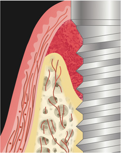
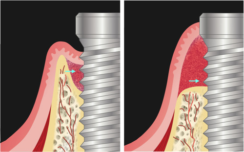
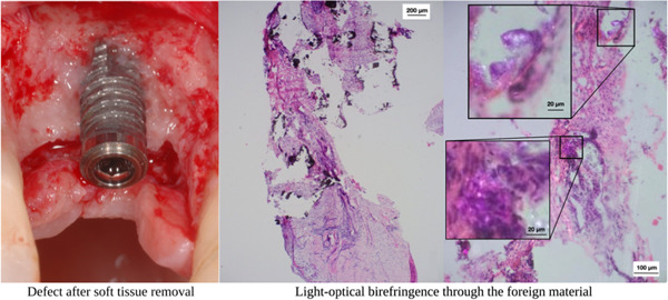
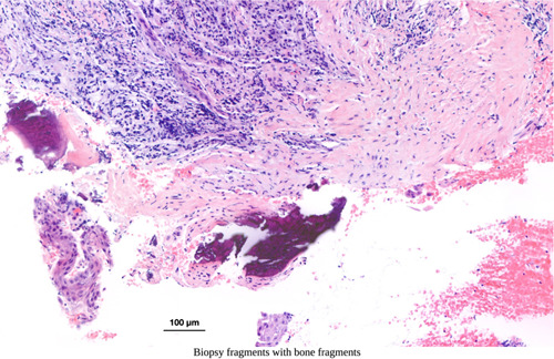
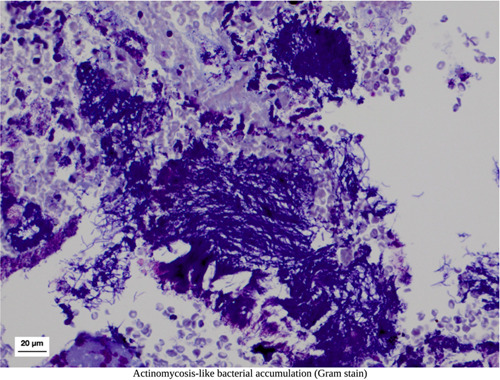
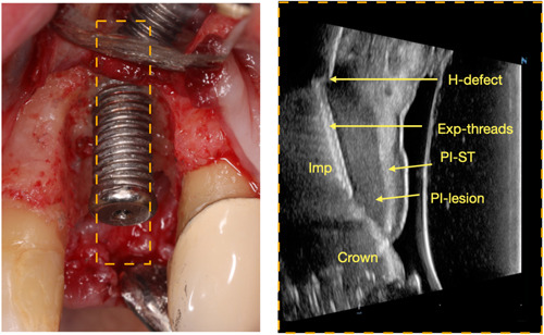
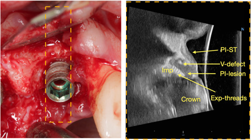
Results: The four interrelated negative conditions hampering effective reconstruction are: inferior tissue perfusion, unfavorable bone topography, ineffective surface treatment, and unstable wound. First, peri-implant tissues resemble scars with reduced cellularity and vascularity, coupled with the absence of the periodontal ligament plexuses and the avascular implant and biomaterials, maintaining primary closure is a challenge, which is critical for regeneration. Second, defect morphology and bone topography surrounding implants determine the reconstructive potential. Unfortunately, noncontained defects are frequently encountered, with a combination of suprabony (horizontal bone loss) and infrabony (vertical usually involving circumferential bone loss) defects. Third, current attempts for implant surface decontamination are insufficient due to inaccessible macrostructure and rough surfaces in the micro-scale. Histologic evaluation has shown bacteria aggregation and calcified deposits around implants. Lastly, wound stability is difficult to achieve due to inherent soft tissue biomechanical quality and quantity deficiencies and mobile bone particulates. Opportunities to tackle the abovementioned challenges include the use of novel imaging technologies, such as high-frequency dental ultrasound and laser speckle imaging to evaluate tissue perfusion, soft tissue quality/quantity, and bone topography pre-surgically. The use of the operating microscope could allow better visualization and removal of etiologic factors. Strategies to improve soft tissue quality may include preoperative control of soft tissue inflammation and the potential use of biologics. Methods such as fixation to stabilize the biomaterials could be beneficial.
Conclusions: A more nuanced understanding of the current challenges and opportunities can lead to more effective preoperative and postoperative care protocols, ultimately improving the success rate of reconstructive procedures.
Keywords: microsurgery; peri-implantitis; ultrasonography; wound healing.
© 2023 The Authors. Clinical and Experimental Dental Research published by John Wiley & Sons Ltd.
Conflict of interest statement
The authors declare no conflict of interest.
Figures
Similar articles
-
Surgical reconstruction of peri-implantitis with adjunctive antimicrobial photodynamic therapy: A case report with 5-year follow-up.Clin Adv Periodontics. 2024 Sep;14(3):185-191. doi: 10.1002/cap.10275. Epub 2023 Nov 29. Clin Adv Periodontics. 2024. PMID: 38029379
-
Operating microscope-assisted reconstructive strategy for peri-implantitis: A case series report.Clin Adv Periodontics. 2024 Sep;14(3):149-156. doi: 10.1002/cap.10265. Epub 2023 Sep 19. Clin Adv Periodontics. 2024. PMID: 37724638
-
Bone regeneration as treatment of peri-implant disease: A narrative review.Clin Implant Dent Relat Res. 2023 Aug;25(4):696-709. doi: 10.1111/cid.13209. Epub 2023 May 17. Clin Implant Dent Relat Res. 2023. PMID: 37199027 Review.
-
Hard and soft tissue regeneration of severe peri-implantitis defects with the laser-assisted peri-implant defect regeneration technique: 3-year results.Int J Implant Dent. 2023 Feb 5;9(1):3. doi: 10.1186/s40729-023-00467-1. Int J Implant Dent. 2023. PMID: 36739596 Free PMC article.
-
Selecting biomaterials in the reconstructive therapy of peri-implantitis.Periodontol 2000. 2024 Feb;94(1):192-212. doi: 10.1111/prd.12523. Epub 2023 Sep 20. Periodontol 2000. 2024. PMID: 37728141 Review.
Cited by
-
Enhanced peri-implantitis management through purple-LED irradiation coupled with silver ion application and calcium phosphate gene transfection carrier coating.Sci Rep. 2025 Apr 21;15(1):13759. doi: 10.1038/s41598-025-96075-7. Sci Rep. 2025. PMID: 40258901 Free PMC article.
-
Evaluation of peri-implant perfusion in patients who underwent avascular augmentation or microvascular reconstruction using laser Doppler flowmetry and tissue spectrophotometry: a prospective comparative clinical study.Clin Oral Investig. 2024 Jul 17;28(8):431. doi: 10.1007/s00784-024-05825-w. Clin Oral Investig. 2024. PMID: 39017918 Free PMC article.
-
Optical imaging guidance in oncologic surgery and interventional oncology.Pharmacol Res. 2025 Feb;212:107612. doi: 10.1016/j.phrs.2025.107612. Epub 2025 Jan 17. Pharmacol Res. 2025. PMID: 39826822 Free PMC article. Review.
-
Comparative evaluation of the effect of impregnated retraction cord versus laser on gingival attachment level and pain perception following retraction for subgingival margins - A prospective, split-mouth, controlled, clinical study.J Indian Prosthodont Soc. 2024 Apr 1;24(2):136-143. doi: 10.4103/jips.jips_437_23. Epub 2024 Apr 23. J Indian Prosthodont Soc. 2024. PMID: 38650338 Free PMC article.
-
Challenges of Gingival Surgery Approaches in the Treatment of Peri-Implantitis: A Systematic Review.J Pharm Bioallied Sci. 2025 May;17(Suppl 1):S159-S162. doi: 10.4103/jpbs.jpbs_405_25. Epub 2025 Apr 29. J Pharm Bioallied Sci. 2025. PMID: 40511003 Free PMC article. Review.
References
-
- Alghamdi, H. S. , & Jansen, J. A. (2020). The development and future of dental implants. Dental Materials Journal, 39(2), 167–172. - PubMed
-
- Bosshardt, D. D. , Chappuis, V. , & Buser, D. (2017). Osseointegration of titanium, titanium alloy and zirconia dental implants: Current knowledge and open questions. Periodontology 2000, 73(1), 22–40. - PubMed
-
- Burkhardt, R. , & Lang, N. P. (2010). Role of flap tension in primary wound closure of mucoperiosteal flaps: A prospective cohort study. Clinical Oral Implants Research, 21(1), 50–54. - PubMed
Publication types
MeSH terms
Substances
Grants and funding
LinkOut - more resources
Full Text Sources
