. 2023 Nov 1;31(11):3243-3258.
doi: 10.1016/j.ymthe.2023.09.013. Epub 2023 Sep 20.

Human macrophage migration inhibitory factor potentiates mesenchymal stromal cell efficacy in a clinically relevant model of allergic asthma

Affiliations

Human macrophage migration inhibitory factor potentiates mesenchymal stromal cell efficacy in a clinically relevant model of allergic asthma

Ian J Hawthorne et al. Mol Ther. .

Abstract

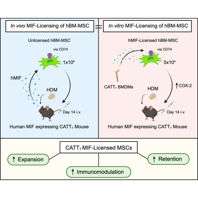
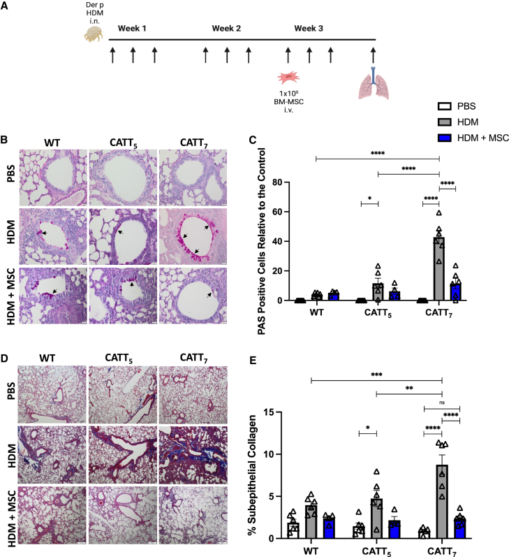
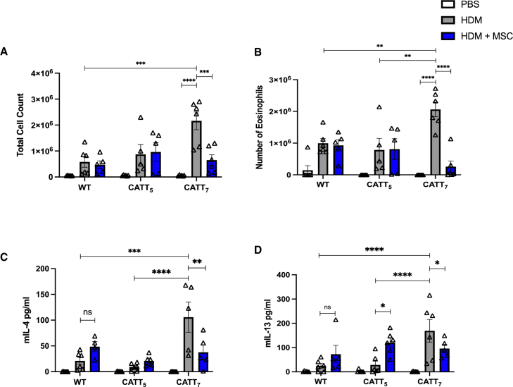
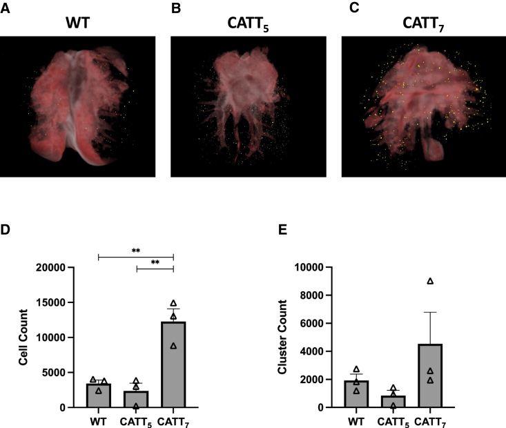
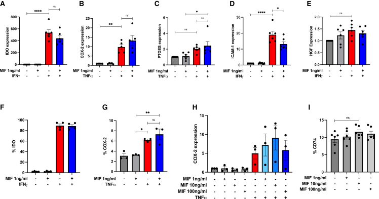
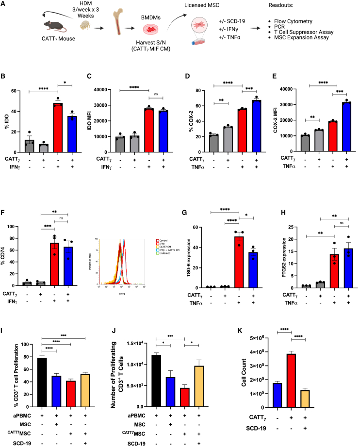
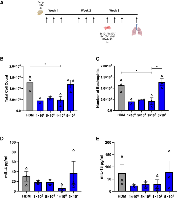
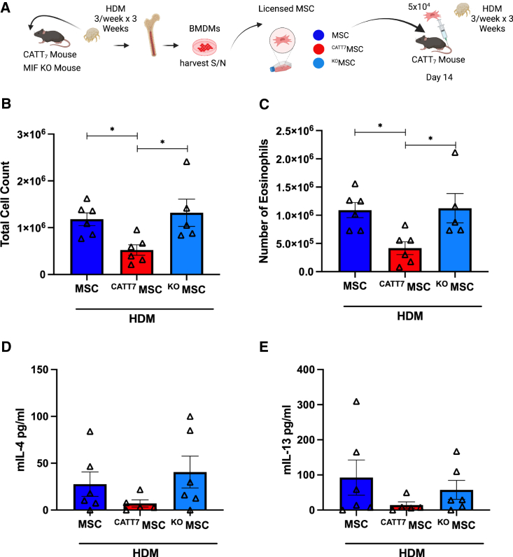
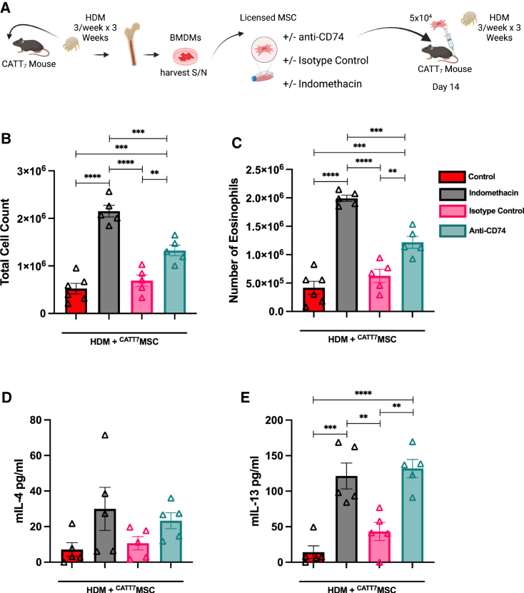
Current asthma therapies focus on reducing symptoms but fail to restore existing structural damage. Mesenchymal stromal cell (MSC) administration can ameliorate airway inflammation and reverse airway remodeling. However, differences in patient disease microenvironments seem to influence MSC therapeutic effects. A polymorphic CATT tetranucleotide repeat at position 794 of the human macrophage migration inhibitory factor (hMIF) gene has been associated with increased susceptibility to and severity of asthma. We investigated the efficacy of human MSCs in high- vs. low-hMIF environments and the impact of MIF pre-licensing of MSCs using humanized MIF mice in a clinically relevant house dust mite (HDM) model of allergic asthma. MSCs significantly attenuated airway inflammation and airway remodeling in high-MIF-expressing CATT7 mice but not in CATT5 or wild-type littermates. Differences in efficacy were correlated with increased MSC retention in the lungs of CATT7 mice. MIF licensing potentiated MSC anti-inflammatory effects at a previously ineffective dose. Mechanistically, MIF binding to CD74 expressed on MSCs leads to upregulation of cyclooxygenase 2 (COX-2) expression. Blockade of CD74 or COX-2 function in MSCs prior to administration attenuated the efficacy of MIF-licensed MSCs in vivo. These findings suggest that MSC administration may be more efficacious in severe asthma patients with high MIF genotypes (CATT6/7/8).

Keywords: allergic asthma; cyclooxygenase; house dust mite; macrophage migration inhibitory factor; mesenchymal stromal cells.


Conflict of interest statement

Declaration of interests The authors declare no competing interests.

Figures

None
Graphical abstract
Figure 1
Figure 1
Human BM-MSCs significantly reduce goblet cell metaplasia and collagen deposition in CATT7 mice challenged with HDM (A) PBS and HDM groups received PBS or HDM i.n. 3 times a week for 3 consecutive weeks. 1 × 106 human BM-MSCs were administered i.v. to the HDM+MSC groups on day 14. Mice were sacrificed on day 21 (schematic created with BioRender). (B) Representative images of lung tissue from WT, CATT5, and CATT7 mice stained with periodic acid-Schiff (PAS) at 20× magnification; scale bar, 20 μm. Arrows show examples of mucin-containing goblet cells. (C) Goblet cell hyperplasia was investigated through the quantitation of PAS-positive cells. (D) Representative images of lung tissue stained with Masson’s trichome at 4× magnification; scale bar, 200 μm. (E) Quantitation of the percentage of subepithelial collagen. Data are presented as mean ± SEM; n = 6 per group. Human BM-MSC donors 001-177 and 003-310 were used (RoosterBio). Statistical analysis was carried out using one-way ANOVA followed by the post hoc Tukey’s multiple-comparisons test: ∗p < 0.05, ∗∗∗p < 0.001, ∗∗∗∗p < 0.0001; ns, non-significant.
Figure 2
Figure 2
Human BM-MSCs significantly reduce levels of Th2 cytokines in the BALF of CATT7 mice challenged with HDM PBS and HDM groups received PBS or HDM i.n. 3 times a week for 3 consecutive weeks. 1 × 106 human BM-MSCs were administered i.v. to the HDM+MSC groups on day 14. BAL was performed 4 h post final HDM challenge on day 18. (A) Total cell count recovered from the BALF. (B) BALF eosinophil count, determined by differential staining of cytospins. (C and D) Cytokine levels of (C) IL-4 and (D) IL-13 in the BALF, determined by ELISA. White bars, PBS; gray bars, HDM blue bars, HDM+MSC. Data are presented as mean ± SEM; n = 5–6 per group. Human BM-MSC donors 001-177 and 003-310 were used (RoosterBio). Statistical analysis was carried out using one-way ANOVA followed by the post hoc Tukey’s multiple-comparisons test: ∗p < 0.05, ∗∗p < 0.01, ∗∗∗p < 0.001, ∗∗∗∗p < 0.0001, ns, non-significant.
Figure 3
Figure 3
High levels of hMIF significantly enhance BM-MSC retention in an HDM model of allergic asthma HDM were administered i.n. 3 times a week for 2 weeks. On day 14, 1 × 106 Qtracker 625-labeled hMSCs were administered i.v. to WT, CATT5, or CATT7 mice. 24 h later the lungs were harvested, embedded in OCT compound and frozen at −80. Tissue blocks were sectioned and imaged using the CryoViz (BioInvision) imaging system. (A–C) 3D images show representative lung images from (A) WT, (B) CATT5, and (C) CATT7 mice, with detected MSCs shown in yellow. (D and E) Total number of MSCs detected in the lungs (D) and number of clusters (E) were quantified using CryoViz quantification software. Data are presented as mean ± SEM; n = 3 per group. Human BM-MSC donor 001-177 was used (RoosterBio). Statistical analysis was carried out using one-way ANOVA followed by the post hoc Tukey’s multiple-comparisons test: ∗∗p < 0.01.
Figure 4
Figure 4
Influence of rhMIF licensing on MSC expression of immunomodulatory factors in vitro (A–E) Gene expression of IDO, COX-2, PTGES, ICAM-1, and HGF by hBM-MSCs after stimulation with rhMIF (1 ng/mL), human TNF-α or human IFNγ for 24 h. Data are presented as mean ± SEM and are representative of 3 independent experiments. Human BM-MSC donors 001-177, 003-310, and 003-307 were used (RoosterBio). Statistical analysis was carried out using one-way ANOVA: ∗p < 0.05, ∗∗p < 0.01, ∗∗∗∗p < 0.0001, ns, non-significant.
Figure 5
Figure 5
CATT7 MIF licensing enhances MSC expansion and immunosuppressive function in vitro (A) Schematic (created using BioRender) depicting the generation of CATT7 MIF CM and experimental design. (B–E) Percentage or mean fluorescence intensity (MFI) of IDO or COX-2 expression in human BM-MSCs, measured by flow cytometry after cells were stimulated with CATT7 MIF CM, human TNF-α, or human IFNγ for 24 h. (F) Percentage expression and representative histogram plots of CD74 surface expression on human MSCs, measured by flow cytometry after cells were stimulated with CATT7 MIF CM and human IFNγ for 24 h. (G and H) Relative gene expression of TSG-6 and PTGS2 by hBM-MSCs after cells were stimulated with endogenous hMIF (CATT7 CM) and human TNF-α for 6 h. (I and J) Licensing of MSCs with supernatants generated from BMDMs from CATT7 HDM-challenged mice enhances MSC suppression of (I) frequency (percent) and (J) absolute number of CD3+ T cells proliferating. Blockade of MIF using SCD-19 (100 μM) in the BMDM supernatants 1 h before addition to MSCs abrogates the enhanced effect of MIF on MSC suppression of T cell proliferation. (K) Licensing of MSCs with CATT7 MIF CM enhances MSC expansion in vitro. Addition of the MIF inhibitor SCD-19 (100 μM) to CATT7 MIF CM 1 h before MSC licensing prevents MIF-enhanced MSC expansion. Data are presented as mean ± SEM and are representative of 3 independent experiments. Human BM-MSC donors 001-177, 003-310, and 003-307 were used (RoosterBio). Statistical analysis was carried out using a one-way ANOVA or unpaired t test: ∗p < 0.05, ∗∗p < 0.01, ∗∗∗p < 0.001, ∗∗∗∗p < 0.0001.
Figure 6
Figure 6
Titration of BM-MSC doses in CATT7 mice challenged with HDM (A) To determine the point where MSCs lose efficacy in CATT7 mice, a range of doses were administered on day 14. BAL was performed 4 h post final HDM challenge on day 18 (schematic created with BioRender). (B) Total cell count recovered from the BALF. (C) Number of eosinophils obtained from the BALF. (D and E) Cytokine levels of (D) IL-4 and (E) IL-13 in the BALF, determined by ELISA. Data are presented as mean ± SEM; n = 2–3 per group. Human BM-MSC donors 001-177 and 003-310 were used (RoosterBio). Statistical analysis was carried out using one-way ANOVA followed by the post hoc Tukey’s multiple-comparisons test: ∗p < 0.05.
Figure 7
Figure 7
MIF licensing restores MSC efficacy at low doses in CATT7 mice (A) 5 × 104 MSCs were administered to HDM-challenged CATT7 mice on day 14. CATT7MSCs were licensed with CATT7 BMDM supernatant for 24 h prior to i.v. administration. The control group KOMSCs were generated by licensing MSCs with BMDM supernatant from MIF KO mice 24 h prior to i.v. administration. BAL was performed 4 h post final HDM challenge on day 18 (schematic created with BioRender). (B and C) Total number of cells in the BALF were determined (B), and differential cell counts were performed on the collected cells to determine the numbers of eosinophils (C). (D and E) Cytokine levels of (D) IL-4 and (E) IL-13 in the BALF determined by ELISA. Data are presented as mean ± SEM; n = 5–6 per group. Human BM-MSC donors 001-177 and 003-310 were used (RoosterBio). Statistical analysis was carried out using one-way ANOVA followed by the post hoc Tukey’s multiple-comparisons test: ∗p < 0.05.
Figure 8
Figure 8
MIF-Licensed MSCs mediate their protective effects in HDM-induced allergic airway inflammation in a CD74- and COX-2-dependent manner in CATT7 mice (A) 5 × 104 MSCs were exposed to the COX-2 inhibitor indomethacin, an anti-CD74 neutralizing antibody, or an isotype control antibody for 24 h in vitro. All MSCs were licensed with CATT7 BMDM supernatant for 24 h prior to i.v. administration. BAL was performed 4 h post final HDM challenge on day 18 (schematic created with BioRender). (B and C) Total number of cells in the BALF were determined (B), and differential cell counts were performed on the collected cells to determine the numbers of eosinophils (C). (D and E) Cytokine levels of (D) IL-4 and (E) IL-13 in the BALF, determined by ELISA. Data are presented as mean ± SEM; n = 5–6 per group. Human BM-MSC donors 001-177 and 003-310 were used (RoosterBio). Statistical analysis was carried out using one-way ANOVA followed by the post hoc Tukey’s multiple-comparisons test: ∗∗p < 0.01, ∗∗∗ p < 0.001, ∗∗∗∗p < 0.0001.

Comment in

Similar articles

Cited by

References

    1. Sverrild A., Hansen S., Hvidtfeldt M., Clausson C.M., Cozzolino O., Cerps S., Uller L., Backer V., Erjefält J., Porsbjerg C. The effect of tezepelumab on airway hyperresponsiveness to mannitol in asthma (UPSTREAM) Eur. Respir. J. 2022;59 - PubMed
    1. Bleecker E.R., FitzGerald J.M., Chanez P., Papi A., Weinstein S.F., Barker P., Sproule S., Gilmartin G., Aurivillius M., Werkström V., et al. Efficacy and safety of benralizumab for patients with severe asthma uncontrolled with high-dosage inhaled corticosteroids and long-acting β2-agonists (SIROCCO): a randomised, multicentre, placebo-controlled phase 3 trial. Lancet Lond. Engl. 2016;388:2115–2127. - PubMed
    1. Corren J., Castro M., O’Riordan T., Hanania N.A., Pavord I.D., Quirce S., Chipps B.E., Wenzel S.E., Thangavelu K., Rice M.S., et al. Dupilumab efficacy in patients with uncontrolled, moderate-to-severe allergic asthma. J. Allergy Clin. Immunol. Pract. 2020;8:516–526. - PubMed
    1. Ortega H.G., Yancey S.W., Mayer B., Gunsoy N.B., Keene O.N., Bleecker E.R., Brightling C.E., Pavord I.D. Severe eosinophilic asthma treated with mepolizumab stratified by baseline eosinophil thresholds: a secondary analysis of the DREAM and MENSA studies. Lancet Respir. Med. 2016;4:549–556. - PubMed
    1. Chan R., Lipworth B. Efficacy of biologic therapy on airway hyperresponsiveness in asthma. Ann. Allergy Asthma Immunol. Off Publ. Am. Coll. Allergy Asthma Immunol. 2023:00121–00127. - PubMed

Publication types

Substances