. 2023 Sep 13:31:497-508.
doi: 10.1016/j.bioactmat.2023.08.019. eCollection 2024 Jan.

Silver-integrated EDM processing of TiAl6V4 implant material has antibacterial capacity while optimizing osseointegration

Affiliations

Silver-integrated EDM processing of TiAl6V4 implant material has antibacterial capacity while optimizing osseointegration

Hilmar Büssemaker et al. Bioact Mater. .

Abstract

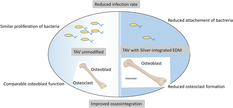
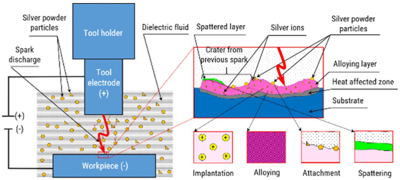
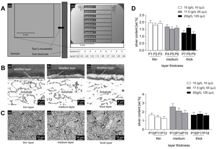
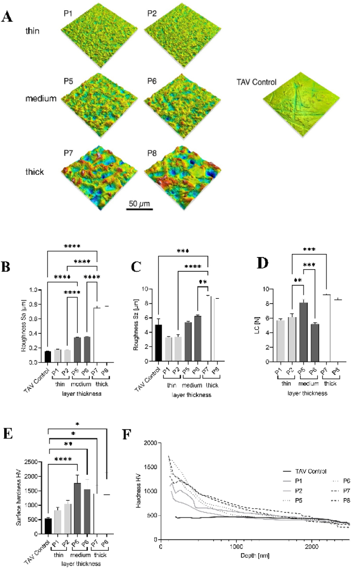
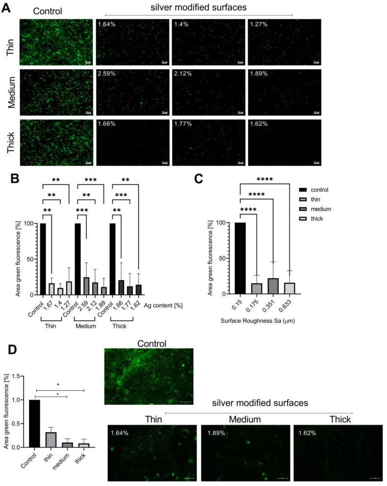
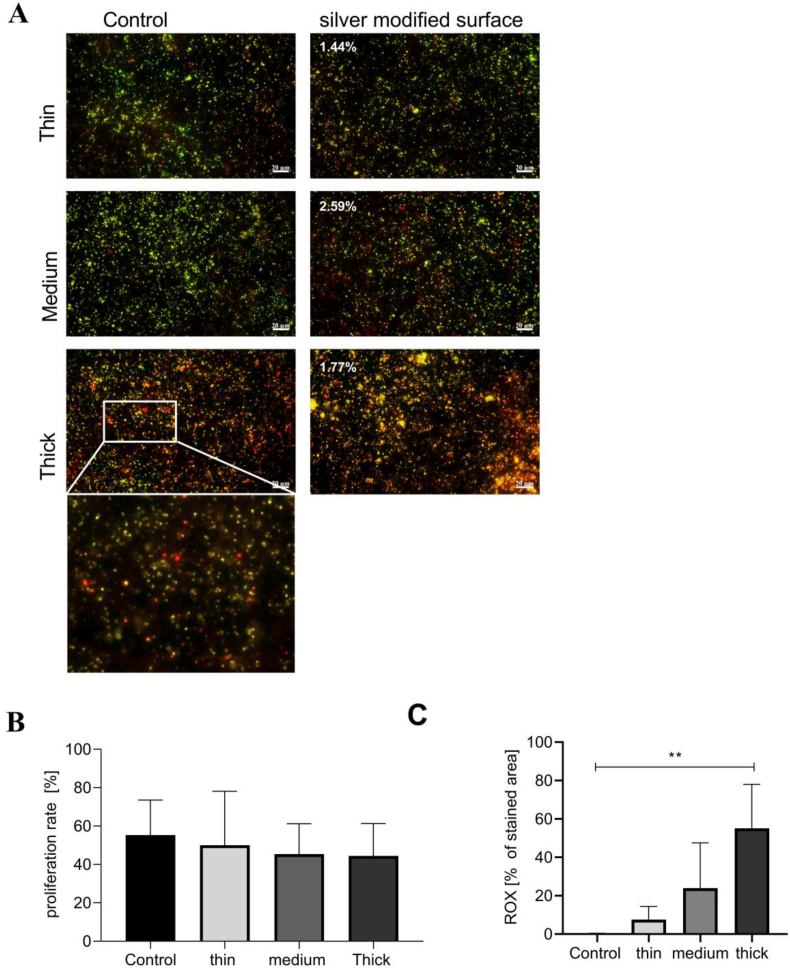
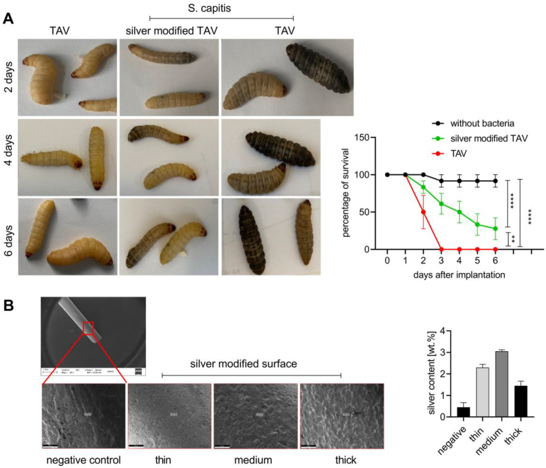
Periprosthetic joint infections (PJI) are a common reason for orthopedic revision surgeries. It has been shown that the silver surface modification of a titanium alloy (Ti-6Al-4V) by PMEDM (powder mixed electrical discharge machining) exhibits an antibacterial effect on Staphylococcus spp. adhesion. Whether the thickness of the silver-modified surface influences the adhesion and proliferation of bacteria as well as the ossification processes and in-vivo antibacterial capacity has not been investigated before. Therefore, the aim of this work is to investigate the antibacterial effect as well as the in vitro ossification process depending on the thickness of PMEDM silver modified surfaces. The attachment of S. aureus on the PMEDM modified surfaces was significantly lower than on comparative control samples, independently of the tested surface properties. Bacterial proliferation, however, was not affected by the silver content in the surface layer. We observed a long-term effect of antibacterial capacity in vitro, as well as in vivo. An induction of ROS, as indicator for oxidative stress, was observed in the bacteria, but not in osteoblast-like cells. No influence on the in vitro osteoblast function was observed, whereas osteoclast formation was drastically reduced on the silver surface. No changes in cell death, the metabolic activity and oxidative stress was measured in osteoblasts. We show that already small amounts of silver exhibit a significant antibacterial capacity while not influencing the osteoblast function. Therefore, PMEDM using silver nano-powder admixed to the dielectric represents a promising technology to shape and concurrently modify implant surfaces to reduce infections while at the same time optimizing bone ingrowth of endoprosthesis.


Conflict of interest statement

The authors have no financial or personal interest or belief that could affect their objectivity with regard to the present manuscript.

Figures

Image 1
Graphical abstract
Fig. 1
Fig. 1
Principle of the PMEDM surface modification [26].
Fig. 2
Fig. 2
(A) Distribution of deposited silver on the (10 × 10) mm2 modified area: (a) top view image of the sample surface; (b) analyzed silver contents of EDS spectra; sample modified using 17.5 g/l powder concentration and 55 μJ discharge energy. (B) SEM images showing the modified layers in cross-section after PMEDM using (a) 15 g/l, 10 μJ; (b) 17.5 g/l, 55 μJ and (c) 20 g/l and 125 μJ. (C) Top view SEM images of the modified surfaces using a backscattered electrons analysis: (a) 15 g/l, 10 μJ; (b) 17.5 g/l, 55 μJ and (c) 20 g/l and 125 μJ. Arrows point on microcracks. (D) Average silver contents (Mean ± SD) of the investigated sample surfaces modified by applying different powder concentrations (g/l) and discharge energies (μJ): upper panel: samples with (10 × 10) mm2 modified area size; Lower panel: samples with (3 × 4) mm2 modified area size.
Fig. 3
Fig. 3
(A)Representative 3D topographic images of the surfaces B) measured arithmetic average roughness (Sa) C) average maximum profile height (Sz) D) LC values from layer adhesion scratch-test (n = 3 ± SD) scratch test D) average indentation hardness constant load cycle (n = 6 ± SD) E) dynamic measurements in sinus mode with depth profiles (n = 6). The statistical analyses was performed using an ordinary one-way ANOVA with a Dunett's post-hoc test. *p < 0.05, **p < 0.01,***p < 0.001, ****p < 0.0001.
Fig. 4
Fig. 4
Significant reduction of S. aureus attachment to silver modified surface layer independent of layer thickness. A) Representative images of the S. aureus/pSB2035 were taken, every green dot represents one bacterium on the surface. The pictures were evaluated depending on the layer thickness and the surface roughness (μm). The pictures were taken with a magnification of 630 fold (scale bar: 20 μm). (B) For the quantification of the fluorescent area the values are normalized to the unmodified TAV control for each experimental setup (Mean ± SD). 5 images were taken per sample and the experiment was repeated in five independent setups (N = 5: n = 25). For the statistical evaluation a Kruskal Wallis test with Benjamini, Krieger and Yekultieri posthoc testwas used (C) Quantification of fluorescent area representing SH1000/pSB2035 attachment to silver modified surface layers evaluated depending on the surface roughness (Sa values). The values are normalized to the unmodified TAV control for each experimental setup (Mean ± SD). 5 images were taken per sample and the experiment was repeated in five independent setups (N = 5: n = 25). D.) Representative images of the S. capitis were taken, every green dot represents one bacterium on the surface. The pictures were evaluated depending on the layer thickness and the surface roughness (μm). The pictures were taken with a magnification of 630 fold (scale bar: 50 μm). For the quantification of the fluorescent area, the values are normalized to the unmodified TAV control for each experimental setup (Mean ± SD). 5 images were taken per sample and the experiment was repeated in five independent setups (N = 3: n = 15). For the statistical evaluation a Friedmann test with Benjamini, Krieger and Yekultieri posthoc test was used; *p < 0.05, **p < 0.01,***p < 0.001,****p < 0.0001.
Fig. 5
Fig. 5
Silver does not inhibit the proliferation of attaching S. aureus (A) Representative images of SH1000/pMD303 on control or silver modified surfaces. Every red dot represents a photo converted bacterium and every green dot shows a proliferating bacterium. The pictures were evaluated according to the layer thickness. The pictures were taken with a magnification of 630 fold (scale bar: 20 μm). (B) For the quantification, we analyzed the proliferation rate of SH1000/pMD303 on different silver modified based on the layer thickness compared to unmodified TAV controls (Mean ± SD). 5 images were taken per sample and the experiment was repeated in five independent setups (N = 5: n = 25). The statistical evaluation was performed using a One-way ANOVA with Dunnett's posthoc test. C) ROX assay for quantification of oxidative stress in adhering bacteria. For the quantification, we analyzed the ROX stained area on different silver modified based on the layer thickness compared to TAV controls (Mean ± SD). 5 images were taken per sample and the experiment was repeated in five independent setups (N = 5: n = 25). The statistical evaluation was performed using a One-way ANOVA with Dunnett's posthoc test (F (3, 16) = 10.53; p = 0.0005). *p < 0.05, **p < 0.01,***p < 0.001,****p < 0.0001.
Fig. 6
Fig. 6
Reduced osteoclast formation, but no influence of silver modified surface layers on in vitro ossification. (A) The graph shows the statistical difference between the number of osteoclasts on a TAV control and the silver-containing samples; silver-containing samples are summarized under silver; N = 3; values are given with Mean ± SD, the statistical evaluation was performed using the Welch's t-test; **p = 0.028. Representative images of osteoclasts from BMMs on TAV controls. The cells were stained with Phalloidin (red) to visualize the cytoskeleton and DAPI (blue) for the cell nuclei; a magnification 400x (scale bar: 50 μm) was used. B.) Quantitative RT-PCR of TRAP expression in wt murine osteoclasts after 11 days of differentiation on silver-modified surfaces or the unmodified TAV control. GAPDH was used as housekeeping gene. The values are normalized to the control and analyzed using an unpaired t-test for statistical significance. C) Quantitative RT-PCR of cathepsin K expression in wt murine osteoclasts after 11 days of differentiation on silver-modified surfaces or the unmodified TAV control. GAPDH was used as housekeeping gene. The values are normalized to the control and analyzed using an unpaired t-test for statistical significance. D.) WST Assay for metabolic activity of SaOs-2 cell on the different silver modified samples. The experiment was repeated in five independent setups (N = 5). No difference in the metabolic activity was observed. The statistical evaluation was performed using a One-way ANOVA with Dunnett's posthoc test (F (3, 19) = 0.42; p = 0.74). D.) The intensity of DHE staining was evaluated for each silver condition. 5 images were taken per sample and the experiment was repeated in five independent setups (N = 5: n = 25). The statistical evaluation was performed using a One-way ANOVA with Dunnett's posthoc test (F (3, 16) = 3.078, p = 0.058). Representative images are shown for the positive control as well as the negative control (DAPI: blue, DHE: red). (F) Alizarin Red Ossification Assay of SaOs 2 cells was performed to calculate the calcification. No significant differences between the TAV control and the silver coatings were analyzed on different silver modified surfaces clustered based on the layer thickness and silver content compared to unmodified TAV controls (one-way ANOVA: with Dunnett's post-hoc was performed; ns > 0.05). *p < 0.05, **p < 0.01,***p < 0.001,****p < 0.0001.
Fig. 7
Fig. 7
Silver modified surfaces exhibit a marked in vivo antibacterial capacity (A) Representative images of Galleria mellonella larvae at the respective time points during the in vivo implant infection model (day 2, 4 and 6). The dark brown larvae are dead, whereas the light brown larvae are alive. A beginning darkening of larvae (melanisation) at the implantation site shows the spreading of infection and induction of immune response. For the quantification, we analyzed the number of living larvae each day in three independent experiments (Mean ± SD). (N = 3: n = 6–12). The statistical evaluation was performed using a two-way ANOVA with Tukey posthoc test. (B) SEM images showing the implant pin (top left). The red square indicates the area for SEM EDX analyses of the modified layers in a top view. The average silver contents (Mean ± SD) of the investigated sample surfaces are given in the graph. p = 0.0005). *p < 0.05, **p < 0.01,***p < 0.001,****p < 0.0001.
Fig. S1
Fig. S1

Similar articles

Cited by

References

    1. Wetters N.G., Murray T.G., Moric M., Sporer S.M., Paprosky W.G., Della Valle C.J. Risk factors for dislocation after revision total hip arthroplasty. Clin. Orthop. Relat. Res. 2013;471(2):410–416. - PMC - PubMed
    1. Zimmerli ATaW. Diagnosis and treatment of implant-associated septic arthritis and osteomyelitis. Curr. Infect. Dis. Rep. 2008;10:394–403. - PubMed
    1. Fehring T.K.O.S., Caltin T.F., Mason J.B. Articulating versus static spacers in revision total knee arthroplasty for sepsis. Orthopaed. Relat. Res. 2000;380:9–16. - PubMed
    1. Goldberg Alfred L., K I.C., Furuno Koji, Fagan Julie M., Baracos Vickie. Activation of protein breakdown and prostaglandin E2 production in rat skeletal muscle in fever is signaled by a macrophage product distinct from interleukin 1 or other known monokines. J. Clin. Invest. 1988;81:1378–1383. - PMC - PubMed
    1. Smucny M., Menendez M.E., Ring D., Feeley B.T., Zhang A.L. Inpatient surgical site infection after shoulder arthroplasty. J. Shoulder Elbow Surg. 2015;24(5):747–753. - PubMed

LinkOut - more resources