Review
. 2023 Sep 16:17:2705-2718.
doi: 10.2147/OPTH.S392665. eCollection 2023.

Keratoconus Diagnosis and Treatment: Recent Advances and Future Directions

Affiliations
Review

Keratoconus Diagnosis and Treatment: Recent Advances and Future Directions

Anh D Bui et al. Clin Ophthalmol. .

Abstract

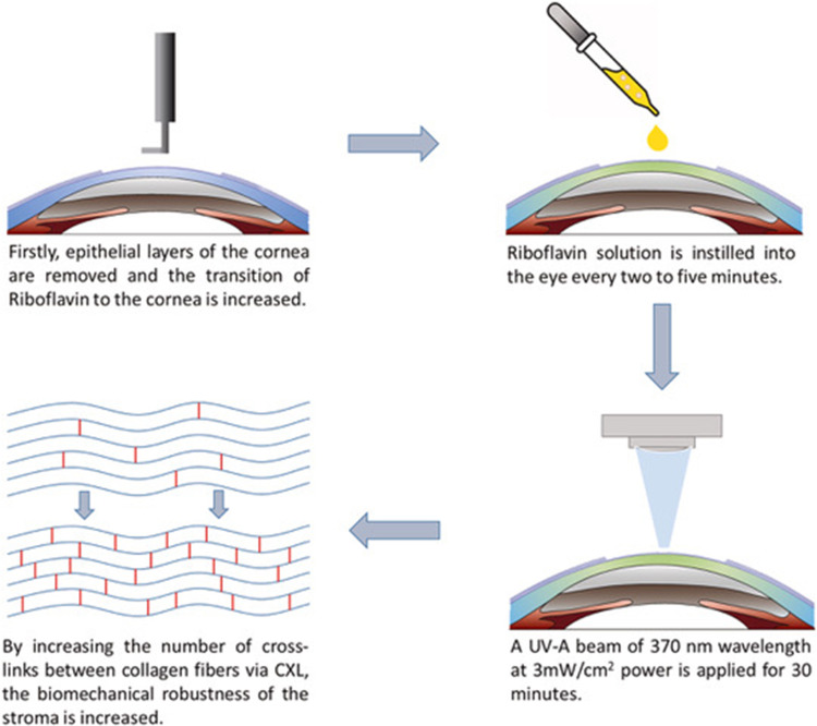
Keratoconus is a disorder characterized by progressive corneal thinning and steepening that may result in significant visual impairment secondary to high astigmatism, corneal scarring, or even corneal perforation. Early detection and screening of keratoconus are essential for effective management and treatment. Several screening methods, such as corneal topography and tomography, corneal biomechanics, and genetic testing, are being developed to detect keratoconus at an early stage. Once detected, prevention of progression is the mainstay of keratoconus management. Corneal collagen cross-linking is a minimally invasive treatment option that can slow or halt the progression of keratoconus. Additionally, recent studies have investigated the potential use of copper sulfate eye drops (IVMED-80) and extracellular vesicles to prevent the progression of keratoconus as non-invasive treatment options. For visual rehabilitation, currently available treatments include scleral lenses, intracorneal ring segments, corneal allogenic intrastromal ring segments, and deep anterior lamellar keratoplasty. The safety and efficacy of these emerging treatment options for keratoconus are currently being investigated.

Keywords: IVMED-80; corneal cross-linking; deep anterior lamellar keratoplasty; extracellular vesicles; intracorneal ring segments; keratoconus.


Conflict of interest statement

Dr Neel Pasricha reports personal fees from Iota Biosciences, Vanda Pharmaceuticals and Zeiss, outside the submitted work. The authors report no other conflicts of interest in this work.

Figures

Figure 1
Figure 1
(A) Scheimpflug image in a keratoconus patient. (B) Axial (power) map, anterior and posterior elevation maps, and global pachymetry map in a keratoconus patient.
Figure 2
Figure 2
Schematic depiction of the Dresden protocol: removal of corneal epithelium, instillation of riboflavin solution, and application of a UV-A beam of 370 nm wavelength.
Figure 3
Figure 3
Intracorneal ring segments implanted within the cornea.
Figure 4
Figure 4
(A and B) Introduction of a CAIRS into an intrastromal channel. The arrows highlight the CAIRS it is being inserted (C and D) Two CAIRS within the intrastromal channel.
Figure 5
Figure 5
Schematic representation of the anterior lamellar keratoplasty technique with varying depths of recipient cornea removal and donor corneal transplantation. (a and b) One-third of the anterior cornea is removed and replaced with a similarly sized donor cornea. (c and d) Larger amount of anterior cornea is removed and replaced. (e and f) Deep anterior lamellar keratoplasty where corneal tissue is removed up to the bare Descemet membrane and donor cornea without Descemet membrane is transplanted.

References

    1. Santodomingo-Rubido J, Carracedo G, Suzaki A, Villa-Collar C, Vincent SJ, Wolffsohn JS. Keratoconus: an updated review. Cont Lens Anterior Eye. 2022;45(3):101559. doi:10.1016/j.clae.2021.101559 - DOI - PubMed
    1. Naderan M, Shoar S, Rezagholizadeh F, Zolfaghari M, Naderan M. Characteristics and associations of keratoconus patients. Cont Lens Anterior Eye. 2015;38(3):199–205. doi:10.1016/j.clae.2015.01.008 - DOI - PubMed
    1. Hashemi H, Heydarian S, Hooshmand E, et al. The prevalence and risk factors for keratoconus: a systematic review and meta-analysis. Cornea. 2020;39(2):263–270. doi:10.1097/ICO.0000000000002150 - DOI - PubMed
    1. Galvis V, Sherwin T, Tello A, Merayo J, Barrera R, Acera A. Keratoconus: an inflammatory disorder? Eye. 2015;29(7):843–859. doi:10.1038/eye.2015.63 - DOI - PMC - PubMed
    1. Armstrong BK, Smith SD, Romac Coc I, Agarwal P, Mustapha N, Navon S. Screening for keratoconus in a high-risk adolescent population. Ophthalmic Epidemiol. 2021;28(3):191–197. doi:10.1080/09286586.2020.1804593 - DOI - PubMed