

Fused Imidazopyrazine-Based Tubulin Polymerization Inhibitors Inhibit Neuroblastoma Cell Function
- PMID: 37736192
- PMCID: PMC10510670
- DOI: 10.1021/acsmedchemlett.3c00298
Fused Imidazopyrazine-Based Tubulin Polymerization Inhibitors Inhibit Neuroblastoma Cell Function
Abstract
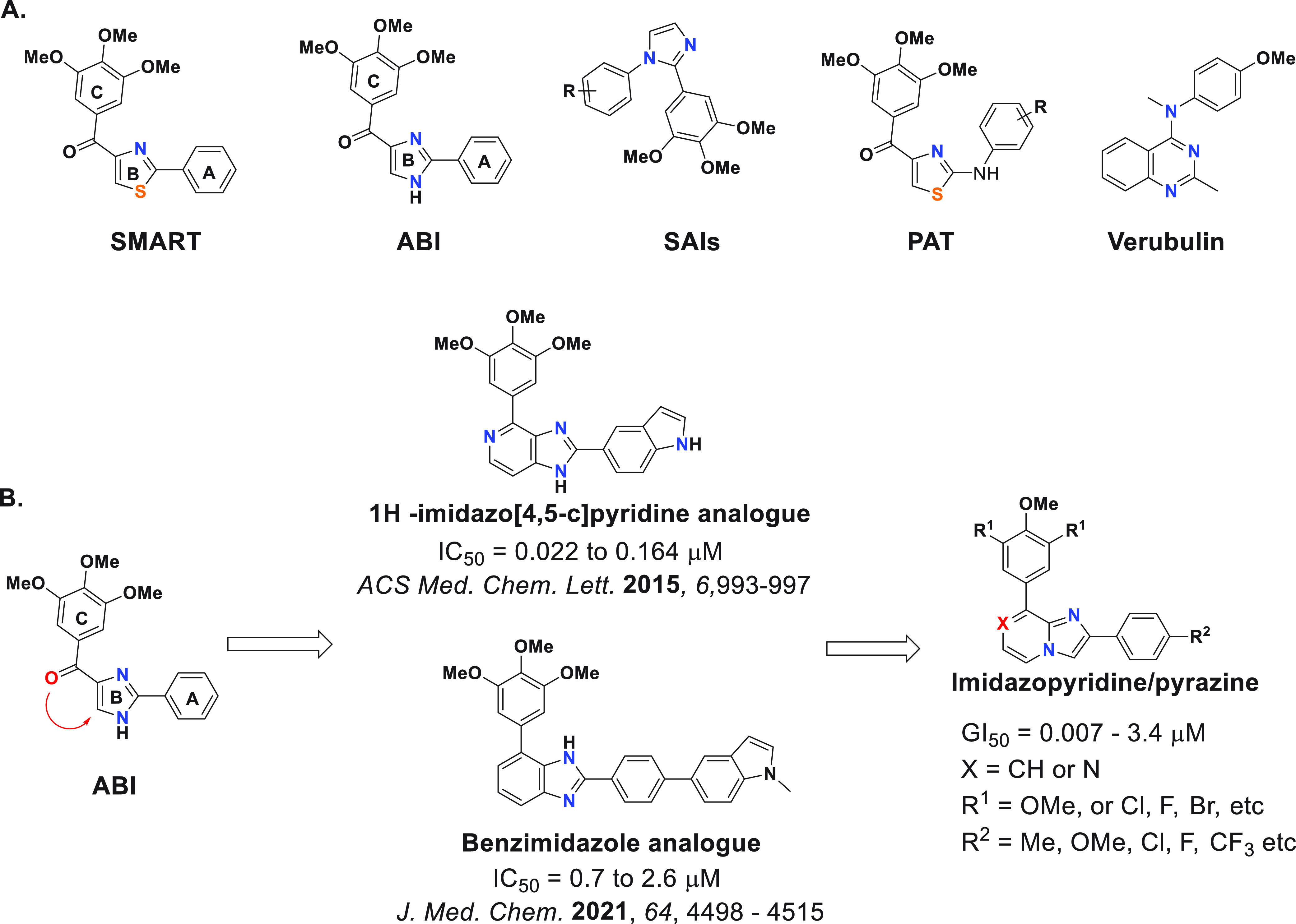
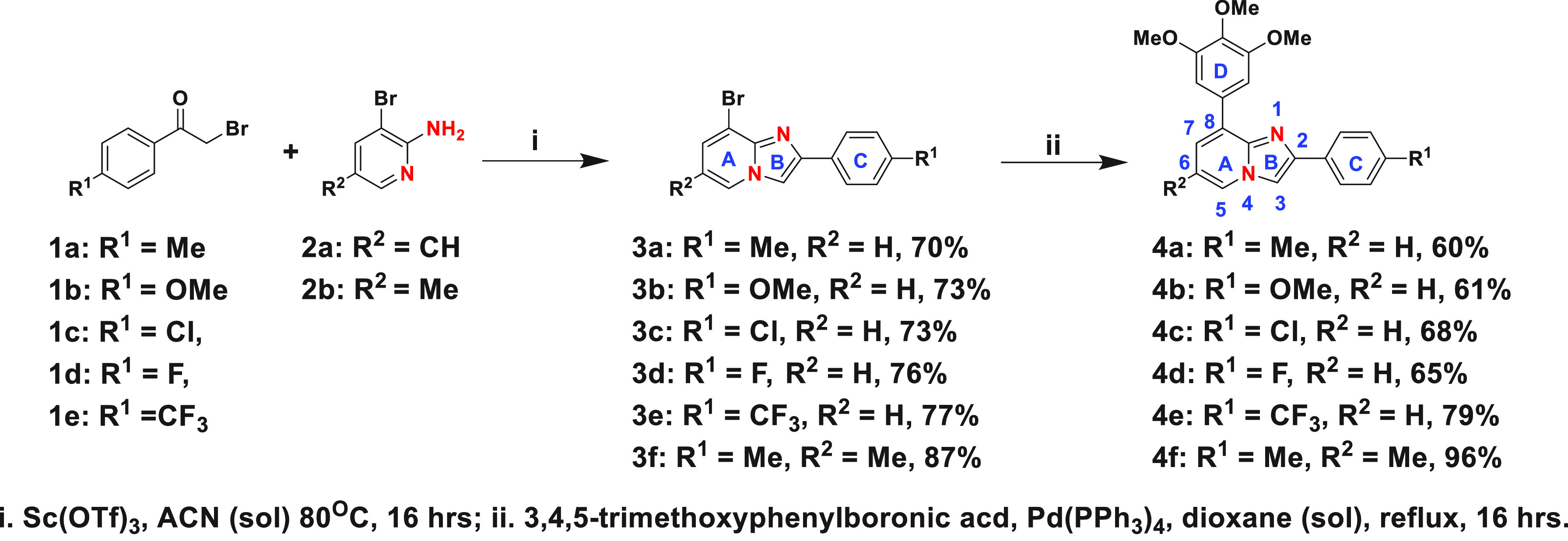
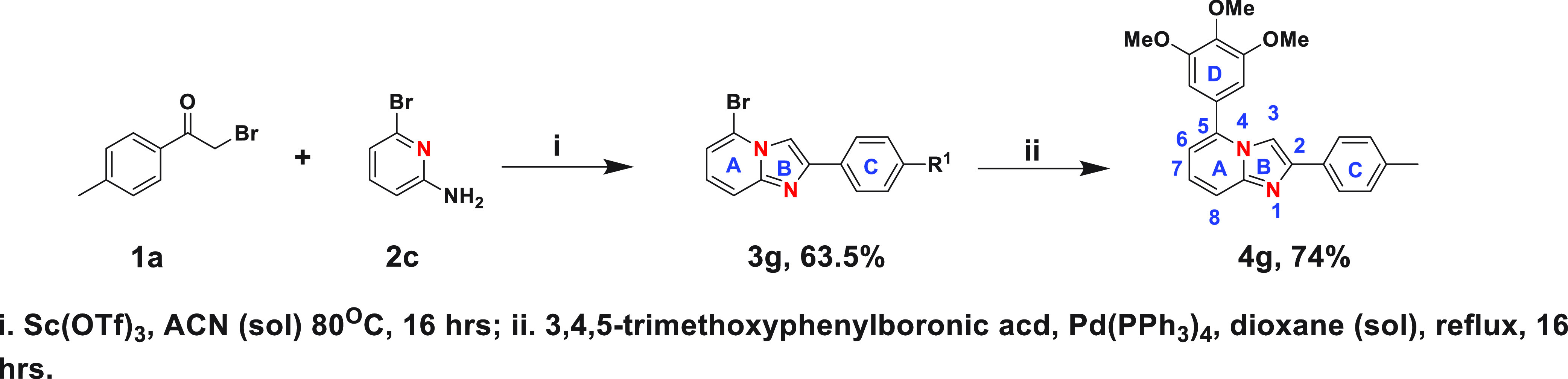
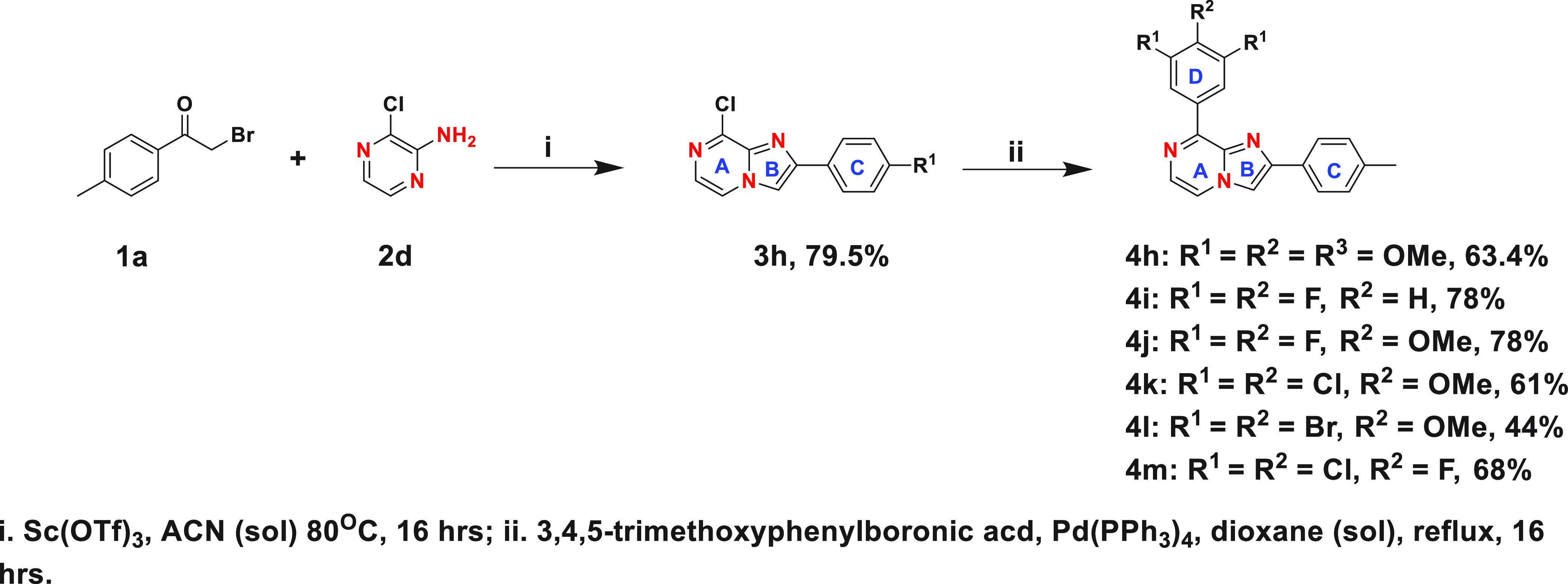
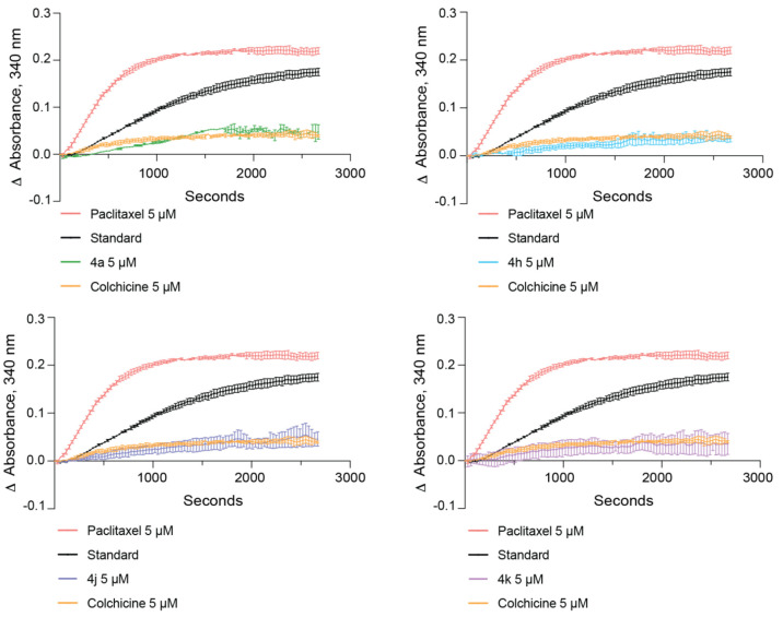
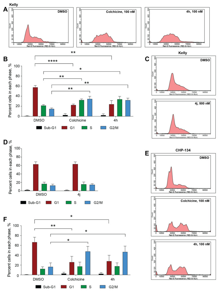
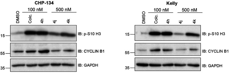
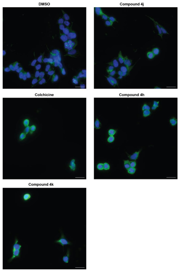
Targeting the colchicine binding site on tubulin is a promising approach for cancer treatment to overcome the limitations of current tubulin polymerization inhibitors. New classes of colchicine binding site inhibitors (CBSIs) are continually being uncovered; however, balancing metabolic stability and cellular potency remains an issue that needs to be resolved. Therefore, we designed and synthesized a series of novel fused imidazopyridine and -pyrazine CBSIs and evaluated their cellular activity, metabolic stability, and tubulin-binding properties. Evidence shows that the imidazo[1,2-a]pyrazine series are effective against neuroblastoma cell lines marked by MYCN amplification. Further assessment shows that a combination of an imidazo[1,2-a]pyrazine core with a trimethoxyphenyl ring D results in the highest cellular activity and binding characteristics compared with a dichloromethoxyphenyl or difluoromethoxyphenyl ring D. However, the metabolic stability of compounds with a dichloromethoxyphenyl or difluoromethoxyphenyl ring D is significantly higher than that of those containing a trimethoxyphenyl ring D, suggesting that improved metabolic stability is achieved with a moderate impact on potency.
© 2023 American Chemical Society.
Conflict of interest statement
The authors declare no competing financial interest.
Figures
Similar articles
-
Design and synthesis of (2-(phenylamino)thieno[3,2-d]pyrimidin-4-yl)(3,4,5-trimethoxyphenyl)methanone analogues as potent anti-tubulin polymerization agents.Eur J Med Chem. 2019 Dec 1;183:111679. doi: 10.1016/j.ejmech.2019.111679. Epub 2019 Sep 5. Eur J Med Chem. 2019. PMID: 31541870
-
Discovery of 6-aryl-2-(3,4,5-trimethoxyphenyl)thiazole[3,2-b][1,2,4]triazoles as potent tubulin polymerization inhibitors.Eur J Med Chem. 2023 Aug 5;256:115402. doi: 10.1016/j.ejmech.2023.115402. Epub 2023 Apr 23. Eur J Med Chem. 2023. PMID: 37182330
-
Design, synthesis, and bioevaluation of imidazo [1,2-a] pyrazine derivatives as tubulin polymerization inhibitors with potent anticancer activities.Bioorg Med Chem. 2022 Dec 15;76:117098. doi: 10.1016/j.bmc.2022.117098. Epub 2022 Nov 19. Bioorg Med Chem. 2022. PMID: 36455508
-
An overview of tubulin inhibitors that interact with the colchicine binding site.Pharm Res. 2012 Nov;29(11):2943-71. doi: 10.1007/s11095-012-0828-z. Epub 2012 Jul 20. Pharm Res. 2012. PMID: 22814904 Free PMC article. Review.
-
Tubulin inhibitors targeting the colchicine binding site: a perspective of privileged structures.Future Med Chem. 2017 Oct;9(15):1765-1794. doi: 10.4155/fmc-2017-0100. Epub 2017 Sep 20. Future Med Chem. 2017. PMID: 28929799 Review.
Cited by
-
Colchicine Binding Site Tubulin Inhibitors Impair Vincristine-Resistant Neuroblastoma Cell Function.Molecules. 2025 May 16;30(10):2186. doi: 10.3390/molecules30102186. Molecules. 2025. PMID: 40430358 Free PMC article.
References
-
- Gaspari R.; Prota A. E.; Bargsten K.; Cavalli A.; Steinmetz M. O. Structural Basis of cis- and trans-Combretastatin Binding to Tubulin. Chem 2017, 2 (1), 102–113. 10.1016/j.chempr.2016.12.005. - DOI
-
- Arnst K. E.; Banerjee S.; Wang Y.; Chen H.; Li Y.; Yang L.; Li W.; Miller D. D.; Li W. X-ray Crystal Structure Guided Discovery and Antitumor Efficacy of Dihydroquinoxalinone as Potent Tubulin Polymerization Inhibitors. ACS Chem. Biol. 2019, 14 (12), 2810–2821. 10.1021/acschembio.9b00696. - DOI - PubMed
-
- Cui M.-T.; Jiang L.; Goto M.; Hsu P.-L.; Li L.; Zhang Q.; Wei L.; Yuan S.-J.; Hamel E.; Morris-Natschke S. L.; Lee K. H.; Xie L. In Vivo and Mechanistic Studies on Antitumor Lead 7-Methoxy-4-(2-methylquinazolin-4-yl)-3,4-dihydroquinoxalin-2(1H)-one and Its Modification as a Novel Class of Tubulin-Binding Tumor-Vascular Disrupting Agents. J. Med. Chem. 2017, 60 (13), 5586–5598. 10.1021/acs.jmedchem.7b00273. - DOI - PMC - PubMed
Grants and funding
LinkOut - more resources
Full Text Sources
