

A Case of Bilateral Papilledema With Improved Clinical Symptoms by Venous Stenting for Superior Sagittal Sinus Stenosis
- PMID: 37736463
- PMCID: PMC10509490
- DOI: 10.7759/cureus.43828
A Case of Bilateral Papilledema With Improved Clinical Symptoms by Venous Stenting for Superior Sagittal Sinus Stenosis
Abstract
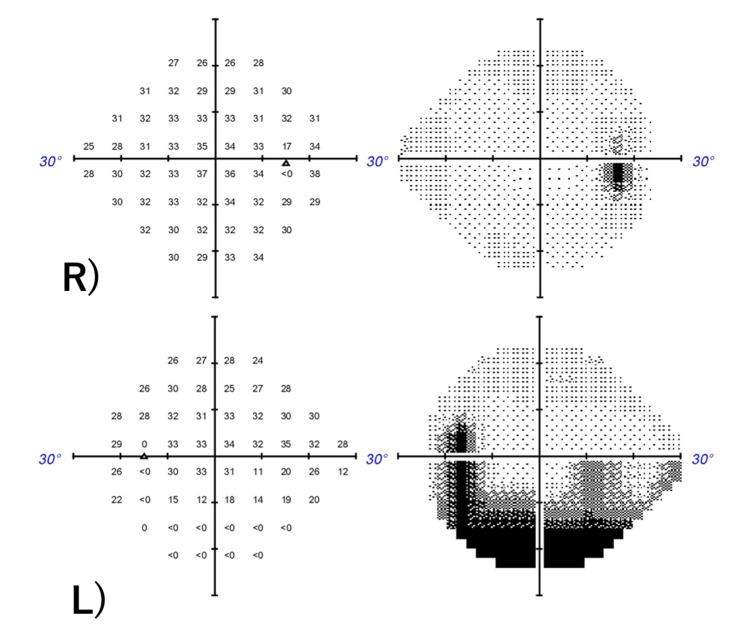
Superior sagittal sinus (SSS) obstruction causes intracranial hypertension, often requiring surgical stenting. Consensus on treating brain venous sinus stenosis, another cause, is lacking. This study reports a case of SSS stenosis and intracranial hypertension treated with venous stenting, improving bilateral papilledema. A 51-year-old with a headache and visual disturbance had papilledema and visual field loss. MR venography showed SSS stenosis, leading to a neurosurgery referral. Lumbar puncture confirmed intracranial hypertension (>35 cmH2O), prompting venous stenting. Post-procedure, papilledema, headache, and visual field loss improved. Venous stenting could be effective for SSS stenosis with clinically proven or recurrent pressure differences. Further cases are needed for standardization.
Keywords: intracranial hypertension; papilledema; sss; superior sagittal sinus stenosis; venous stenting.
Copyright © 2023, Miyoshi et al.
Conflict of interest statement
The authors have declared that no competing interests exist.
Figures


References
-
- EFNS guideline on the treatment of cerebral venous and sinus thrombosis. Einhäupl K, Bousser MG, de Bruijn SF, Ferro JM, Martinelli I, Masuhr F, Stam J. Eur J Neurol. 2006;13:553–559. - PubMed
-
- Cerebral venous thrombosis: a practical guide. Ulivi L, Squitieri M, Cohen H, Cowley P, Werring DJ. Pract Neurol. 2020;20:356–367. - PubMed
-
- Causes and predictors of death in cerebral venous thrombosis. Canhão P, Ferro JM, Lindgren AG, Bousser MG, Stam J, Barinagarrementeria F. Stroke. 2005;36:1720–1725. - PubMed
-
- Diagnosis and management of cerebral venous thrombosis: a statement for healthcare professionals from the American Heart Association/American Stroke Association. Saposnik G, Barinagarrementeria F, Brown RD Jr, et al. Stroke. 2011;42:1158–1192. - PubMed
-
- Imaging of cerebral venous thrombosis: current techniques, spectrum of findings, and diagnostic pitfalls. Leach JL, Fortuna RB, Jones BV, Gaskill-Shipley MF. Radiographics. 2006;26 Suppl 1:0–3. - PubMed
Publication types
LinkOut - more resources
Full Text Sources