Review
. 2023 Sep 6:11:1268922.
doi: 10.3389/fcell.2023.1268922. eCollection 2023.

A Rab-bit hole: Rab40 GTPases as new regulators of the actin cytoskeleton and cell migration

Affiliations
Review

A Rab-bit hole: Rab40 GTPases as new regulators of the actin cytoskeleton and cell migration

Andrew J Neumann et al. Front Cell Dev Biol. .

Abstract

The regulation of machinery involved in cell migration is vital to the maintenance of proper organism function. When migration is dysregulated, a variety of phenotypes ranging from developmental disorders to cancer metastasis can occur. One of the primary structures involved in cell migration is the actin cytoskeleton. Actin assembly and disassembly form a variety of dynamic structures which provide the pushing and contractile forces necessary for cells to properly migrate. As such, actin dynamics are tightly regulated. Classically, the Rho family of GTPases are considered the major regulators of the actin cytoskeleton during cell migration. Together, this family establishes polarity in the migrating cell by stimulating the formation of various actin structures in specific cellular locations. However, while the Rho GTPases are acknowledged as the core machinery regulating actin dynamics and cell migration, a variety of other proteins have become established as modulators of actin structures and cell migration. One such group of proteins is the Rab40 family of GTPases, an evolutionarily and functionally unique family of Rabs. Rab40 originated as a single protein in the bilaterians and, through multiple duplication events, expanded to a four-protein family in higher primates. Furthermore, unlike other members of the Rab family, Rab40 proteins contain a C-terminally located suppressor of cytokine signaling (SOCS) box domain. Through the SOCS box, Rab40 proteins interact with Cullin5 to form an E3 ubiquitin ligase complex. As a member of this complex, Rab40 ubiquitinates its effectors, controlling their degradation, localization, and activation. Because substrates of the Rab40/Cullin5 complex can play a role in regulating actin structures and cell migration, the Rab40 family of proteins has recently emerged as unique modulators of cell migration machinery.

Keywords: Rab40 GTPases; Rho GTPases; SOCS box; actin; cell migration; cytoskeleton; ubiquitination.


Conflict of interest statement

The authors declare that the research was conducted in the absence of any commercial or financial relationships that could be construed as a potential conflict of interest.

Figures
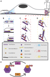
FIGURE 1
FIGURE 1
Formation and regulation of actin cytoskeletal structures during cell migration. (A) Migrating cells display distinct polarization. At the lagging end of the cell, RhoA is active and stimulates the formation of actin-myosin stress fibers. At the leading edge of the cell RhoA, Cdc42, and Rac1 are active. RhoA activity is localized to the very front edge of a migrating cell where it is proposed to stimulate leading edge ruffling. Cdc42 activity is at the front of the leading edge and stimulates filopodia formation. Rac1 activity is present throughout the whole leading edge and contributes to lamellipodia extension. (B) At the lagging edge, RhoA activates mDIA formins to make linear actin filaments. These filaments are joined together with alternating polarity by non-muscle myosin 2 motors to form stress fibers. Stress fibers are anchored to the cell and extracellular surface by focal adhesions. Myosin 2 activity pulls the actin filaments in stress fibers together, providing a contractile force on the rear of the cell. The contractile force breaks the connection between focal adhesions located at the rear of the cell and the extracellular surface. This results in stress fiber contraction pulling the lagging end of the cell forward, causing cell body translocation. (C) At the leading edge, Rac1 activity triggers lamellipodia extension by activating WRC (not pictured). WRC activates the Arp2/3 complex, allowing it to bind previously formed actin filaments. Actin monomers use the Arp2/3 complex as a site of nucleation, allowing a new actin filament to polymerize from roughly a 70⁰ angle to that of the previously existing filament. Continuous Arp2/3 complex induced branched actin polymerization results in a pushing force being applied to a large area of the membrane. This causes the membrane to extend, forming a lamellipodia. (D) Cdc42 activity at the leading edge can activate FMNL formins. FMNL formins bind to free barbed ends of branched actin filaments, causing the linear extension of that actin filament. These linear actin filaments provide a pushing force on a small surface area of the membrane, resulting in skinny membrane protrusions known as filopodia. The coordination of all these actin cytoskeletal structures provides the forces necessary for cellular locomotion. (E) GTPases are active when bound to GTP and inactive when GDP bound. GAPs function to inactive GTPases by converting GTP to GDP while GEFs activate GTPases by replacing GDP with GTP.
FIGURE 2
FIGURE 2
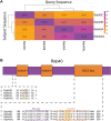
Protein structure of the Rab40 family. (A) Sequences of Homo sapiens Rab40 proteins were BLASTED against each other. Percent identity of the query sequence compared to the subject sequences was recorded and a heatmap was made using the Pheatmap function in R. (B) Diagram of Rab40 protein domains (not to scale) and alignment of regions of Homo sapiens Rab40 amino acid sequence. Upstream of Switch-I is an unexpected glycine residue in Rab40B and Rab40C (red highlight) involved in GEF binding that has reverted back to a serine residue in Rab40A and Rab40AL (yellow highlight). The SOCS box domain contains both a BC-box that facilitates Elongin B and C binding (purple outline) and a Cul-box that binds Cullin (orange outline). The LPLP motif in the Cul-box is conserved across all 4 Rab40 family members and creates specificity for Cullin5 binding (orange highlight).
FIGURE 3
FIGURE 3
The role of the Rab40 family of proteins in cell migration and actin cytoskeleton regulation. (A) The Rab40 proteins bind to Cullin5 to function as part of a Cullin-RING E3 ubiquitin ligase complex. This complex consists of the scaffold protein Cullin5, a SOCS box containing protein (Rab40), the adaptor proteins Elongin B and Elongin C, and the RING protein Rbx1/2. The complex functions to ubiquitinate (Ub) its substrates. (B) The known mechanisms and substrates of the Rab40 family of proteins [colors correspond to Rab40 name in (A)] and the Cullin-RING E3 ubiquitin ligase complex as it relates to cell migration and the actin cytoskeleton. * Signifies a migration related function of Rab40 thought to be independent of the Cullin-RING E3 ubiquitin ligase complex.

References

    1. Albiges-Rizo C., Destaing O., Fourcade B., Planus E., Block M. R. (2009). Actin machinery and mechanosensitivity in invadopodia, podosomes and focal adhesions. J. Cell. Sci. 122 (17), 3037–3049. 10.1242/jcs.052704 - DOI - PMC - PubMed
    1. Bachir Alexia I., Zareno J., Moissoglu K., Plow Edward F., Gratton E., Horwitz Alan R. (2014). Integrin-associated complexes form hierarchically with variable stoichiometry in nascent adhesions. Curr. Biol. 24 (16), 1845–1853. 10.1016/j.cub.2014.07.011 - DOI - PMC - PubMed
    1. Bianco A. M., Faletra F., Vozzi D., Girardelli M., Knowles A., Tommasini A., et al. (2015). Two-gene mutation in a single patient: biochemical and functional analysis for a correct interpretation of exome results. Mol. Med. Rep. 12 (4), 6128–6132. 10.3892/mmr.2015.4215 - DOI - PubMed
    1. Bisi S., Disanza A., Malinverno C., Frittoli E., Palamidessi A., Scita G. (2013). Membrane and actin dynamics interplay at lamellipodia leading edge. Curr. Opin. Cell. Biol. 25 (5), 565–573. 10.1016/j.ceb.2013.04.001 - DOI - PubMed
    1. Block J., Breitsprecher D., Kühn S., Winterhoff M., Kage F., Geffers R., et al. (2012). FMNL2 drives actin-based protrusion and migration downstream of Cdc42. Curr. Biol. 22 (11), 1005–1012. 10.1016/j.cub.2012.03.064 - DOI - PMC - PubMed