Review
. 2023 Sep 22;15(1):44.
doi: 10.1038/s41368-023-00249-w.

Oral squamous cell carcinomas: state of the field and emerging directions

Affiliations
Review

Oral squamous cell carcinomas: state of the field and emerging directions

Yunhan Tan et al. Int J Oral Sci. .

Abstract

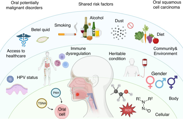
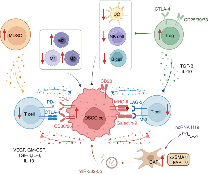
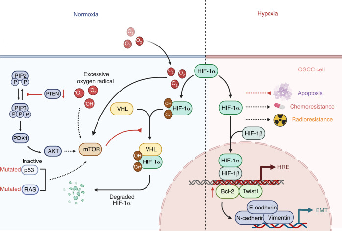
Oral squamous cell carcinoma (OSCC) develops on the mucosal epithelium of the oral cavity. It accounts for approximately 90% of oral malignancies and impairs appearance, pronunciation, swallowing, and flavor perception. In 2020, 377,713 OSCC cases were reported globally. According to the Global Cancer Observatory (GCO), the incidence of OSCC will rise by approximately 40% by 2040, accompanied by a growth in mortality. Persistent exposure to various risk factors, including tobacco, alcohol, betel quid (BQ), and human papillomavirus (HPV), will lead to the development of oral potentially malignant disorders (OPMDs), which are oral mucosal lesions with an increased risk of developing into OSCC. Complex and multifactorial, the oncogenesis process involves genetic alteration, epigenetic modification, and a dysregulated tumor microenvironment. Although various therapeutic interventions, such as chemotherapy, radiation, immunotherapy, and nanomedicine, have been proposed to prevent or treat OSCC and OPMDs, understanding the mechanism of malignancies will facilitate the identification of therapeutic and prognostic factors, thereby improving the efficacy of treatment for OSCC patients. This review summarizes the mechanisms involved in OSCC. Moreover, the current therapeutic interventions and prognostic methods for OSCC and OPMDs are discussed to facilitate comprehension and provide several prospective outlooks for the fields.


Conflict of interest statement

The authors declare no competing interests.

Figures

Fig. 1
Fig. 1
Risk factors of OPMDs and OSCC. The initiation and development of OPMDs and OSCC share similar risk factors, including smoking, alcohol abuse, betel quid (BQ) chewing, human papillomavirus (HPV) infection, nutritional insufficiency, immune deficiency, and hereditary conditions. OPMDs oral potentially malignant disorders, OSCC oral squamous cell carcinoma, PAH polycyclic aromatic hydrocarbons, ROS reactive oxygen species, TSNA tobacco-specific nitrosamines
Fig. 2
Fig. 2
Genetic alterations in OSCC. Genetic alteration in the TP53/RB, p16/Cyclin D1/Rb, EGFR, Wnt/β-catenin, JAK/STAT, NOTCH, PI3K/AKT/mTOR, MET, RAS/RAF/MASK signaling pathways contribute to OSCC progression. EGFR epidermal growth factor receptors, JAK Janus-activated kinase, MAPK mitogen-activated protein kinase, OSCC oral squamous cell carcinoma, RB retinoblastoma, Rb retinoblastoma tumor suppressor protein, STAT signal transducer and activator of the transcription, TP53 tumor protein p53
Fig. 3
Fig. 3
Immunosuppressive TME in OSCC. The tumor microenvironment (TME) contains various immunomodulatory cells, including cancer-associated fibroblasts (CAFs), regulatory T cells (Tregs), tumor-associated macrophages (TAMs), and myeloid-derived suppressor cells (MDSCs). The interaction between programmed death 1 (PD-1) and programmed death ligand-1 (PD-L1) leads to T-cell suppression and adaptive immunity tolerance, whereas cytotoxic T-lymphocyte-associated antigen 4 (CTLA-4) competes with CD28 by interacting with CD80/86 on OSCC cells. Other immune checkpoints also play a role, including LAG-3 and TIM-3. OSCC cells produce immune suppressive factors such as vascular endothelial growth factor (VEGF), granulocyte-macrophage colony-stimulating factor (GM-CSF), transforming growth factor (TGF)-β, interleukin (IL)-6, and IL-10, which block effector cells. Tregs generate TGF-β and IL-10 to diminish the functions of T cells. CAFs express α-smooth muscle actin (α-SMA) and fibroblast activation protein (FAP) and promote tumor growth by overexpressing miR-385-5p in their exosomes. In the TME, there is a greater proportion of M2 macrophages to M1 macrophages, with M2 TAMs possessing carcinogenic properties. OSCC oral squamous cell carcinoma
Fig. 4
Fig. 4
Hypoxia in OSCC. Under normoxic conditions, VHL degrades the HIFα subunits. In the condition of hypoxia, HIFα becomes stable and binds to HIFβ within the nucleus, adhering to hypoxia HREs to enable tumor adaptation. Hypoxia also promotes Bcl-2/Twist1 interaction by enhancing Bcl-2 attachment to Twist. Particularly, the mTOR pathway has been demonstrated to increase the level of HIFα in tumor regions that do not experience significant hypoxia. Several oncogenic mechanisms, such as inactive p53 mutations, RAS mutations, excessive oxygen radical accumulation, suppression of PTEN, and the infective HIF-1α degradation by VHL mutation, have been identified as contributing to this development. In addition, HIF-1α stimulates Twist1 to transactivate EMT-related genes, including Vimentin, N-cadherin, and E-cadherin, in order to drive EMT. Moreover, HIF-1α blocks apoptosis and confers higher resistance to chemotherapy and radiotherapy on OSCC. EMT epithelial–mesenchyme transition, HERs hypoxia response elements, HIF hypoxia-inducible factors, OSCC oral squamous cell carcinoma, PTEN phosphatase and tensin homolog, VHL Von Hippel-Lindau
Fig. 5
Fig. 5
Dysbiosis in OSCC. a A correlation exists between dysbiosis and the occurrence of various cancers. OSCC could be caused by Lactobacillus, Porphyomonas gingivalis, Fusobacterium nucleatum, and Prevotella intermedia. OSCC patients have elevated levels of Prevotella melanogenic, Streptococcus mitis, and Capnocytophaga gingivalis in their saliva, while Rothia, Leptotrichia, Haemophilus, Aggregatibacter, and Neisseria are diminished. b P. gingivalis stimulates the expression of B7-H1 receptors on OSCC cells. B7-H1 activates the development of Tregs, thereby inhibiting effector T cells. S. mitis has been demonstrated to trigger mutations and epithelial hyperplasia by generating acetaldehyde. OSCC oral squamous cell carcinoma, Tregs regulatory T cells
Fig. 6
Fig. 6
Treatments of OSCC. a Oncogene-targeted drugs facilitate OSCC chemotherapy. Cetuximab is an antibody that suppresses the EGFR pathway. NVP-BEZ235 is a PI3K/AKT/mTOR pathway inhibitor. Flavopereirine silences the JAK/STAT pathway and upregulates LASP1 expression. FLI-06 inactivates the Notch pathway. In the Wnt/β-catenin pathway, OMP-18R5 inhibits Fzd receptors; PRI-724 interrupts the interaction between β-catenin and CBP; and LGK974 targets PORCN. b Immunotherapy is an alternative treatment. Monoclonal antibodies pembrolizumab, nivolumab, and lgG4 have been authorized to target PD-1. LAG-3 and TIM-3 are blocked by their antibodies. IRX-2, comprising IL-2, IL-1β, IFN-γ, and TNF-α, is proven effective against inflammatory immune suppression cytokines in OSCC. Gemtuzmab ozogamicin promotes the differentiation of MDSCs into mature phenotypes, thereby reducing their immunosuppressive properties. EGFR epidermal growth factor receptors, PD-1 programmed death 1, OSCC oral squamous cell carcinoma

References

    1. Vigneswaran N, Williams MD. Epidemiologic trends in head and neck cancer and aids in diagnosis. Oral Maxillofac. Surg. Clin. 2014;26:123–141. - PMC - PubMed
    1. Ng JH, Iyer NG, Tan M-H, Edgren G. Changing epidemiology of oral squamous cell carcinoma of the tongue: a global study. Head Neck. 2017;39:297–304. - PubMed
    1. Mody MD, Rocco JW, Yom SS, Haddad RI, Saba NF. Head and neck cancer. Lancet. 2021;398:2289–2299. - PubMed
    1. Romano A, et al. Noninvasive imaging methods to improve the diagnosis of oral carcinoma and its precursors: state of the art and proposal of a three-step diagnostic process. Cancers. 2021;13:2864. - PMC - PubMed
    1. Safi A-F, et al. Clinicopathological parameters affecting nodal yields in patients with oral squamous cell carcinoma receiving selective neck dissection. J. Cranio-Maxillofac. Surg. 2017;45:2092–2096. - PubMed

Publication types

MeSH terms