. 2023 Sep 22;14(1):5894.
doi: 10.1038/s41467-023-41484-3.

CREB1-driven CXCR4hi neutrophils promote skin inflammation in mouse models and human patients

Affiliations

CREB1-driven CXCR4hi neutrophils promote skin inflammation in mouse models and human patients

Jiaoling Chen et al. Nat Commun. .

Abstract

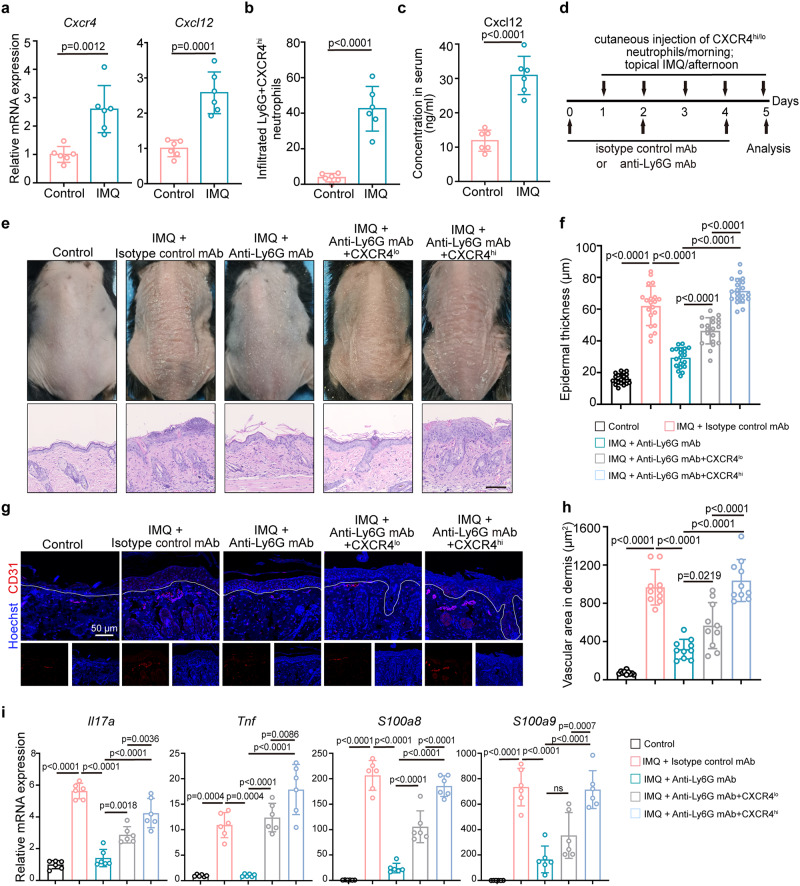
Neutrophils have a pathogenic function in inflammation via releasing pro-inflammatory mediators or neutrophil extracellular traps (NETs). However, their heterogeneity and pro-inflammatory mechanisms remain unclear. Here, we demonstrate that CXCR4hi neutrophils accumulate in the blood and inflamed skin in human psoriasis, and correlate with disease severity. Compared to CXCR4lo neutrophils, CXCR4hi neutrophils have enhanced NETs formation, phagocytic function, neutrophil degranulation, and overexpression of pro-inflammatory cytokines and chemokines in vitro. This is accompanied by a metabolic shift in CXCR4hi neutrophils toward glycolysis and lactate release, thereby promoting vascular permeability and remodeling. CXCR4 expression in neutrophils is dependent on CREB1, a transcription factor activated by TNF and CXCL12, and regulated by de novo synthesis. In vivo, CXCR4hi neutrophil infiltration amplifies skin inflammation, whereas blockade of CXCR4hi neutrophils through CXCR4 or CXCL12 inhibition leads to suppression of immune responses. In this work, our study identifies CREB1 as a critical regulator of CXCR4hi neutrophil development and characterizes the contribution of CXCR4hi neutrophils to vascular remodeling and inflammatory responses in skin.


Conflict of interest statement

The authors declare no competing interests.

Figures

Fig. 1
Fig. 1. CD15+CXCR4hi neutrophils are increased in the peripheral blood and inflamed psoriatic lesions.
a The mean fluorescence intensity (MFI) of CXCR4 in peripheral neutrophils from healthy controls (n = 24) and psoriasis patients (n = 54). b Correlation of the CXCR4 MFI on peripheral neutrophils with PASI in psoriasis patients (n = 25). c The proportion of circulating CD15+CXCR4hi neutrophils in healthy controls (n = 20) and psoriasis patients (n = 25). d Correlation of the proportions of CXCR4hi neutrophils with PASI in psoriasis patients. The adjusted R2 and P-values were plotted in the graph. e Representative immunoblots of total CXCR4 in circulating neutrophils from healthy controls and psoriasis patients. The relative multiple expression was counted. Blots for each antigen were processed in the same experiment in parallel. The result was repeated twice independently with similar results. f Immunofluorescence staining of CD15 (green) and CXCR4 (red) in normal and inflamed psoriatic skin. Scale bar = 50 µm. n = 10 biologically independent samples. g The proportion of CXCR4lo vs. CXCR4hi neutrophils in inflamed psoriatic skin (n = 30 fields from 10 patient samples). MFI of CXCR4 on peripheral neutrophils (h) and serum protein levels of CXCL12, IL-17A, and MPO (i) before and after treatment with anti-IL-17 inhibitor for 12 weeks. n = 5 biologically independent samples. Data are mean ± SD. Analyses: unpaired Student’s t-test in (a) and (c); The Spearman method in (b) and (d); Paired Student’s t-test in (h) and (i). The paired and unpaired Student’s t-test were conducted as two-sided tests. FMO, Fluorescence Minus One; HC, healthy control; MFI, mean fluorescence intensity; Pre, pre-treatment; Pso, psoriasis patients. Source data are provided as a Source Data file.
Fig. 2
Fig. 2. CXCR4hi neutrophils display enhanced pro-inflammatory functions.
a Representative images and quantification of different nucleus morphology of CXCR4lo and CXCR4hi neutrophils from healthy controls and psoriasis patients. Scale bar = 5 µm. b Flow cytometry analysis of key immune markers between peripheral CXCR4lo and CXCR4hi neutrophils from healthy controls and psoriasis patients (scale: log-transformed MFI). c Assessment of CXCR4hi neutrophil survival by Annexin V and 7-AAD staining after 24 h of culture. d Measurement of ROS production by DHE fluorescence. e Quantification of NETs in CXCR4lo and CXCR4hi neutrophils from healthy controls and psoriasis patients. Phagocytosis of pHrodo Green E. coli by neutrophils was evaluated by immunofluorescence staining (f) and flow cytometry (g). Scale bar = 5 µm. h Degranulation of CXCR4lo or CXCR4hi neutrophils as assessed by CD63 expression. ELISA quantification (i) and representative immunoblots (j) for mature MMP-9 in the supernatant of CXCR4lo and CXCR4hi neutrophils from healthy controls and psoriasis patients that cultured for 24 h. GAPDH was analyzed from corresponding neutrophils. Blots for each antigen were processed in the same experiment in parallel. k Relative mRNA expression of pro-inflammatory mediators in CXCR4lo and CXCR4hi neutrophils from healthy controls and psoriasis patients. Data are mean ± SD (n = 6 biologically independent samples). The immunofluorescence staining was repeated three times independently with similar results. Two-way ANOVA with Tukey’s post hoc test was performed as two-sided analyses and adjusted for multiple comparisons in the statistical analyses. ns, not significant. HC, healthy control; MFI, mean fluorescence intensity; MMP-9, matrix metalloprotein 9; NETs; neutrophil extracellular traps; neu, neutrophils; Pso, psoriasis patients. Source data are provided as a Source Data file.
Fig. 3
Fig. 3. RNA sequencing reveals the immune signature of CXCR4hi neutrophils.
a Volcano plot showing the number of differentially expressed genes (DEGs) between CXCR4lo and CXCR4hi neutrophils from healthy controls (n = 7) and psoriasis patients (n = 6). b Heatmap of DEGs between psoriatic CXCR4lo and CXCR4hi neutrophils related to neutrophil and immune function. c Enriched GO terms between CXCR4lo and CXCR4hi psoriatic neutrophils. d Gene set enrichment analysis showing enrichment of genes involved in glycolysis in psoriatic CXCR4hi neutrophils. e Flow cytometry of glycolytic markers in CXCR4lo and CXCR4hi neutrophils from healthy controls and psoriasis patients. f Uptake of glucose (2-NBDG) in CXCR4lo and CXCR4hi neutrophils from healthy controls and psoriasis patients. g Extracellular lactate production in CXCR4lo and CXCR4hi neutrophils from healthy controls and psoriasis patients. Mean ± SD (n = 6 biologically independent samples/group). Two-way ANOVA with Tukey’s post hoc test was performed as two-sided analyses and adjusted for multiple comparisons in the statistical analyses. ns, not significant. HC, healthy control; MFI, mean fluorescence intensity; Pso, psoriasis patients; RNA-seq, RNA sequencing. Source data are provided as a Source Data file.
Fig. 4
Fig. 4. Lactate released by CXCR4hi neutrophils induces vascular remodeling and permeability.
a Representative images of adherent CXCR4lo and CXCR4hi neutrophils co-cultured with HMEC-1 cells. White arrows indicate HMEC-1 cells; red arrows indicate adherent neutrophils. Scale bar = 10 µm. b Relative mRNA expression of adhesion molecules in HMEC-1 cells with indicated treatment. Representative immunoblots (c) and qRT-PCR analysis (d) of tight junctions in HMEC-1 cells co-cultured with indicated treatment. e Stimulation of HMEC-1 cells with CXCR4lo and CXCR4hi neutrophils for 6 h, followed by analysis of leaked fluorescence intensity of FITC-dextran in a Transwell system. Representative immunoblots of HMEC-1 cells pre-incubated with LDHA inhibitor for 30 min following co-culture with psoriatic CXCR4hi neutrophils (f) and qRT-PCR assessment of tight junction genes (g). h Analysis of released FITC-dextran in a Transwell system with HMEC-1 cells, pre-incubated with LDHA inhibitor for 30 min, followed by co-culture with psoriatic CXCR4hi neutrophils. i Confocal images of CD31 (green) and GPR81 (purple) in psoriatic lesions (n = 6) with CD15+CXCR4hi neutrophils adjacent to GPR81+ vascular ECs. Scale bar = 50 µm, 20 µm. The result was repeated three times independently with similar results. HMEC-1 cells were transfected with GPR81 siRNA and subjected to indicated treatment, followed by Western blot (j), qRT-PCR (k), and FITC-dextran leakage (l). The immunoblotting samples shown are from the same experiment (c, f and j) and blots were processed in parallel. Mean ± SD (n = 6 biologically independent samples/group). Analyses: two-way ANOVA with Tukey’s post hoc test in (b), (d), (e), (h) and (l); One-way ANOVA with Tukey’s post hoc test in (g) and (k). One or two-way ANOVA tests were performed as two-sided analyses and adjusted for multiple comparisons in the statistical analyses. ns, not significant. HC, healthy control; HMEC-1 cells, human microvascular endothelial cells; Pso, psoriasis patients. Source data are provided as a Source Data file.
Fig. 5
Fig. 5. CXCR4 expression in neutrophils is regulated by psoriasis-related mediators.
a Expression of CXCR4 on neutrophils stimulated with IL-25, CXCL12, and TNF for 2 h was measured by flow cytometry. b Serum level of CXCL12 in healthy controls (n = 24) and psoriasis patients (n = 30) was detected by ELISA. c Expression of CXCR4 on neutrophils with indicated treatment was measured by flow cytometry. d Representative immunofluorescence staining of CD31 (yellow), Vimentin (green), and CXCL12 (purple) in inflamed psoriatic skin (n = 6). Scale bar = 50 µm, 20 µm. e Representative immunofluorescence staining of CD31 (yellow), CXCR4 (green), and CXCL12 (purple) in inflamed psoriatic skin (n = 6). Scale bar = 50 µm, 20 µm. QRT-PCR analysis (f) and flow cytometry analysis (g) of CXCR4 in healthy neutrophils with indicated treatments. h Representative immunofluorescence co-staining of CXCR4 (red) and LAMP1 (green) in neutrophils. Scale bar = 2 µm. i Examples of low colocalization values in a sample of CXCR4hi and CXCR4lo neutrophils of psoriasis patients stained for LCN2 (red) and CD63 (yellow) was visualized by ImageStream analysis. Images are from one representative experiment out of six. The scale bar indicates 7 μm. Isolated neutrophils were used for (a), (c), (f), and (g). Mean ± SD (n = 6 biologically independent samples/group). The immunofluorescence staining was repeated three times independently with similar results. Analyses: one-way ANOVA with Tukey’s post hoc test in (a) and (c); two-way ANOVA with Tukey’s post hoc test in (f) and (g); Unpaired Student’s t-test in (b). The unpaired Student’s t-test was conducted as two-sided tests. One or two-way ANOVA tests were performed as two-sided analyses and adjusted for multiple comparisons in the statistical analyses. ns, not significant. FMO, Fluorescence Minus One; HC, healthy control; LCN2, lipocalin 2; MFI, mean fluorescence intensity; Pso, psoriasis patients. Source data are provided as a Source Data file.
Fig. 6
Fig. 6. CREB1 promotes the development of CXCR4hi neutrophils.
a Conceptual network diagram of genes showing the relationship between CREB1 and enrichment pathways, and heatmap of top transcription factors in CXCR4hi neutrophils. The mRNA expression of CREB1 (b) and protein levels of p-CREB1 (s133) (c) in CXCR4lo and CXCR4hi neutrophils. d Flow cytometry analysis of p-CREB1 (s133) in neutrophils treated with indicated stimulation. e Co-localization of CXCR4 (green) and p-CREB1 (red) in neutrophils from healthy controls and psoriasis patients. Scale bar = 2 µm. Representative staining (f) and Western blot (g) of CBP (green) and p-CREB1 (red) in neutrophils treated with indicated stimulation for 2 h. h, i Healthy neutrophils were pre-treated with CREB1 inhibitor (KG-501, 300 μM) for 1 h, followed by indicated stimulation for 2 h. Flow cytometry analysis of the expression of membrane molecules (h), as well as glycolysis (i). j Representative co-staining of DNA (Hoechst), Cit-H3 (citrullinated histone-3, red), and MPO (myeloperoxidase, green) to assess NETs formation in psoriatic neutrophils in vitro after pretreatment with KG-501 (300 μM) for 1 h. Scale bar = 20 µm. k Western blot of dHL-60 cells transfected with CREB1 siRNA and subjected to the indicated treatment. l Depiction of the CREB1 binding site in the CXCR4 promoter for ChIP assay. m The luciferase activities of wild type CXCR4 and CXCR4 with the CREB1-binding site mutant were determined by luciferase reporter gene assays in dHL-60 cells. Isolated neutrophils were used for (b), (d), (h), and (i), and whole blood was used for (c). Mean ± SD (n = 6 biologically independent samples). The immunofluorescence staining was repeated three times independently with similar results. Blots for each antigen were processed in the same experiment in parallel. Two-way ANOVA with Tukey’s post hoc test (b, c, h, i, and m) and one-way ANOVA with Tukey’s post hoc test (d) were performed as two-sided analyses and adjusted for multiple comparisons in the statistical analyses. ns, not significant. HC, healthy control; MFI, mean fluorescence intensity; NETs, neutrophil extracellular traps; Pso, psoriasis patients. Source data are provided as a Source Data file.
Fig. 7
Fig. 7. CXCR4hi neutrophils contribute to psoriasis-like inflammation in vivo.
a Relative mRNA expressions of CXCR4 and CXL12 in mice tissues was evaluated by qRT-PCR. n = 6 mice. b Proportion of Ly6G+CXCR4hi neutrophils in mice skin was determined by flow cytometry. n = 6 mice. c Serum level of CXCL12 in control and IMQ-treated mice was detected by ELISA. n = 6 mice. d Schematic diagram of mouse experimental protocol. IMQ mice were injected intraperitoneally with an anti-Ly6G antibody every other day and then injected subcutaneously with fresh isolated homologous Ly6G+CXCR4lo or Ly6G+CXCR4hi neutrophils daily. e Phenotype and representative H&E staining of IMQ-treated mice in indicated groups on day 5. Images are representative of six individual mouse per group. Control group was topically applied with Vaseline cream. Bar = 200 µm. n = 6 mice. f Epidermal thickness was assessed by H&E. n = 20 vision fields from 6 mice. g Representative immunofluorescence staining of CD31 (red) in inflamed skin. Scale bar = 50 µm. n = 6 mice. h Quantification of the dermal vascular area in the H&E-stained sections. n = 10 vision fields from 6 mice. i Relative mRNA expressions of inflammatory cytokines in mice tissues. n = 6 mice. The immunofluorescence staining was repeated three times independently with similar results. Mean ± SD. Analyses: unpaired Student’s t-test in (a), (b), and (c); One-way ANOVA with Tukey’s post hoc test in (f), (h), and (i). The unpaired Student’s t-test was conducted as two-sided tests. One-way ANOVA test was performed as two-sided analyses and adjusted for multiple comparisons in the statistical analyses. ns, not significant; IMQ, imiquimod. Source data are provided as a Source Data file.
Fig. 8
Fig. 8. Targeting CXCR4/CXCL12 axis alleviates IMQ-induced psoriasiform inflammation.
a Phenotype and H&E staining of IMQ-treated mice in different treatment groups. Images are representative of six individual mice per group. The mice in the control group was topically applied with vaseline cream. Bar = 200 µm. n = 6 mice. b Epidermal thickness as assessed by H&E staining. n = 20 vision fields from 6 mice. c Proportion of Ly6G+CXCR4hi neutrophils in inflamed skin was determined by flow cytometry. n = 6 mice. d Representative immunofluorescence staining of CD31 (red) in lesional skin of indicated groups. Scale bar = 50 µm. n = 6 mice. e Quantification of the dermal vascular area in the H&E-stained sections. n = 10 vision fields from 6 mice. f Quantification of extracted Evans blue dye in inflamed skin in indicated groups. n = 6 mice. g Relative mRNA expressions of inflammatory cytokines in mice tissues. n = 6 mice. The immunofluorescence staining was repeated three times independently with similar results. Mean ± SD. One-way ANOVA with Tukey’s post hoc test was performed as two-sided analyses and adjusted for multiple comparisons in the statistical analyses. ns, not significant; IMQ, imiquimod. Source data are provided as a Source Data file.
Fig. 9
Fig. 9. Proposed mechanisms of CXCR4hi neutrophil phenotype in skin inflammation.
This study provides insights into the development and role of CXCR4hi neutrophils in skin inflammation. Compared to CXCR4lo neutrophils, CXCR4hi neutrophils exhibit increased capacity for NETs formation, phagocytosis, and degranulation, as well as higher expression of pro-inflammatory mediators, supported by activated glycolysis. CXCR4 expression on neutrophils is induced by CXCL12, IL-25, and TNF, which requires de novo mRNA and protein synthesis and intracellular protein transport, but is not stored in neutrophil granules and regulated by degranulation. CREB1 is activated by phosphorylation on Ser-133 upon stimulation, enabling interaction with its coactivator protein CBP to initiate transcription of CREB-responsive genes, thereby contributing to CXCR4 expression and CXCR4hi neutrophils in skin inflammation. Our data further emphasize the crucial role of CXCR4hi neutrophils in promoting vascular remodeling through lactate and MMP-9 release (B), facilitating immune cell infiltration into tissues, and increasing inflammatory responses (A). Moreover, we identify the CXCR4/CXCL12 axis as a potential therapeutic target for inflammatory skin diseases, with a focus on CREB1, CXCR4/CXCL12, or the formation of NETs. CBP, CREB-binding protein; EC, endothelial cell; MMP9, matrix metallopeptidase 9; NETs; neutrophil extracellular traps; PAD4 i, PAD4 inhibitor. Created with BioRender.com.

References

    1. Liew PX, Kubes P. The Neutrophil’s role during health and disease. Physiol. Rev. 2019;99:1223–1248. - PubMed
    1. Burn GL, Foti A, Marsman G, Patel DF, Zychlinsky A. The Neutrophil. Immunity. 2021;54:1377–1391. - PubMed
    1. Herster F, et al. Neutrophil extracellular trap-associated RNA and LL37 enable self-amplifying inflammation in psoriasis. Nat. Commun. 2020;11:105. - PMC - PubMed
    1. Jarrot PA, et al. Neutrophil extracellular traps are associated with the pathogenesis of diffuse alveolar hemorrhage in murine lupus. J. Autoimmun. 2019;100:120–130. - PubMed
    1. O’Neil LJ, et al. Neutrophil-mediated carbamylation promotes articular damage in rheumatoid arthritis. Sci. Adv. 2020;6:eabd2688. - PMC - PubMed

Publication types