Review
. 2023 Sep 1;9(5):1649-1659.
doi: 10.3390/tomography9050132.

The State of Intraoperative OCT in Vitreoretinal Surgery: Recent Advances and Future Challenges

Affiliations
Review

The State of Intraoperative OCT in Vitreoretinal Surgery: Recent Advances and Future Challenges

Nicolò Ciarmatori et al. Tomography. .

Abstract

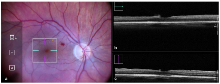
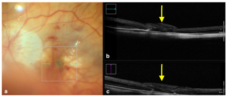
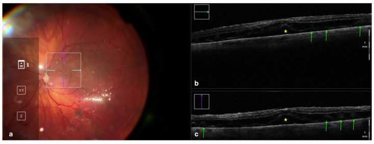
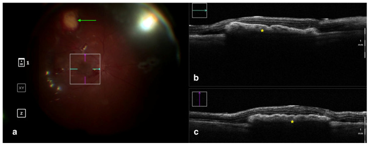
Since its first introduction more than 30 years ago, optical coherence tomography (OCT) has revolutionized ophthalmology practice, providing a non-invasive in vivo cross-sectional view of the structures of the eye. Mostly employed in the clinical setting due to its tabletop configuration requiring an upright patient positioning, the recent advent of microscope-integrated systems now allows ophthalmologists to perform real-time intraoperative OCT (iOCT) during vitreoretinal surgical procedures. Numerous studies described various applications of this tool, such as offering surgeons feedback on tissue-instrument interactions in membrane peeling, providing structural images in macular hole repair, and showing residual subretinal fluid or perfluorocarbon in retinal detachment surgery. This narrative review aims at describing the state of the art of iOCT in vitreoretinal procedures, highlighting its modern role and applications in posterior segment surgery, its current limitations, and the future perspectives that may improve the widespread adoption of this technology.

Keywords: epiretinal membrane; intraoperative OCT; macular hole; retinal detachment; subretinal surgery; vitreoretinal surgery.


Conflict of interest statement

The authors declare no conflict of interest.

Figures

Figure 1
Figure 1
Rescan 700 iOCT showing on the left the en face ophthalmoscopic surgical view after staining and peeling. The digital overlay indicates the scanning area (a), and the horizontal and vertical scans are found in the upper and lower right portion of the image, respectively (b,c). The iOCT shows the surgeon the release of traction on the fovea and the complete removal of the ERM in the scanned area. Abbreviations: ERM, epiretinal membrane; iOCT, intraoperative optical coherence tomography.
Figure 2
Figure 2
Rescan 700 iOCT showing an inverted ILM flap performed in a case of myopic full-thickness macular hole. The display simultaneously shows, on the left, the en face ophthalmoscopic surgical view with a digital overlay indicating the scanning area (a) and, in the upper and lower right portions of the image, the horizontal and vertical scans, respectively (b,c). The iOCT indicates to the surgeon the architecture of the hole and shows the positioning of the ILM flap (yellow arrows). Abbreviations: ILM, internal limiting membrane; iOCT, intraoperative optical coherence tomography.
Figure 3
Figure 3
Rescan 700 iOCT showing a case of RRD after perfluorocarbon flattening. On the left is the en face ophthalmoscopic surgical view with a digital overlay indicating the scanning area (a) and in the upper and lower right portions of the image are the horizontal and vertical scans, respectively (b,c). The iOCT shows the surgeon the successful reattachment of the retina, although with persistence of subfoveal fluid (yellow asterisk) and the presence of outer retinal folds (green arrows) Abbreviations: iOCT, intraoperative optical coherence tomography; RRD, rhegmatogenous retinal detachment.
Figure 4
Figure 4
Rescan 700 iOCT showing a case of RPE transplantation. On the left is the en face ophthalmoscopic surgical view with a digital overlay indicating the scanning area (a). The green arrow indicates the sites used for harvesting the graft. In the upper and lower right portions of the image are the horizontal and vertical scans, respectively (b,c). The iOCT shows the surgeon the correct positioning and orientation of the graft under the retina (indicated by the yellow asterisk). Abbreviations: iOCT, intraoperative optical coherence tomography; RPE, retinal pigment epithelium.

Similar articles

Cited by

References

    1. Fercher A.F., Hitzenberger C.K., Drexler W., Kamp G., Sattmann H. In vivo optical coherence tomography. Am. J. Ophthalmol. 1993;116:113–114. doi: 10.1016/S0002-9394(14)71762-3. - DOI - PubMed
    1. Dayani P.N., Maldonado R., Farsiu S., Toth C.A. Intraoperative Use of Handheld Spectral Domain Optical Coherence Tomography Imaging in Macular Surgery. Retina. 2009;29:1457–1468. doi: 10.1097/IAE.0b013e3181b266bc. - DOI - PMC - PubMed
    1. [(accessed on 31 May 2023)]. Available online: https://www.spiritmedical.cz/uploads/pdf/bioptigen-envisu.pdf.
    1. [(accessed on 31 May 2023)]. Available online: https://www.crvmedical.it/wp-content/uploads/bsk-pdf-manager/b-optovue-m....
    1. Binder S., Falkner-Radler C.I., Hauger C., Matz H., Glittenberg C.G. Clinical Applications of Intrasurgical SD-Optical Coherence Tomography. Investig. Ophthalmol. Vis. Sci. 2010;51:268.

LinkOut - more resources