Generating Synthetic Radiological Images with PySynthMRI: An Open-Source Cross-Platform Tool
- PMID: 37736990
- PMCID: PMC10514862
- DOI: 10.3390/tomography9050137
Generating Synthetic Radiological Images with PySynthMRI: An Open-Source Cross-Platform Tool
Abstract
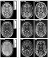
Synthetic MR Imaging allows for the reconstruction of different image contrasts from a single acquisition, reducing scan times. Commercial products that implement synthetic MRI are used in research. They rely on vendor-specific acquisitions and do not include the possibility of using custom multiparametric imaging techniques. We introduce PySynthMRI, an open-source tool with a user-friendly interface that uses a set of input images to generate synthetic images with diverse radiological contrasts by varying representative parameters of the desired target sequence, including the echo time, repetition time and inversion time(s). PySynthMRI is written in Python 3.6, and it can be executed under Linux, Windows, or MacOS as a python script or an executable. The tool is free and open source and is developed while taking into consideration the possibility of software customization by the end user. PySynthMRI generates synthetic images by calculating the pixelwise signal intensity as a function of a set of input images (e.g., T1 and T2 maps) and simulated scanner parameters chosen by the user via a graphical interface. The distribution provides a set of default synthetic contrasts, including T1w gradient echo, T2w spin echo, FLAIR and Double Inversion Recovery. The synthetic images can be exported in DICOM or NiFTI format. PySynthMRI allows for the fast synthetization of differently weighted MR images based on quantitative maps. Specialists can use the provided signal models to retrospectively generate contrasts and add custom ones. The modular architecture of the tool can be exploited to add new features without impacting the codebase.
Keywords: magnetic resonance imaging; software tool; synthetic imaging.
Conflict of interest statement
Matteo Cencini and Michela Tosetti received a research grant from GE Healthcare. The other authors (Luca Peretti, Graziella Donatelli, Paolo Cecchi, Guido Buonincontri, Mirco Cosottini, Mauro Costagli) declare no conflict of interest.
Figures


References
-
- Andica C., Hagiwara A., Hori M., Kamagata K., Koshino S., Maekawa T., Suzuki M., Fujiwara H., Ikeno M., Shimizu T., et al. Review of synthetic MRI in pediatric brains: Basic principle of MR quantification, its features, clinical applications, and limitations. J. Neuroradiol. 2019;46:268–275. doi: 10.1016/j.neurad.2019.02.005. - DOI - PubMed
-
- Tanenbaum L.N., Tsiouris A.J., Johnson A.N., Naidich T.P., DeLano M.C., Melhem E.R., Quarterman P., Parameswaran S.X., Shankaranarayanan A., Goyen M., et al. Synthetic MRI for Clinical Neuroimaging: Results of the Magnetic Resonance Image Compilation (MAGiC) Prospective, Multicenter, Multireader Trial. Am. J. Neuroradiol. 2017;38:1103–1110. doi: 10.3174/ajnr.A5227. - DOI - PMC - PubMed
Publication types
MeSH terms
Substances
LinkOut - more resources
Full Text Sources

