. 2023 Nov;80(11):2357-2371.
doi: 10.1161/HYPERTENSIONAHA.123.21241. Epub 2023 Sep 22.

E2F1 Mediates SOX17 Deficiency-Induced Pulmonary Hypertension

Affiliations

E2F1 Mediates SOX17 Deficiency-Induced Pulmonary Hypertension

Dan Yi et al. Hypertension. 2023 Nov.

Abstract

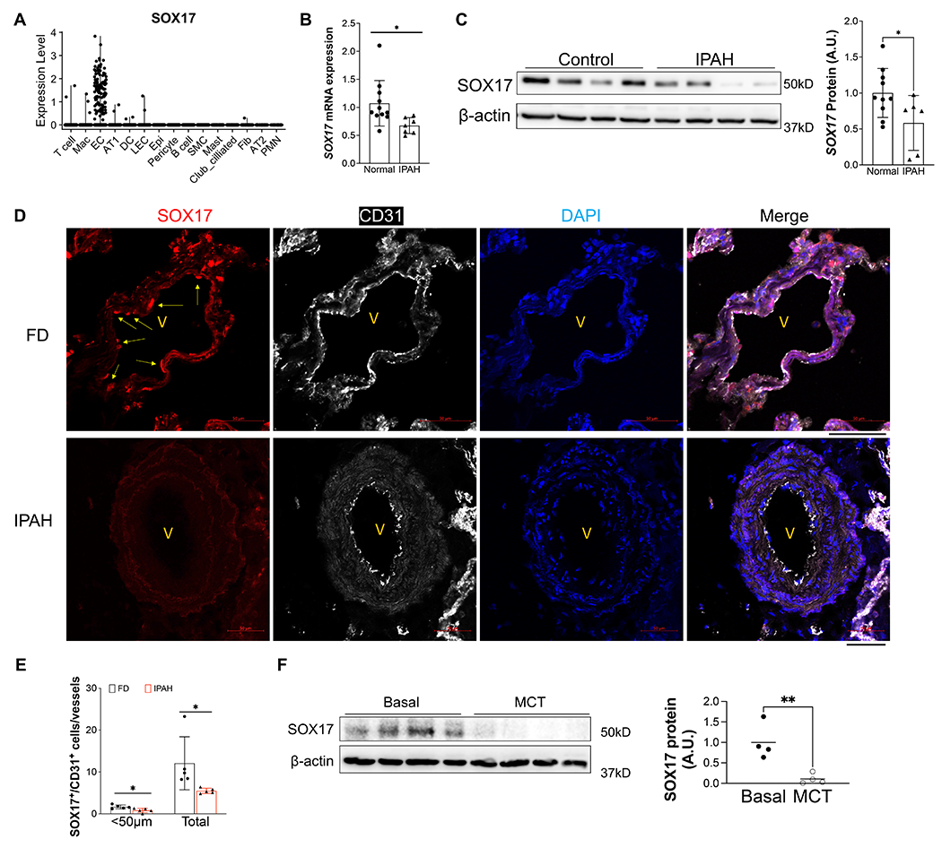
Background: Rare genetic variants and genetic variation at loci in an enhancer in SOX17 (SRY-box transcription factor 17) are identified in patients with idiopathic pulmonary arterial hypertension (PAH) and PAH with congenital heart disease. However, the exact role of genetic variants or mutations in SOX17 in PAH pathogenesis has not been reported.

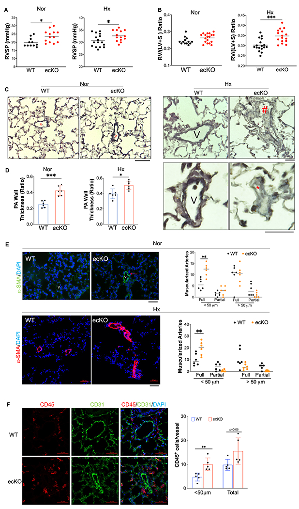
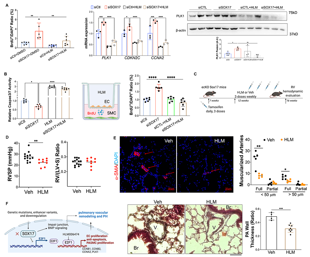
Methods: SOX17 expression was evaluated in the lungs and pulmonary endothelial cells (ECs) of patients with idiopathic PAH. Mice with Tie2Cre-mediated Sox17 knockdown and EC-specific Sox17 deletion were generated to determine the role of SOX17 deficiency in the pathogenesis of PAH. Human pulmonary ECs were cultured to understand the role of SOX17 deficiency. Single-cell RNA sequencing, RNA-sequencing analysis, and luciferase assay were performed to understand the underlying molecular mechanisms of SOX17 deficiency-induced PAH. E2F1 (E2F transcription factor 1) inhibitor HLM006474 was used in EC-specific Sox17 mice.

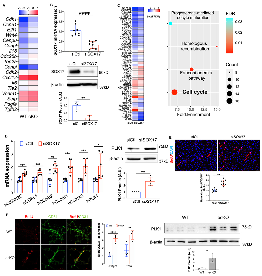
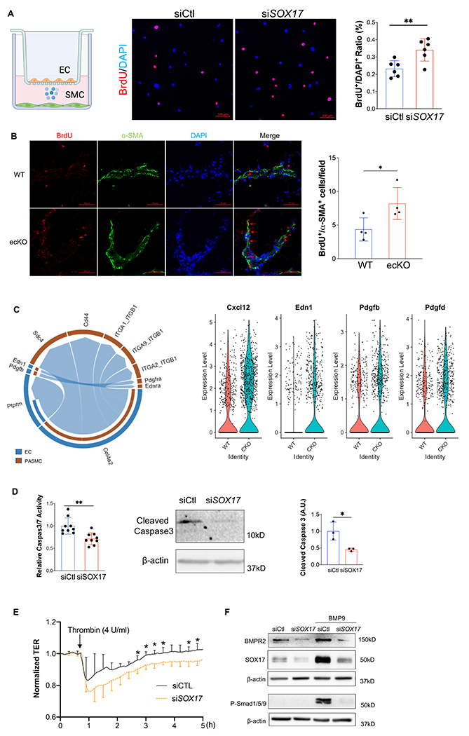
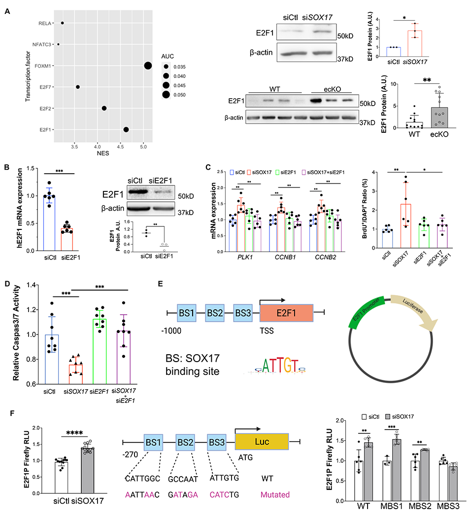
Results: SOX17 expression was downregulated in the lung and pulmonary ECs from patients with idiopathic PAH. Mice with Tie2Cre-mediated Sox17 knockdown and EC-specific Sox17 deletion induced spontaneously mild pulmonary hypertension. Loss of endothelial Sox17 in EC exacerbated hypoxia-induced pulmonary hypertension in mice. Loss of SOX17 in lung ECs induced endothelial dysfunctions including upregulation of cell cycle programming, proliferative and antiapoptotic phenotypes, augmentation of paracrine effect on pulmonary arterial smooth muscle cells, impaired cellular junction, and BMP (bone morphogenetic protein) signaling. E2F1 signaling was shown to mediate the SOX17 deficiency-induced EC dysfunction. Pharmacological inhibition of E2F1 in Sox17 EC-deficient mice attenuated pulmonary hypertension development.

Conclusions: Our study demonstrated that endothelial SOX17 deficiency induces pulmonary hypertension through E2F1. Thus, targeting E2F1 signaling represents a promising approach in patients with PAH.

Keywords: angiogenesis; endothelial cells; hypertension; mice; pulmonary artery.


Conflict of interest statement

Disclosures None.

Figures

Figure 1.
Figure 1.. Downregulation of endothelial SOX17 in the patients with PAH.
(A) A violin plot showing SOX17 is restricted in the ECs of human lungs via scRNA-seq. Mac=macrophage; DC= dendritic cell; LEC=lymphatic EC; Epi=epithelium; SMC= smooth muscle cell; Fib=fibroblast; AT1 or AT2 = alveolar type 1 or 2 epithelium; PMN=neutrophils. (B) qRT-PCR analysis showed that SOX17 mRNA levels were downregulated in the sub-confluent PVECs isolated from IPAH patients (n=11) and normal non-PAH donors (n=7). Each data point represents cells from one human subject including both male and female. (C) Western blotting demonstrated reduction of SOX17 protein expression in the IPAH PVECs (n=10) compared with normal non-PAH donors (n=6). Each data point represents cells from one human subject including both male and female. (D, E) Immunostaining against SOX17 showing diminished SOX17 expression in the ECs of remodeling lesions from IPAH patients. Arrows indicate SOX17 positive ECs in non-PAH failed donors (FD). SOX17+/CD31+ cell number was quantified and normalized by vessels number. Each dot represents one subject. (F) SOX17 is decreased in the lungs of established PH rats at 4 weeks post MCT (33mg/kg subcutaneously) treatment. Student t test (B, C, E, F). *, P< 0.05; **, P< 0.01. A.U. = arbitrary units; Scale bar, 50μm.
Figure 2.
Figure 2.. Endothelial SOX17 deficiency induced PH in mice.
(A) ecKO Sox17 mice exhibited increase of RVSP at both normoxic (Nor) and hypoxic (Hx) condition. (B) ecKO Sox17 mice exhibited hypoxia-induced right heart hypertrophy compared with WT mice. (C) Representative micrographs of Russell-Movat pentachrome staining showing increased medial thickness in ecKO Sox17 mice compared with WT mice in normoxic and hypoxic condition. ecKO Sox17 mice also developed occlusive vascular lesion in response to hypoxia. V=vessel, # indicates narrower vessel, * indicates occlusive vessel. (D) Quantification of pulmonary artery wall thickness. Wall thickness was calculated by the distance between internal wall and external wall divided by the distance between external wall and the center of lumen. (E) Representative micrographs and quantification of muscularization of distal pulmonary vessels showed that markedly enhanced muscularization of distal pulmonary vessels in ecKO Sox17 mice compared with WT mice under normoxic and hypoxic condition. Lung sections were immunostained with anti–α-SMA (green). Red arrow indicates a-SMA+ distal pulmonary vessels. α-SMA+ vessels were quantified in 20 field at 10X magnification per mouse. (F) Immunostaining against CD45 (Red) demonstrated that there was upregulated accumulation of inflammatory cells in the perivascular bed of ecKO Sox17 mice. Student t test (A, B, D, E, F). *, P< 0.05; **, P< 0.01, ***, P< 0.001. Scale bar, 50μm.
Figure 3.
Figure 3.. Loss of SOX17 induced EC proliferation.
(A) scRNA transcriptomics showed that Sox17 deficiency ECs expressed higher levels of proliferation genes compared to WT ECs. scRNA-seq analysis was performed on the whole lung of WT and cKO mice. Lung ECs transcriptomics were analyzed. (B) qRT-PCR analysis showing efficient knockdown of SOX17 via siRNA against SOX17 in HPVECs assessed by QPCR and western blot. (C)A representative heatmap of RNA-sequencing analysis of SOX17 knockdown in HPVECs. HPVECs were transfected with control siRNA (siCtl) or SOX17 siRNA for 48 hours. Equal amount of RNA from three replicates per group were pooled for RNA-seq. KEGG pathway enrichment analysis of upregulated genes in SOX17 deficient lung ECs demonstrating that cell cycle pathway is the top upregulated signaling induced by loss of SOX17. (D) qRT-PCR analysis confirmed the upregulation of cell proliferation related genes including CKDN2C, CDKL1, CCNB2, CCNB1, CCNA2, and PLK1. Western Blotting analysis demonstrated induction of PLK1 protein expression by SOX17 deficiency. (E) BrdU incorporation assay demonstrated increased of EC proliferation in SOX17 deficient HPVECs. At 48 hours post-transfection, HPVECs were starved in serum/growth factors free medium for 12 hours. BrdU was added in the medium at 4 hours prior to cells harvest. BrdU was stained with anti-BrdU antibodies. Red indicated BrdU positive cells. Nucleus were co-stained with DAPI. (F) In vivo BrdU incorporation assay showed upregulation of lung ECs proliferation in ecKO Sox17 mice during hypoxia condition. WT and ecKO Sox17 mice were incubated in hypoxia (10% O2) for 10 days. BrdU (25 mg/kg) was injected i.p. between day 7 to day 9. Lung sections were stained with anti-BrdU and anti-CD31. BrdU+/CD31+ cells were quantified. Augmentation of cell proliferation marker PLK1 expression in the lung of ecKO Sox17 (ecKO) mice compared to WT mice. β-actin level was used as an internal control. Student t test (B, D, E,F). *, P< 0.05; **, P< 0.01. ***, P< 0.001. Scale bar, 50μm.
Figure 4.
Figure 4.. Loss of endothelial SOX17 promoted EC dysfunction.
(A) SOX17 deficiency in lung ECs promoted PASMCs proliferation assessed by Transwell co-culture and BrdU assay. PASMCs were seeded on the cover slides on the lower chamber. SOX17 deficiency or control HPVECs were seeded on the top chamber for 48 hours. PASMCs were starved overnight, then co-cultured with HPVECs. BrdU was added in the lower chamber at 8 hours prior to cells harvest. BrdU was stained with anti-BrdU antibodies. Red indicated BrdU positive cells. Nucleus were co-stained with DAPI. (B) In vivo BrdU incorporation assay showed upregulation of PASMCs proliferation in ecKO Sox17 mice during hypoxia condition. WT and ecKO Sox17 mice were incubated in hypoxia (10% O2) for 10 days. BrdU (25 mg/kg) was injected i.p. between day 7 to day 9. Lung sections were stained with anti-BrdU and anti-α-SMA. BrdU+/α-SMA+ cells were quantified. (C) CellChat prediction using scRNA-seq dataset showed the upregulation of ligand and receptor pairs (Pdgfb-Pdgfra, Edn1-Ednra) in cKO mice. ScRNA-seq analysis showed the increase of EC derived cytokines including Cxcl12, Edn1, Pdgfb, Pdgfd. (D) SOX17 deficiency promoted anti-apoptotic phenotype of HPVECs during starvation assessed by Caspase 3/7 activities. (J) Western blotting analysis demonstrated reduction of cleaved Caspase 3 in SOX17 deficient HPVECs. (E) Impairment of endothelial barrier function in SOX17 deficient HPVECs. At 60 hours post-transfection, TER was monitored for up to 5 hours. Thrombin (4U/ml) was added to disrupt the cellular junction. (n=4). (F) Sox17 deficiency reduced BMPR2 expression and impaired BMPR2 activity via assessing P-Smad1/5/9 expression. Student t test (A, B, D, E). *, P< 0.05; **, P< 0.01. Scale bar, 50μm.
Figure 5.
Figure 5.. E2F1 mediated SOX17 deficiency-induced dysfunction.
(A) iRegulon analysis demonstrated that E2F1 is the top enriched transcriptional factors potentially governing cell cycle programming in SOX17 deficient HPVECs. Upregulation of E2F1 protein expression by SOX17 knockdown. Increased of E2F1 expression in the lung of ecKO Sox17 mice compared to WT mice. (B) E2F1 siRNA markedly reduced E2F1 mRNA and protein expression. (C) QRT-PCR analysis demonstrated that E2F1 knockdown blocked the genes associated with proliferation including PLK1, CCNB1, and CCNB2 in the presence of SOX17 deficiency. BrdU incorporation assay demonstrated that E2F1 knockdown normalized cell proliferation induced by loss of SOX17. (D) E2F1 knockdown restored EC apoptosis which was inhibited by SOX17 deficiency. Studies were repeated at least 3 times. (E) A diagram shows that there are 3 putative SOX17 binding sites in the proximal promoter region of human E2F1 gene and a representative map for pLV-E2F1P/Luc plasmid. (F) Loss of SOX17 increased E2F1 promoter activities assessed by luciferase assay. HPVECs were transfected with control of SOX17 siRNA for 12 hours, followed by infected with pLV-E2F1P/luc lentivirus for 48 hours. A diagram showing that the SOX17 putative binding sites in E2F1 promoter/luciferase constructs were mutated. Purple highlight letters indicate mutated DNA sequences of the SOX17 putative binding sites in the E2F1 promoter. Binding site 3 mutation blocked SOX17 deficiency-induced E2F1 promoter activation. MBS1/2/3 indicate mutated binding site 1/2/3. HPVECs were transfected with control of SOX17 siRNA for 12 hours, followed by infected with WT or mutated pLV-E2F1P/luc lentiviruses for 48 hours. Studies were repeated at least 3 times. One-way ANOVA with Tukey post hoc analysis (C and D). Student t test (A, B, F). *, P< 0.05; **, P< 0.01, ***, P< 0.001, ****, P< 0.0001.
Figure 6.
Figure 6.. Pharmacological inhibition of E2F1 reduced EC dysfunction and PH development in ecKO Sox17 mice.
(A) E2F1 inhibition reduced EC proliferation measured by BrdU incorporation assay. At 48 hours post-transfection of siRNA against SOX17 or control siRNA, HPVECs were treated with DMSO or HLM (40 μM) for 12 hours in serum/growth factors free medium. 2.5% FBS and BrdU were added in the medium at 4 hours prior to cells harvest. qRT-PCR analysis demonstrated normalization of the expression of genes related to cell proliferation after E2F1 inhibition in HPVECs. At 48 hours post-transfection, HPVECs were treated with DMSO or HLM for 12 hours in serum/growth factors free medium. 2.5% FBS were added in the medium at 4 hours prior to RNA isolation. E2F1 inhibition reduced cell proliferation marker PLK1 expression in SOX17 deficiency in HPVECs. (B) Pharmacological inhibition of E2F1 increased EC apoptosis in SOX17 deficient HPVECs. At 48 hours post-transfection, HPVECs were treated with DMSO or HLM for 12 hours in serum/growth factors free medium, followed by measurement of Caspase 3/7 activities. (C) A diagram showing the strategy of E2F1 inhibition in ecKO Sox17 mice. (D) RVSP was attenuated by E2F1 inhibition in ecKO Sox17 mice. RV hypertrophy was not altered by E2F1 inhibition. (E) Muscularization of distal pulmonary arteries were reduced by E2F1 inhibition in ecKO Sox17 mice compared to vehicle. α-SMA+ vessels were quantified in 20 field at 10X magnification per mouse. Pentachrome staining showed that E2F1 inhibition by HLM attenuated pulmonary wall thickness. Wall thickness was calculated by the distance between internal wall and external wall divided by the distance between external wall and the center of lumen. Studies were repeated at least 3 times (A and B). One-way ANOVA with Tukey post hoc analysis (A and B) and Student t test (D and E). *, P< 0.05; **, P< 0.01, ***, P< 0.001, ****, P< 0.0001. Scale bar, 50μm.

Update of

References

    1. Humbert M, Kovacs G, Hoeper MM, Badagliacca R, Berger RMF, Brida M, Carlsen J, Coats AJS, Escribano-Subias P, Ferrari P, Ferreira DS, Ghofrani HA, Giannakoulas G, Kiely DG, Mayer E, Meszaros G, Nagavci B, Olsson KM, Pepke-Zaba J, Quint JK, Rådegran G, Simonneau G, Sitbon O, Tonia T, Toshner M, Vachiery JL, Vonk Noordegraaf A, Delcroix M, Rosenkranz S, ESC/ERS Scientific Document Group, Schwerzmann M, Dinh-Xuan AT, Bush A, Abdelhamid M, Aboyans V, Arbustini E, Asteggiano R, Barberà JA, Beghetti M, Čelutkienė J, Cikes M, Condliffe R, De Man F, Falk V, Fauchier L, Gaine S, Galié N, Gin-Sing W, Granton J, Grünig E, Hassoun PM, Hellemons M, Jaarsma T, Kjellström B, Klok FA, Konradi A, Koskinas KC, Kotecha D, Lang I, Lewis BS, Linhart A, Lip GYH, Løchen ML, Mathioudakis AG, Mindham R, Moledina S, Naeije R, Nielsen JC, Olschewski H, Opitz I, Petersen SE, Prescott E, Rakisheva A, Reis A, Ristić AD, Roche N, Rodrigues R, Selton-Suty C, Souza R, Swift AJ, Touyz RM, Ulrich S, Wilkins MR, Wort SJ. 2022 ESC/ERS Guidelines for the diagnosis and treatment of pulmonary hypertension. European Heart Journal. 2022;43:3618–3731. - PubMed
    1. Austin ED, Newman JH, Loyd JE, Phillips JA. Heritable and Idiopathic Forms of Pulmonary Arterial Hypertension. In: Emery and Rimoin’s Principles and Practice of Medical Genetics and Genomics. Elsevier; 2020. p. 439–462.
    1. Evans JDW, Girerd B, Montani D, Wang X-J, Galiè N, Austin ED, Elliott G, Asano K, Grünig E, Yan Y, Jing Z-C, Manes A, Palazzini M, Wheeler LA, Nakayama I, Satoh T, Eichstaedt C, Hinderhofer K, Wolf M, Rosenzweig EB, Chung WK, Soubrier F, Simonneau G, Sitbon O, Gräf S, Kaptoge S, Di Angelantonio E, Humbert M, Morrell NW. BMPR2 mutations and survival in pulmonary arterial hypertension: an individual participant data meta-analysis. The Lancet Respiratory Medicine. 2016;4:129–137. - PMC - PubMed
    1. Southgate L, Machado RD, Gräf S, Morrell NW. Molecular genetic framework underlying pulmonary arterial hypertension. Nature Reviews Cardiology. 2020;17:85–95. - PubMed
    1. Clarke RL, Yzaguirre AD, Yashiro-Ohtani Y, Bondue A, Blanpain C, Pear WS, Speck NA, Keller G. The expression of Sox17 identifies and regulates haemogenic endothelium. Nature Cell Biology. 2013;15:502–510. - PMC - PubMed

MeSH terms