Review
. 2023 Sep 22;30(1):82.
doi: 10.1186/s12929-023-00967-7.

Induced pluripotent stem cells: ex vivo models for human diseases due to mitochondrial DNA mutations

Affiliations
Review

Induced pluripotent stem cells: ex vivo models for human diseases due to mitochondrial DNA mutations

Chao Chen et al. J Biomed Sci. .

Abstract

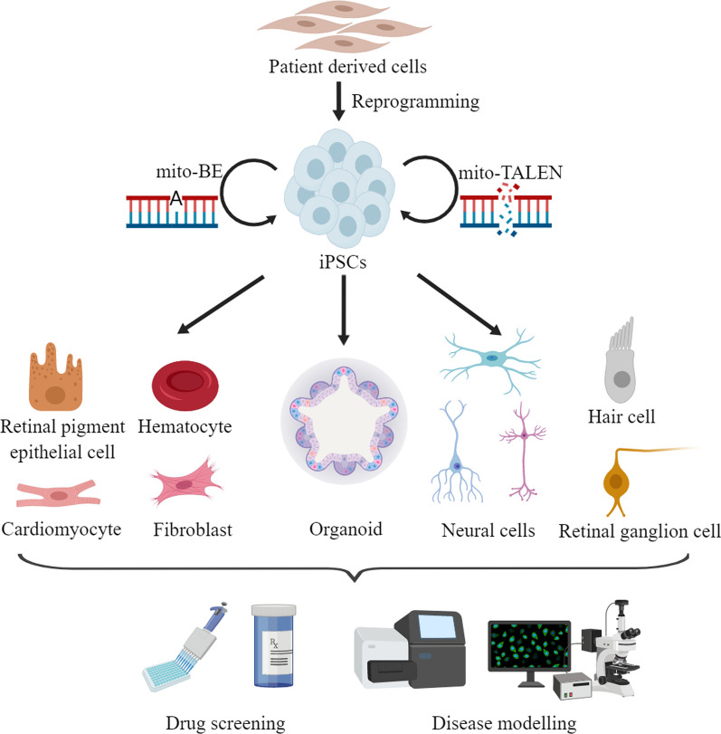
Mitochondria are essential organelles for cellular metabolism and physiology in eukaryotic cells. Human mitochondria have their own genome (mtDNA), which is maternally inherited with 37 genes, encoding 13 polypeptides for oxidative phosphorylation, and 22 tRNAs and 2 rRNAs for translation. mtDNA mutations are associated with a wide spectrum of degenerative and neuromuscular diseases. However, the pathophysiology of mitochondrial diseases, especially for threshold effect and tissue specificity, is not well understood and there is no effective treatment for these disorders. Especially, the lack of appropriate cell and animal disease models has been significant obstacles for deep elucidating the pathophysiology of maternally transmitted diseases and developing the effective therapy approach. The use of human induced pluripotent stem cells (iPSCs) derived from patients to obtain terminally differentiated specific lineages such as inner ear hair cells is a revolutionary approach to deeply understand pathogenic mechanisms and develop the therapeutic interventions of mitochondrial disorders. Here, we review the recent advances in patients-derived iPSCs as ex vivo models for mitochondrial diseases. Those patients-derived iPSCs have been differentiated into specific targeting cells such as retinal ganglion cells and eventually organoid for the disease modeling. These disease models have advanced our understanding of the pathophysiology of maternally inherited diseases and stepped toward therapeutic interventions for these diseases.

Keywords: Maternally inherited diseases; Mitochondria; iPSCs; mtDNA mutations.


Conflict of interest statement

All authors have no proprietary or commercial interest in any of materials discussed in this article.

Figures

Fig. 1
Fig. 1
Mitochondrial genome and pathogenic mtDNA mutations. Human mitochondrial genome is shown as circular, double-stranded DNA molecule with annotations. The ribosomal RNA genes are shown in purple, while tRNA genes are shown in white and annotated with single letter abbreviations. The subunits of complex I (cyan), cytochrome b (green) of complex III, subunits of complex IV (yellow) and complex V (red) are denoted by the position along the mtDNA sequence, with the outer circle as the heavy chain, and inner circle as the light chain. The positions of pathogenic mtDNA mutations are marked by black arrows. KSS Kearns–Sayre syndrome, LHON Leber’s hereditary optic neuropathy, MELAS Mitochondrial Encephalomyopathy, Lactic Acidosis, and Stroke-like episodes, MERRF Myoclonic Epilepsy and Ragged Red Muscle Fibers
Fig. 2
Fig. 2
iPSC models for human diseases due to mtDNA alterations. In modelling mtDNA diseases, patient derived cells are firstly reprogrammed to iPSCs. With mitochondrial base editing and mito-TALEN, the manipulating of mtDNA in iPSCs is achievable. The patient derived iPSCs and genetically corrected iPSCs are differentiated to distinct types of targeting cells to investigate the pathophysiology and to develop the therapeutic intervention approaches for these diseases
Fig. 3
Fig. 3
Therapeutic approaches combining iPSCs with mtDNA gene editing technology. For iPSCs derived from patients carrying heteroplasmic mtDNA mutation, mitochondrial restriction endonucleases (mtREs), mitochondrial-targeted transcription activator-like effector nucleases (mtTALENs), and zinc-finger nucleases (mtZFN) are utilized to eliminate the mutant mtDNA molecules. For iPSCs derived from patients bearing homoplasmic mtDNA mutation, mtDNA base editing technology is preferred to correct the mutations. After gene editing, the corrected iPSCs differentiate to target cells with remodeling cellular functions. The black circles indicate mtDNA. The red dots on black circles denote mtDNA mutations. The green color of mitochondria indicates normal condition, and the pink color of mitochondria show damaged function. Other colors of cells do not indicate any information

References

    1. Wallace DC. Mitochondrial genetic medicine. Nat Genet. 2018;50:1642–1649. - PubMed
    1. Nunnari J, Suomalainen A. Mitochondria: in sickness and in health. Cell. 2012;148(6):1145–1159. - PMC - PubMed
    1. Chan DC. Mitochondria: dynamic organelles in disease, aging, and development. Cell. 2006;125(7):1241–1252. - PubMed
    1. Picard M, Shirihai OS. Mitochondrial signal transduction. Cell Metab. 2022;34(11):1620–1653. - PMC - PubMed
    1. Pfanner N, Warscheid B, Wiedemann N. Mitochondrial proteins: from biogenesis to functional networks. Nat Rev Mol Cell Biol. 2019;20(5):267–284. - PMC - PubMed

Substances