. 2023 Sep 22;13(1):15779.
doi: 10.1038/s41598-023-42708-8.

T cell exhaustion is associated with cognitive status and amyloid accumulation in Alzheimer's disease

Affiliations

T cell exhaustion is associated with cognitive status and amyloid accumulation in Alzheimer's disease

Jason M Grayson et al. Sci Rep. .

Abstract

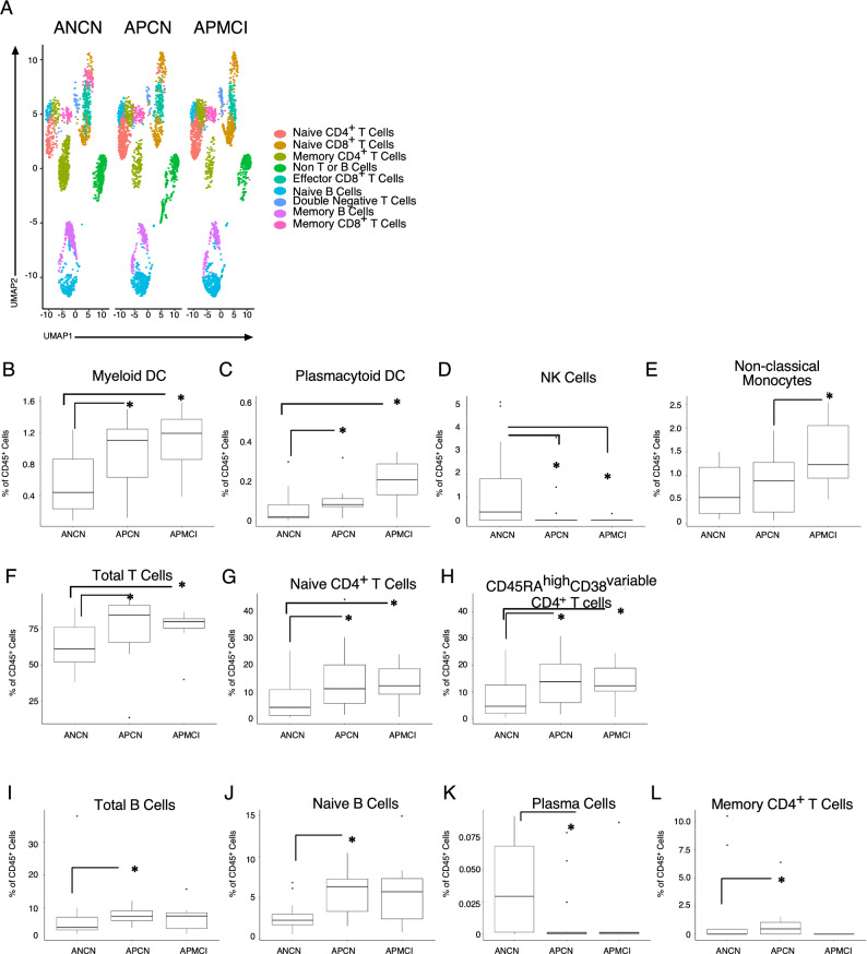
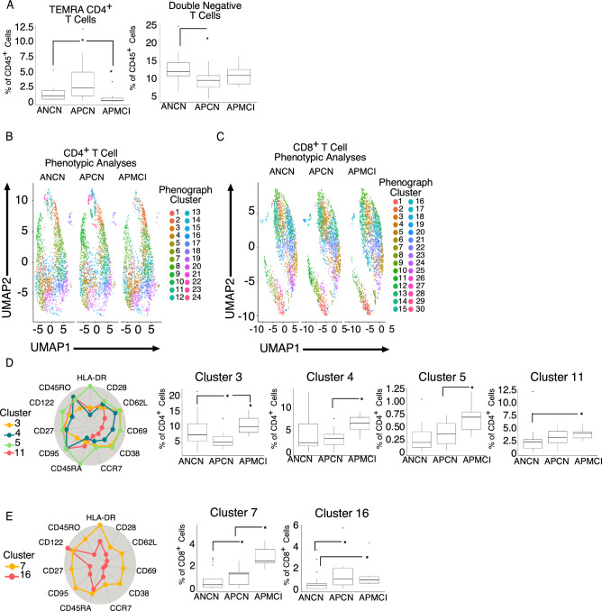
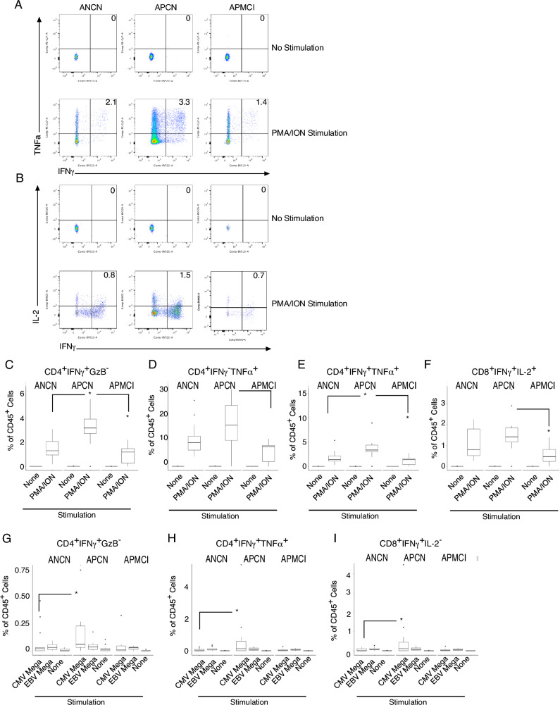
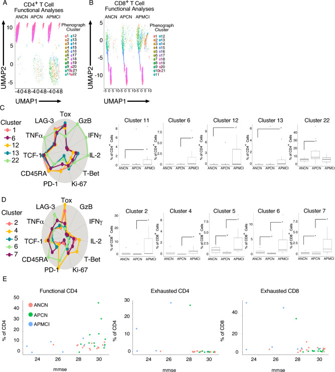
Studies over the last 100 years have suggested a link between inflammation, infectious disease, and Alzheimer's Disease (AD). Understanding how the immune system changes during the development of AD may facilitate new treatments. Here, we studied an aging cohort who had been assessed for AD pathology with amyloid positron emission tomography and cognitive testing, and conducted high dimensional flow cytometry on peripheral blood mononuclear and cerebrospinal fluid cells. Participants were assigned a classification of being amyloid negative cognitively normal, amyloid positive cognitively normal (APCN), or amyloid positive mild cognitive impairment (APMCI), an early stage of AD. We observed major alterations in the peripheral innate immune system including increased myeloid and plasmacytoid dendritic cells in the blood of APMCI participants. When the adaptive immune system was examined, amyloid positive participants, regardless of cognitive status, had increased CD3+ T cells. Further analyses of CD4+ and CD8+ T cells revealed that APMCI participants had an increase in more differentiated phenotype T cells, such as effector memory and effector memory CD45RA expressing (TEMRA), compared to those with normal cognition. When T cell function was measured, we observed that T cells from APCN participants had increased IFNγ+GzB- producing cells compared to the other participants. In contrast, we demonstrate that APMCI participants had a major increase in T cells that lacked cytokine production following restimulation and expressed increased levels of PD-1 and Tox, suggesting these are exhausted cells. Rejuvenation of these cells may provide a potential treatment for AD.


Conflict of interest statement

The authors declare no competing interests.

Figures

Figure 1
Figure 1
Altered innate and adaptive immune cell populations in amyloid positive aging participants. Peripheral blood mononuclear cells (PBMCs) from participants were stained with a pan-immune antibody cocktail and viable, CD45+ events were analyzed and (A) uniform manifold and approximation projection (UMAP) dimensionality reduction and Phenograph clustering was performed. Automated clusters were condensed and annotated by user expertise. In a separate analysis using prior immunological knowledge (B) Myeloid Dendritic cells, (C) Plasmacytoid Dendritic cells, (D) Natural killer cells, (E) Non-classical monocytes, (F) Total T Cells, (G) Naïve CD4+ T cells, (H)CD4+CD45RAhighCD38variable T cells, (I) Total B cells, (J) Naïve B Cells, (K) Plasma B cells were quantitated and the percentage of viable CD45+ cells is plotted with the median and interquartile ranges shown. Cerebrospinal fluid was analyzed (L) and the percentage of memory CD4+ T cells was plotted. The percentage of viable CD45+ cells is plotted with the median and interquartile ranges shown. Thirty-eight participants were analyzed (ANCN:16, APCN:15, ANMCI:7).* indicates p-value ≤ 0.05 in Wilcoxon testing.
Figure 2
Figure 2
Increased T cell activation in amyloid positive aging participants. Peripheral blood monuclear cells (PBMCs) from participants were stained with a T cell differentiation antibody cocktail and viable, CD45+CD3+ events were analyzed and (A) CD4+ TEMRA and (B) CD4-CD8- Double Negative T cells as determined by user expertise were plotted. (C) CD4+ or (D) CD8+ T cells were exported and uniform manifold and approximation projection (UMAP) dimensionality reduction and Phenograph clustering was performed. Automated clusters are indicated in the legend. Clusters with statistically significant differences in the (D) CD4+ or (E) CD8+ T cell compartment are plotted. The percentage of viable CD4+ or CD8+ T cells is plotted with the median and interquartile ranges shown. Forty participants were analyzed (ANCN:17, APCN:15, ANMCI:7). * indicates p-value ≤ 0.05 in Wilcoxon testing.
Figure 3
Figure 3
Increased T cell cytokine production in amyloid positive cognitively normal aging participants. Peripheral blood monuclear cells (PBMCs) from participants were stained with a T cell function antibody cocktail and viable, CD45+CD3+ events were analyzed and gated on (A) CD4+ or (B) CD8+ T cells and the indicated cytokines are plotted. The number in the upper right quadrant is the percent of viable CD45+ cells. CD4+ T cells were examined for cytokine production following PMA and ION stimulation and (C) IFNγ+GranzymeB- (D) IFNγ-TNFα+ or (E) IFNγ+TNFα + as a percentage of viable CD45+ cells is plotted with the median and interquartile ranges shown. (F) CD8+ IFNγ+IL-2+ cells were quantitated after PMA and ION stimulation. (G) CD4+ or (H, I) CD8+ cells were stimulated with the indicated peptide mega-pool and (G) IFNγ+GranzymeB- (H) IFNγ-TNFα+ (I) IFNγ+IL-2- are plotted. Thirty-two participants were analyzed (ANCN:13, APCN:12, ANMCI:7). * indicates p-value ≤ 0.05 in Wilcoxon testing.
Figure 4
Figure 4
Increased T cell exhaustion in amyloid positive mild cognitive impairment aging participants. Peripheral blood mononuclear cells (PBMCs) from participants were stained with a T cell function antibody cocktail and viable, CD45+CD3+ events were analyzed and gated on (A) CD4+ or (B) CD8+ T cells and and uniform manifold and approximation projection (UMAP) dimensionality reduction and Phenograph clustering was performed. Automated clusters are indicated in the legend. Clusters with statistically significant differences in the (C) CD4+ or (D) CD8+ T cell compartment are plotted. The percentage of viable CD4+ or CD8+ cells is plotted with the median and interquartile ranges shown. (E) Exhausted and functional T cell clusters were condensed and plotted against MMSE score for each individual. Thirty-two participants were analyzed (ANCN:13, APCN:12, ANMCI:7). * indicates p-value ≤ 0.05 in Wilcoxon testing.

References

    1. 2021 Alzheimer's disease facts and figures. Alzheimers Dement17, 327-406, doi:10.1002/alz.12328 (2021). - PubMed
    1. Skaria AP. The economic and societal burden of Alzheimer disease: Managed care considerations. Am. J. Manag. Care. 2022;28:S188–S196. doi: 10.37765/ajmc.2022.89236. - DOI - PubMed
    1. Wong W. Economic burden of Alzheimer disease and managed care considerations. Am. J. Manag. Care. 2020;26:S177–S183. doi: 10.37765/ajmc.2020.88482. - DOI - PubMed
    1. Fan L, et al. New insights into the pathogenesis of Alzheimer's disease. Front. Neurol. 2019;10:1312. doi: 10.3389/fneur.2019.01312. - DOI - PMC - PubMed
    1. Atri A. Current and future treatments in Alzheimer's disease. Semin Neurol. 2019;39:227–240. doi: 10.1055/s-0039-1678581. - DOI - PubMed

Publication types

Substances