. 2023 Dec 15;4(4):102586.
doi: 10.1016/j.xpro.2023.102586. Epub 2023 Sep 21.

In vivo functional brain mapping using ultra-high-field fMRI in awake common marmosets

Affiliations

In vivo functional brain mapping using ultra-high-field fMRI in awake common marmosets

Alessandro Zanini et al. STAR Protoc. .

Abstract

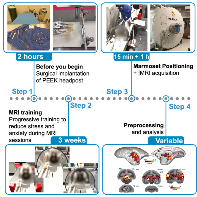
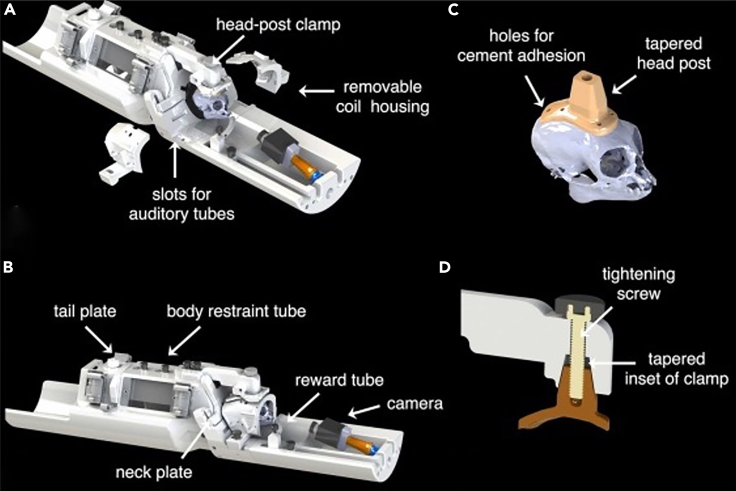
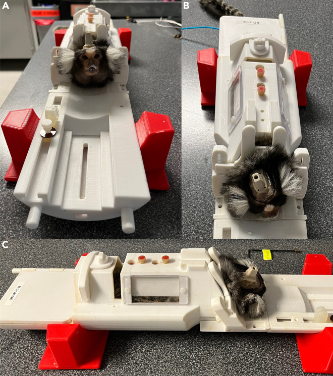
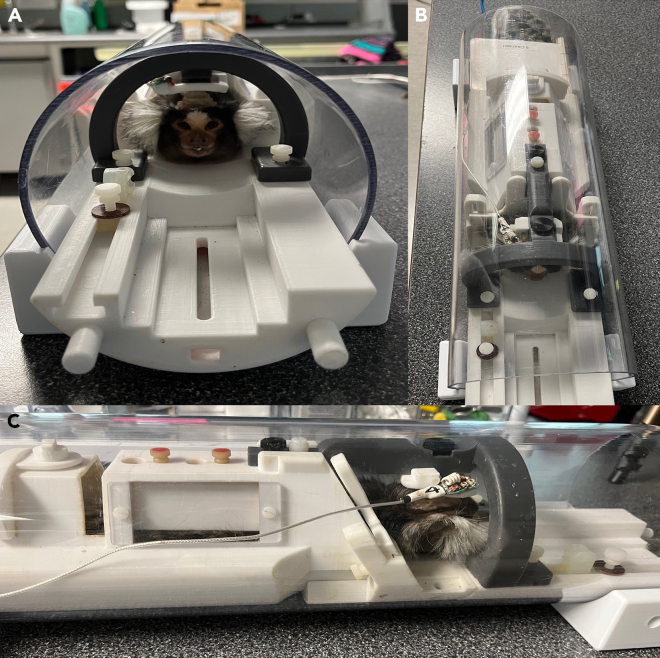
The common marmoset (Callithrix jacchus) is gaining attention in the field of cognitive neuroscience. The development of an effective protocol for fMRI data acquisition in awake marmosets is a key factor in developing reliable comparative studies. Here, we describe a protocol to obtain fMRI data in awake marmosets using auditory and visual stimulation. We describe steps for surgical and anesthesia procedures, MRI training, and positioning the marmosets within an MRI-compatible body restraint. We then detail fMRI scanning and preprocessing of functional images. For complete details on the use and execution of this protocol, please refer to Jafari et al. (2023).1.

Keywords: Behavior; Cognitive Neuroscience; Neuroscience.


Conflict of interest statement

Declaration of interests The authors declare no competing interests.

Figures

None
Graphical abstract
Figure 1
Figure 1
Detailed representation of the body restraint system and the PEEK headpost (A) CAD drawing of the body restraint used in this paper and fully described in Gilbert et al., 2023. Initially secure the marmoset in the body restraint using the neck plate. Subsequently, depending on the size of the animal, place within the body restraint a back plate of variable size and close the tail plate. Finally, screw the clamp first to the head-post and secondly to the body restraint. Once the monkey is fixed, earphones (if necessary) or silicone earplugs to reduce the gradient noise during the fMRI sessions may be positioned. (B) After the steps described in A, screw the two parts of the coil onto the dedicated slots in the frontal part of the body restraint. (C and D) (C) Representation of the position and size of the head post applied on the marmosets’ skull. To fixate the animal’s head to the body restraint, position the clamp onto the head post, and then secure it with a PEEK screw, as shown in D. This procedure will draw the head post into the clamp, which can finally be secured to the body restraint. From ref. with permission.
Figure 2
Figure 2
First week of MRI training: Familiarization with the body restraint (A–C) Frontal (A), superior (B) and lateral (C) views of the setting used for the first week of MRI training. During this week, the marmoset’s head is not fixed to the body restraint and no gradient-coil sounds are played.
Figure 3
Figure 3
Second week of MRI training: Gradient-coil sounds in the mock-MRI scanner (A–C) Frontal (A), superior (B) and lateral (C) views of the setting used for the second week of MRI training. During this week, the marmoset’s head is not fixed, but the body restraint is positioned inside a mock-MRI scanner and gradient-coil sounds are played during the session.
Figure 4
Figure 4
Third week of MRI training: Head fixation (A–C) Frontal (A), superior (B) and lateral (C) views of the setting used for the last week of MRI training. During this week, the marmoset’s head is fixed to the body restraint via the head-post clamp. The body restraint is then positioned inside the mock-MRI tube and gradient-coil sounds are played during the session.
Figure 5
Figure 5
Detailed view of the removable coil housing positioning (A and B) Representation of an erroneous (A) and a correct (B) position of the two removable coil housing parts. The frontal-posterior shifting of the two parts may negatively affect the quality of the signal during the MR scan.
Figure 6
Figure 6
Representation of the temporal SNR produced by a 5-channel (top row) and our 8-channel coil (bottom row), obtained during a two-fold accelerated EPI acquisition with 500-μm isotropic resolution The 8-channel coil produces higher tSNR across the entire brain, with the highest gains in the periphery. White lines delineate the boundaries of an undistorted, co-registered anatomical image. Yellow arrows delineate regions in which the 5-channel coil produced a signal dropout or greater distortion. Adapted with permission from ref.
Figure 7
Figure 7
Functional activations for the comparison between marmosets’ vocalizations and scrambled vocalizations T-maps are thresholded at T = 3.35 (p < 0.01 uncorrected) and displayed on the right fiducial brain surface (lateral and medial view). White lines delineate the brain parcellation based on the Paxinos marmoset’s atlas. Coronal slices of anatomical MR images are used to depict subcortical activations. Adapted from ref. with permissions. TPt: temporoparietal transitional area; MST: medial superior temporal area; FST: fundus of superior temporal sulcus; A1: primary auditory cortex; CL: caudolateral auditory cortex; ML: middle lateral auditory cortex; CPB: caudal parabelt auditory area; RPB: rostral parabelt area; AL: anterolateral auditory area; TPO: temporo-parieto-occipital association area; STR: superior temporal rostral area; 6DR: dorsorostral area 6; IC: inferior colliculus; Hb: habenula; Pul: pulvinar; MG: medial geniculate nucleus; Th: thalamus; Hip: hippocampus.
Figure 8
Figure 8
Functional activations for the comparison between goal-directed and non-goal-directed actions (A and B) Z-maps are thresholded at z = 3.29 (corresponding to p < 0.001) and survived the cluster-size correction (10,000 Monte-Carlo simulations, α = 0.05). Results are displayed on the left (A) and right (B) fiducial brain surfaces (both on lateral and medial view). White lines delineate the brain parcellation based on the Paxinos marmoset’s atlas. From ref. with permission. 6Va: ventral part a of area 6; 6DC: dorsocaudal area 6; 6DR: dorsorostral area 6; FST: fundus of the superior temporal sulcus.

Similar articles

Cited by

References

    1. Jafari A., Dureux A., Zanini A., Menon R.S., Gilbert K.M., Everling S. A vocalization-processing network in marmosets. Cell Rep. 2023;42 doi: 10.1016/j.celrep.2023.112526. - DOI - PubMed
    1. Dureux A., Zanini A., Everling S. Face-Selective Patches in Marmosets Are Involved in Dynamic and Static Facial Expression Processing. J. Neurosci. 2023;43:3477–3494. doi: 10.1523/JNEUROSCI.1484-22.2023. - DOI - PMC - PubMed
    1. Zanini A., Dureux A., Selvanayagam J., Everling S. Ultra-high field fMRI identifies an action-observation network in the common marmoset. Commun. Biol. 2023;6:553–611. doi: 10.1038/s42003-023-04942-8. - DOI - PMC - PubMed
    1. Dureux A., Zanini A., Selvanayagam J., Menon R.S., Everling S. Gaze patterns and brain activations in humans and marmosets in the Frith-Happé theory-of-mind animation task. Elife. 2023;12 doi: 10.7554/eLife.86327. - DOI - PMC - PubMed
    1. Schaeffer D.J., Gilbert K.M., Hori Y., Gati J.S., Menon R.S., Everling S. Integrated radiofrequency array and animal holder design for minimizing head motion during awake marmoset functional magnetic resonance imaging. Neuroimage. 2019;193:126–138. doi: 10.1016/j.neuroimage.2019.03.023. - DOI - PubMed

Publication types