. 2023 Nov 30;35(12):4284-4303.
doi: 10.1093/plcell/koad236.

Evolutionarily conserved protein motifs drive interactions between the plant nucleoskeleton and nuclear pores

Affiliations

Evolutionarily conserved protein motifs drive interactions between the plant nucleoskeleton and nuclear pores

Sarah Mermet et al. Plant Cell. .

Abstract

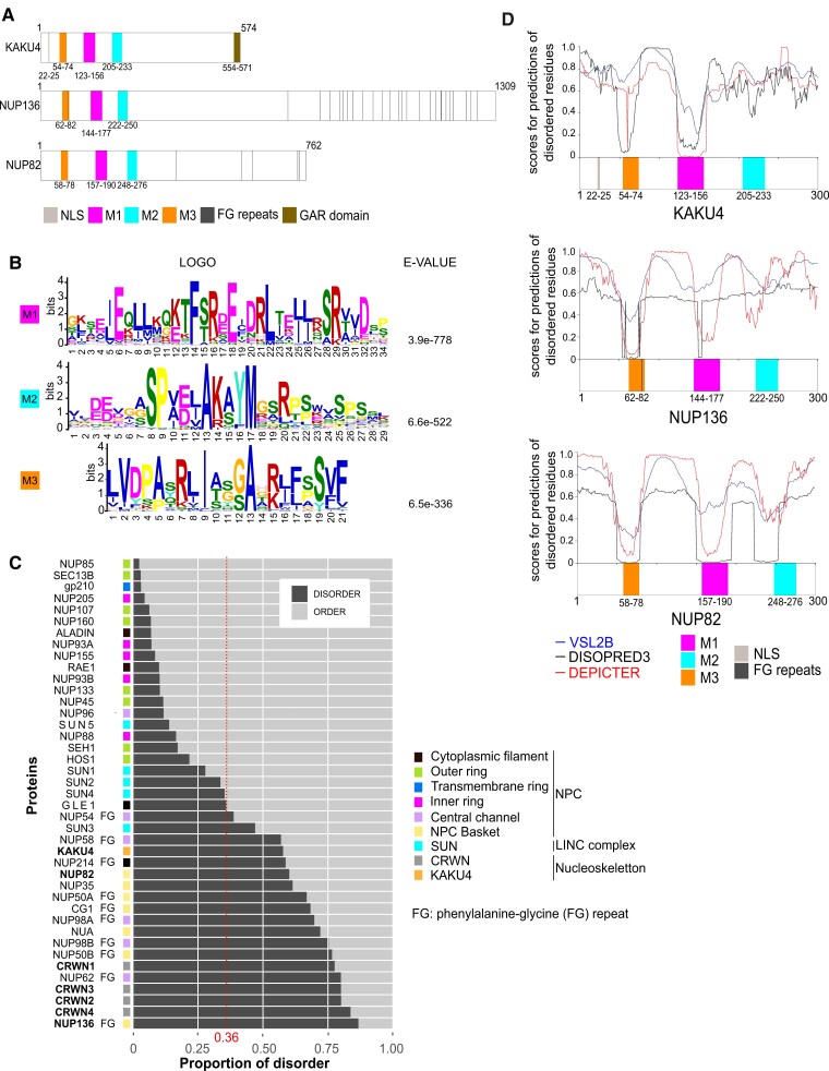
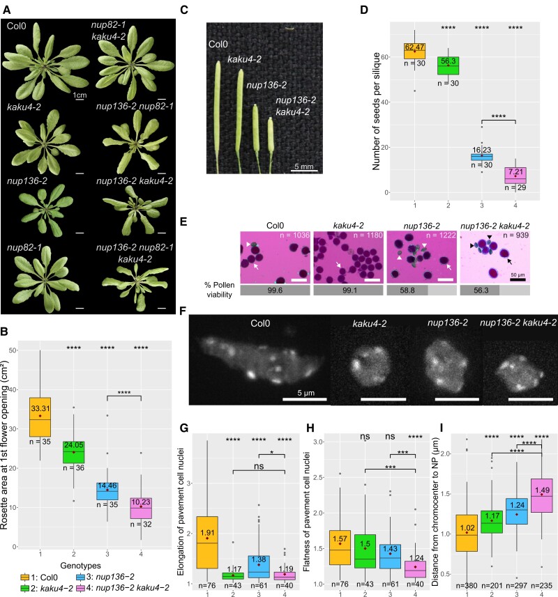
The nucleoskeleton forms a filamentous meshwork under the nuclear envelope and contributes to the regulation of nuclear shape and gene expression. To understand how the Arabidopsis (Arabidopsis thaliana) nucleoskeleton physically connects to the nuclear periphery in plants, we investigated the Arabidopsis nucleoskeleton protein KAKU4 and sought for functional regions responsible for its localization at the nuclear periphery. We identified 3 conserved peptide motifs within the N-terminal region of KAKU4, which are required for intermolecular interactions of KAKU4 with itself, interaction with the nucleoskeleton protein CROWDED NUCLEI (CRWN), localization at the nuclear periphery, and nuclear elongation in differentiated tissues. Unexpectedly, we find these motifs to be present also in NUP82 and NUP136, 2 plant-specific nucleoporins from the nuclear pore basket. We further show that NUP82, NUP136, and KAKU4 have a common evolutionary history predating nonvascular land plants with KAKU4 mainly localizing outside the nuclear pore suggesting its divergence from an ancient nucleoporin into a new nucleoskeleton component. Finally, we demonstrate that both NUP82 and NUP136, through their shared N-terminal motifs, interact with CRWN and KAKU4 proteins revealing the existence of a physical continuum between the nuclear pore and the nucleoskeleton in plants.


Conflict of interest statement

Conflict of interest statement. None declared.

Figures

Figure 1.
Figure 1.
Arabidopsis KAKU4 contains conserved motifs required for PPI. A) Domains and motifs organization of KAKU4: N- (1 to 310) and C-terminal region (311 to 574), nuclear localization signal (NLS, 22 to 25), glycine arginine rich domain (GAR domain, 554 to 571), and 3 conserved motifs named M1, M2, and M3. B) Consensus motifs identified by MEME from 16 species representative of the green lineage (see ‘Materials and methods’ section). Position of M1 (123 to 155), M2 (209 to 237), and M3 (50 to 69) in Arabidopsis KAKU4 and logos corresponding to the probability of each possible amino acid at each position in the peptide sequence. E-values are indicated for the 3 motifs. C) PEP-FOLD3 prediction with the peptide sequences used for prediction (top) and the predicted helices (boxes) as well as the superposition of the 5 best models of the helical organization of the motifs (bottom). D) Schematic representation of KAKU4 constructs on the left deletion constructs (deletion as dashed lines) and on the right constructs expressing the motifs alone. E and F) Summary of Y2H experiments testing interaction of M1, M2, and M3 motifs (white cells) or M1 and M2 deletions (gray cells) and the N-terminal region of KAKU4 E) or the CRWN proteins F) (see also Supplemental Fig. S2 for original data) by growth of yeast cell on restrictive medium (SD/-Leu/-Trp/-Ade/-His). Strong interaction (++), interaction (+), and no interaction (−) are indicated. Empty vectors pGBKT7 and pGADT7 are used, respectively, as bait and prey negative controls.
Figure 2.
Figure 2.
Function of short motifs in nuclear shape and KAKU4 localization in Arabidopsis. A to C) Transient expression assays in N. benthamiana of different KAKU4 constructs expressed as GFP fusions as described in Fig. 1D. For each panel, representative nuclei for the most prominent nuclear phenotypes are shown. The percentage of nuclei displaying membrane overgrowth (white arrowhead) or localization exclusively at the nuclear periphery (NP) is indicated for each construct. A) When highly expressed, full-length KAKU4 can cause membrane overgrowth in N. benthamiana (left). When KAKU4 is expressed at low to moderate levels, localization is observed exclusively at the NP with no membrane overgrowth (right). B and C) Quantification of NP localization and membrane overgrowth for various combinations of B) motifs alone or C) motif deletions. D) Representative nuclei from Arabidopsis anther filaments stained with DAPI in WT (elongated nuclei) or kaku4-2 mutant (rounded nuclei). E) Quantification of nuclear shape using the aspect ratio (major axis/minor axis) calculated with the Fiji plugin Shape Descriptors of DAPI-stained anther filament nuclei in WT and kaku4-2 mutants. F and G) Complementation assay using Arabidopsis transgenic lines expressing different KAKU4 constructs as GFP fusions in kaku4-2 mutant background (representative nuclei in Supplemental Fig. S3). F) Aspect ratio based on the localization of the GFP signal. A dashed line indicates the aspect ratio value (2.06) for kaku4-2 mutant. G) Quantification of the localization of GFP-tagged KAKU4 constructs. Values are expressed as the log2 ratio of fluorescence intensity at the NP versus the nucleoplasm. Statistical significance is indicated for the kaku4-2 mutant relative to WT using Wilcoxon test (E, P-value = 2.02e−11) or relative to p35S-GFP-K4 in kaku4-2F) and G) using the Kruskal–Wallis test with P-value = 7.97e−05 F) and 8.04e−04 G), followed by pairwise comparisons using the Wilcoxon test with Holm correction for F) and G). The box represents the 25 to 75th percentiles, the median is indicated as a horizontal line across the box, and the dark red diamond and value represent the mean. The whiskers are equal to 1.5× the interquartile range. Outliers are represented by dots. Ns, not significant (P-value >0.05), *P-value <0.05, **P-value <0.01, ****P-value <0.0001. Scale bar = 10 µm. n = number of nuclei.
Figure 3.
Figure 3.
The 3 motifs of KAKU4 are conserved in NUP136 and NUP82. A) Schematic representation of Arabidopsis KAKU4, NUP136, and NUP82 proteins including the 3 motifs M1, M2, and M3. B) MEME logos and associated E-values for 3 motifs conserved between KAKU4, NUP82, and NUP136 from 16 species representative of the green lineage (see ‘Materials and methods’ section). C) Proportion of IDRs and ordered regions in NUP, CRWN, KAKU4, and SUN proteins from Arabidopsis. The median value (0.36) of disorder proportion of the Arabidopsis proteome is indicated as a red dashed line. NUPs containing phenylalanine-glycine (FG) repeats and NPC substructures are indicated by FG and with different colors, respectively. D) Profiles of intrinsically disordered residues predicted by VSL2B, DISOPRED3, and DEPICTER within the N-terminal regions (1 to 300) of KAKU4, NUP82, and NUP136 containing M3, M1, and M2 peptide motifs.
Figure 4.
Figure 4.
NUP82 and NUP136 interact with CRWN proteins via their motifs M1 and M2. A, C, and F) Y2H assays showing growth on test (SD/-Leu/-Trp/-Ade/-His) or permissive (SD/-Leu/-Trp) media. Empty vectors pGBKT7 and pGADT7 are used, respectively, as bait and prey negative controls. Interactions (strong interaction [++], interaction [+], and no interaction [−]) among KAKU4, NUP136, and NUP82 proteins A) (Supplemental Fig. S4 for original data) and CRWN proteins C). B) PPI assay in N. benthamiana using BiFC. Different proteins fused to the N-terminal (nY) or C-terminal (cY) half of yellow fluorescent protein (YFP) are coexpressed with mRFP-fibrillarin2 (Fib2) as a positive marker for transformed cells. D) Schematic representation of the PPI between NUP136, NUP82, KAKU4, and CRWNs proteins recorded by BiFC and Y2H in this study. E) Schematic representation of NUP136 and NUP82 constructs with deletions (dashed line) of motifs M1 (magenta) and M2 (cyan) or motifs M1 and M2 only. F) Y2H assay testing the role of M1 and M2 motifs from NUP136 and NUP82 in the interaction with CRWN proteins (Supplemental Fig. S4 for original data). G) Transient coexpression of mRFP-NLS/GFP-CRWN1 and mRFP-NLS/GFP-KAKU4 in Arabidopsis root protoplasts from WT and nup136-2 nup82-1 mutant. Scale bar = 10 µm.
Figure 5.
Figure 5.
Analysis of genetic interactions between NUP136 and KAKU4. A) Photos of WT and single (kaku4-2, nup136-2, and nup82-1), double (nup136-2 kaku4-2, nup82-1 kaku4-2, and nup136-2 nup82-1), and triple mutant (nup136-2 nup82-1 kaku4-2) combinations at the day of first flower opening. Photo scale = 1 cm. B to D) Quantification of different plant morphology parameters in WT and kaku4-2, nup136-2 single mutants and nup136-2 kaku4-2 double mutants. B) Rosette area at the day of first flower opening (see also Supplemental Fig. S5A). C) Size of mature siliques (Supplemental Fig. S5B for quantification). Scale bar = 5 mm. D) Number of seeds per silique. n = number of siliques. E) Microscopy observation of Alexander's stained pollen and quantification of the percentage of viable pollen. n = number of pollen grain scored. Scale bar = 50 µm. Viable pollen appears in dark pink (arrow) and dead pollen in blue (arrowhead). F) Images of pavement cell nuclei from 14-d-old seedlings stained with DAPI in WT and mutants. Scale bar = 5 µm. G to I) Quantification of nuclear flatness G) and nuclear elongation H) computed by NucleusJ 2.0 software and distance from chromocenter to nuclear periphery I) determined by NODeJ 1.0 software. The box represents the 25 to 75th percentiles, the median is indicated as a horizontal line across the box, and the dark red diamond and value represent the mean. The whiskers are equal to 1.5× the interquartile range. Outliers are represented by dots. n = number of nuclei G to H) or chromocenters I). Statistics analyses were done by comparing to the WT or by comparing the specified genotypes using Wilcoxon tests; ns, not significant (P-value >0.05), *P-value <0.05, ***P-value <0.001, ****P-value <0.0001.
Figure 6.
Figure 6.
Phylogenetic relationships among KAKU4, NUP82, and NUP136. A) Presence or absence of NUP136, NUP82, and KAKU4 orthologs in 6 groups of species representative of the green lineage (groups indicated as different colors). The 22 plant species used in this study (left) and numbers of paralogs per species (right) for NUP136, NUP82, and KAKU4 proteins are indicated. B) Maximum likelihood tree constructed with IQ-TREE from a MAFFT alignment of amino acid sequences using orthologs of NUP136, NUP82, and KAKU4 proteins. Clades are distinguished by thin black (NUP136), thick black (NUP82), and thick light gray branches (KAKU4). Evolution rates expressed as substitutions per site are given for each clade at the right of the tree. Tree scale: 1 substitution per site. C) Evolution rates expressed as dN/dS ratio for full length, N-terminal, and C-terminal regions of KAKU4, NUP82, and NUP136. Genetic drift (M0) hypothesis is rejected if P-value <0.5. D) Schematic representation of NUP82, NUP136, and KAKU4 divergence from an ancestral gene that has undergone 2 successive rounds of duplication and subsequent divergence of paralogs. Proteins are represented by a N-terminal region corresponding to the first 300 amino acids (white box with M1, M2, and M3, respectively in magenta, cyan, and orange) and a C-terminal region as a gray box (>300 amino acids). Two hypotheses are proposed to explain KAKU4 divergence (in red): (i) the C-terminal region of KAKU4 derived from the same ancestral gene as NUP82 and NUP136 or (ii) the C-terminal region was acquired through a recombination event from another yet unknown gene. E) Exon structures of KAKU4, NUP82, and NUP136 genes from Arabidopsis (Ath) drawn with the Gene Structure Display Server (GSDS). N- and C-terminal regions of KAKU4, NUP82, and NUP136 are indicated at the bottom of the figure. Exons (rectangles) are drawn to scale, realigned manually to include M1, M2, and M3 motifs and FG repeats; exon scale = 1 kb. Introns are not drawn to scale and are depicted as a black line. NW global alignment for the N-terminal part (exons 1 to 5 or 1 to 6, left) and the C-terminal part (exons 6 to 8 or 7 to 13, right) shown as a similarity matrix among the 3 Arabidopsis proteins with the percentage of identity on the top and NW scores on the bottom of the matrix. Exon 9 of NUP136 containing the FG repeats was analyzed separately (right panel).
Figure 7.
Figure 7.
Model for the plant-specific continuum between nucleoskeleton and nuclear pores. During plant evolution, KAKU4 emerged as a new nucleoskeleton component from an ancestral nucleoporin (NUP). KAKU4 contains the short peptide motifs M1, M2 and M3 shared with the nucleoporins NUP82 and NUP136 from the nuclear pore basket. M1 and M2 from KAKU4, NUP82, and NUP136 are required for interaction with CRWN proteins establishing a continuum between the nucleoskeleton and the nuclear pore basket through direct PPI (arrows).

Similar articles

Cited by

References

    1. Al-Haboubi T, Shumaker DK, Köser J, Wehnert M, Fahrenkrog B. Distinct association of the nuclear pore protein Nup153 with A- and B-type lamins. Nucleus. 2011:2(5):500–509. 10.4161/nucl.2.5.17913 - DOI - PubMed
    1. Allan C, Burel J-M, Moore J, Blackburn C, Linkert M, Loynton S, MacDonald D, Moore WJ, Neves C, Patterson A, et al. . OMERO: flexible, model-driven data management for experimental biology. Nat Methods. 2012:9(3):245–253. 10.1038/nmeth.1896 - DOI - PMC - PubMed
    1. Altschul SF, Gish W, Miller W, Myers EW, Lipman DJ. Basic local alignment search tool. J Mol Biol. 1990:215(3):403–410. 10.1016/S0022-2836(05)80360-2 - DOI - PubMed
    1. Bailey TL, Johnson J, Grant CE, Noble WS. The MEME suite. Nucleic Acids Res. 2015:43(W1):W39–W49. 10.1093/nar/gkv416 - DOI - PMC - PubMed
    1. Barik A, Katuwawala A, Hanson J, Paliwal K, Zhou Y, Kurgan L. DEPICTER: intrinsic disorder and disorder function prediction server. J Mol Biol. 2020:432(11):3379–3387. 10.1016/j.jmb.2019.12.030 - DOI - PubMed

MeSH terms

Substances

LinkOut - more resources