. 2023 Sep 21;83(18):3377-3392.e6.
doi: 10.1016/j.molcel.2023.08.022.

Elucidation of E3 ubiquitin ligase specificity through proteome-wide internal degron mapping

Affiliations

Elucidation of E3 ubiquitin ligase specificity through proteome-wide internal degron mapping

Zhiqian Zhang et al. Mol Cell. .

Erratum in

Abstract

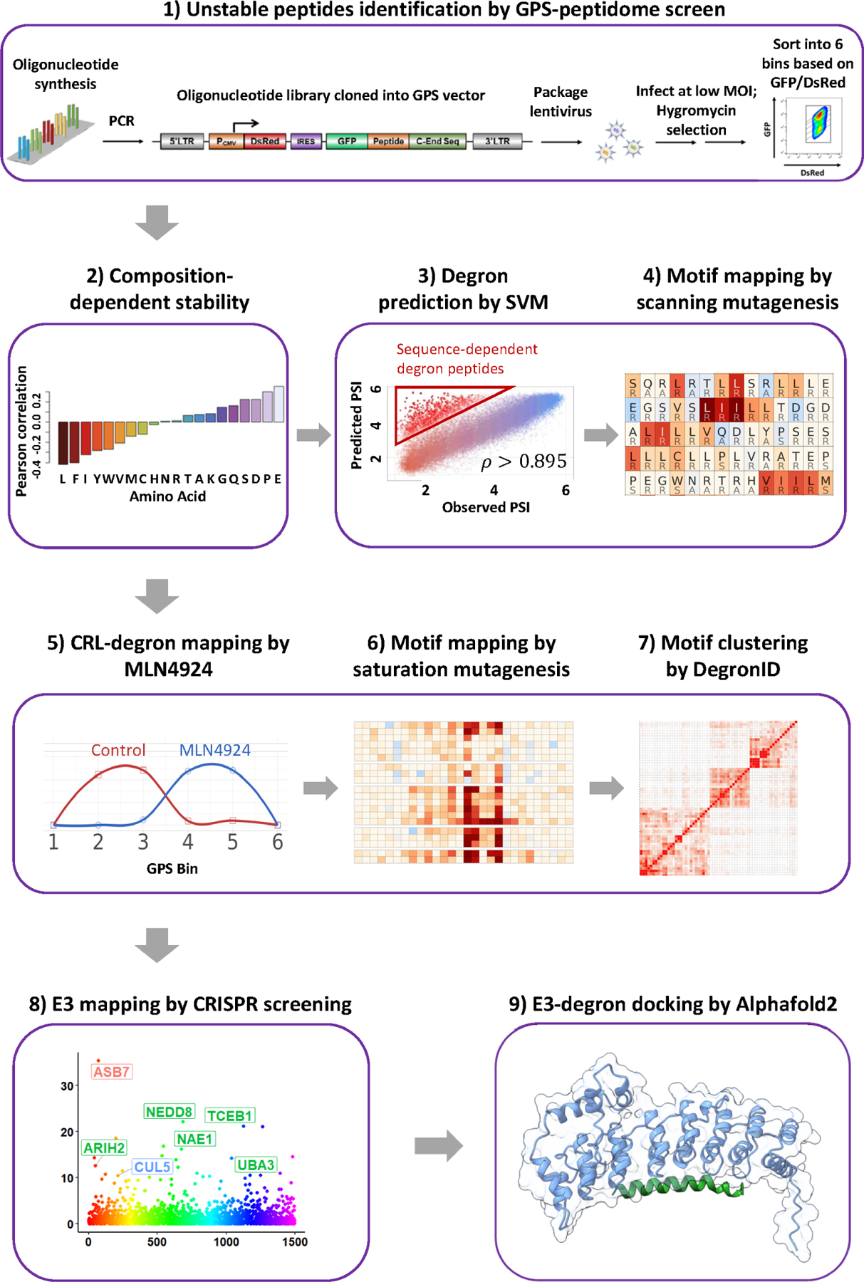
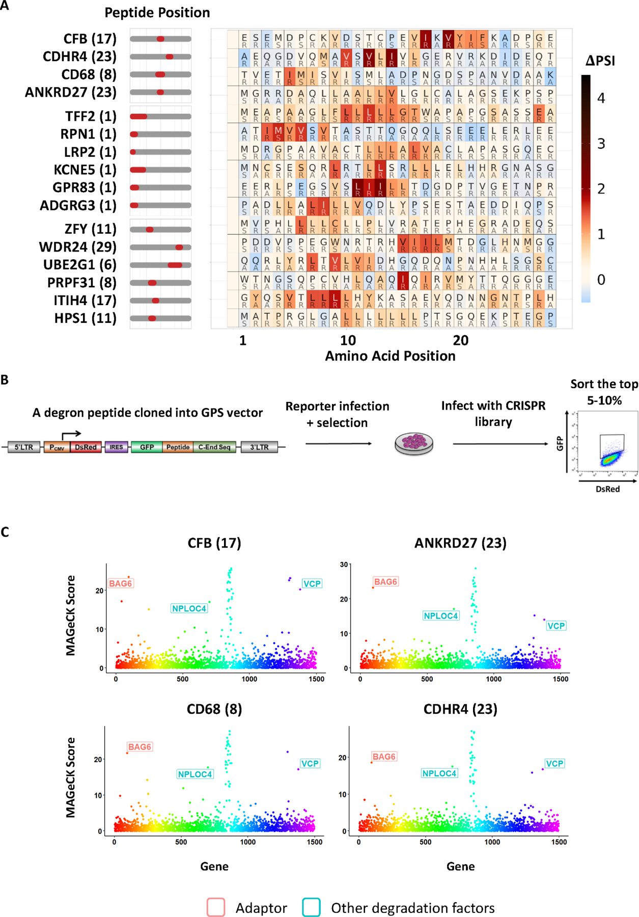
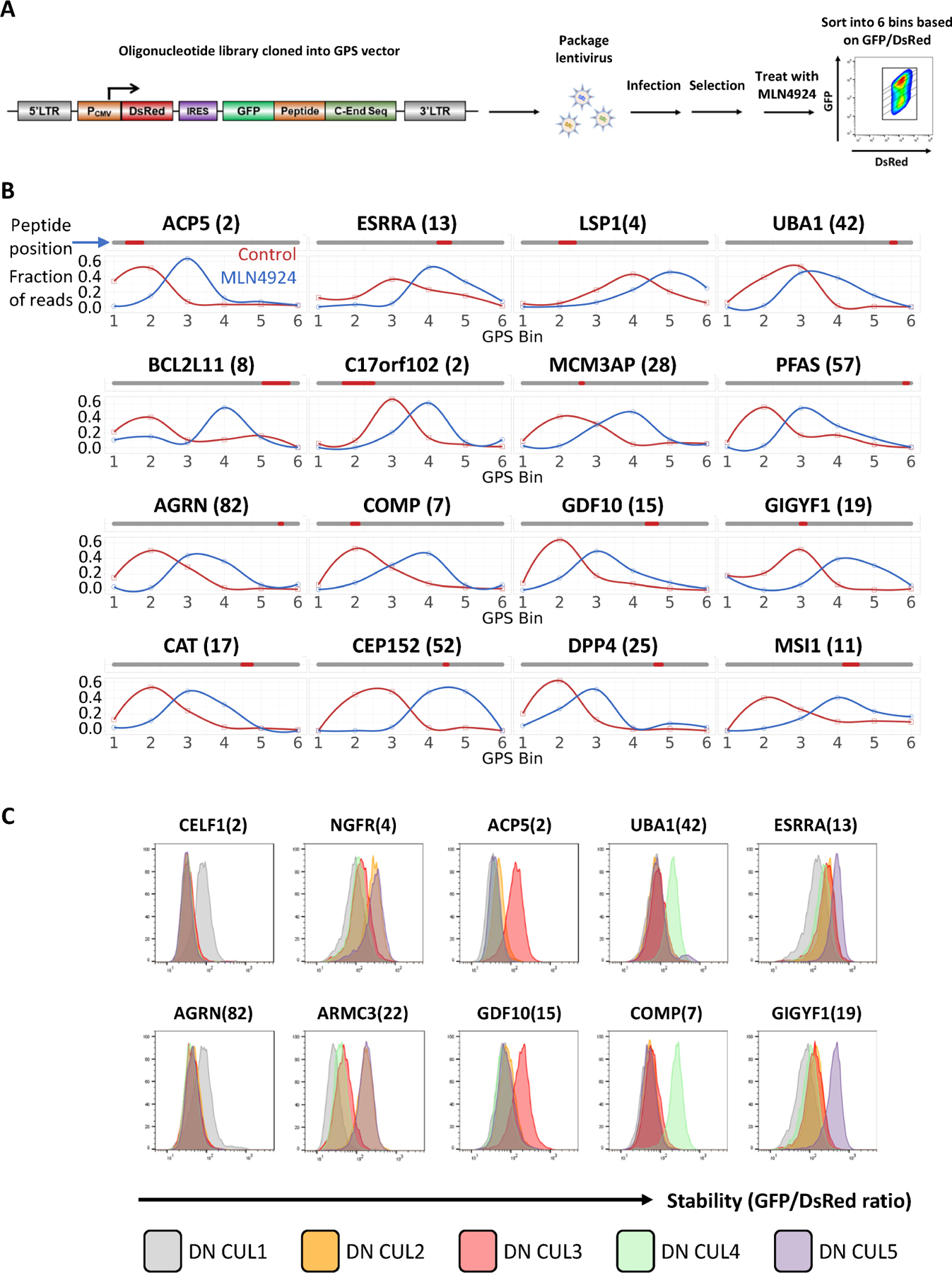
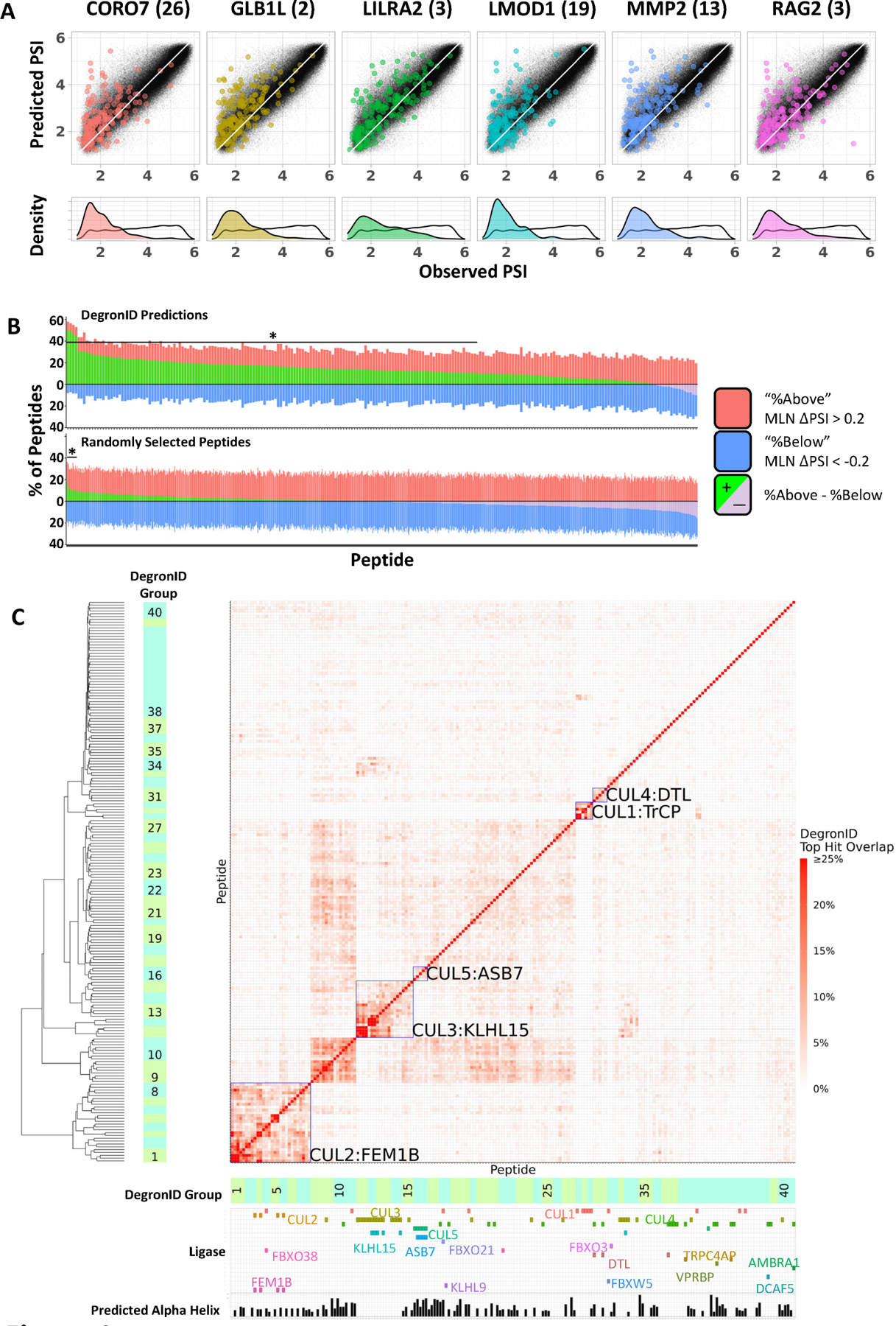
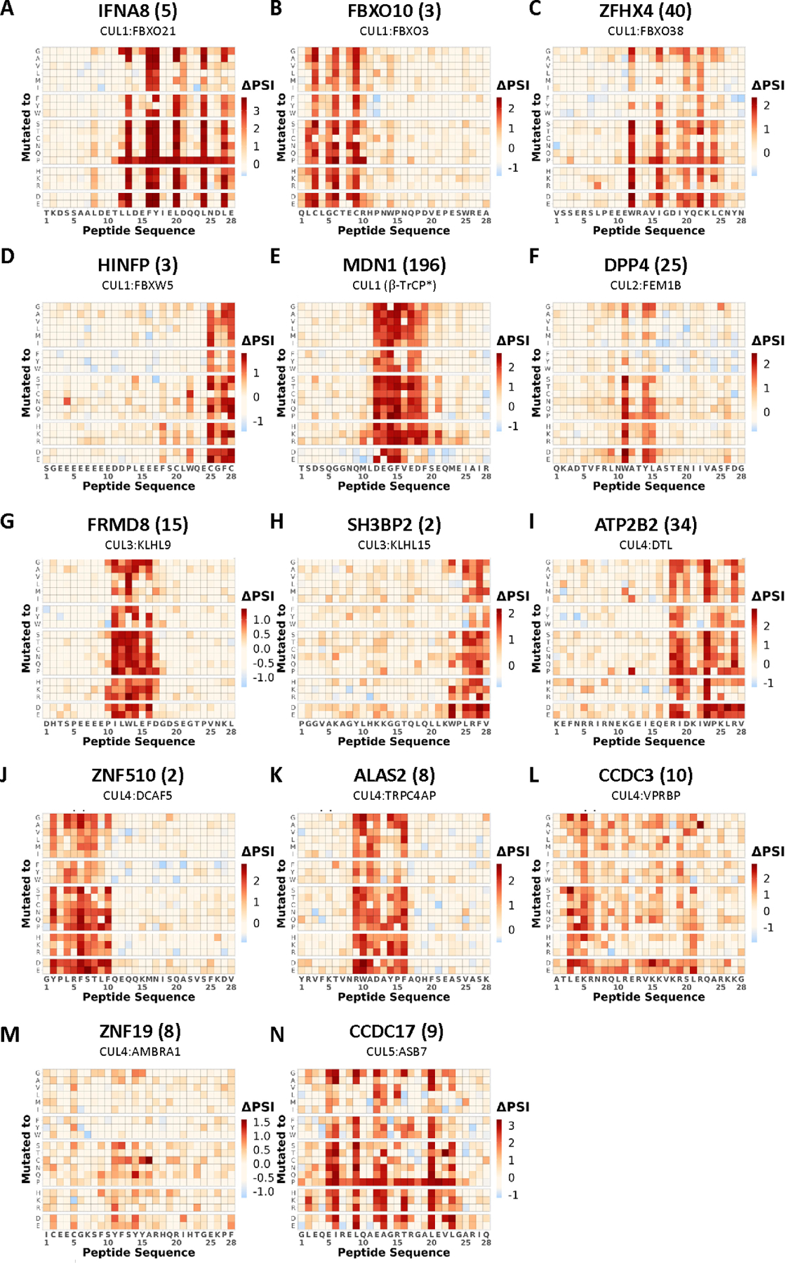
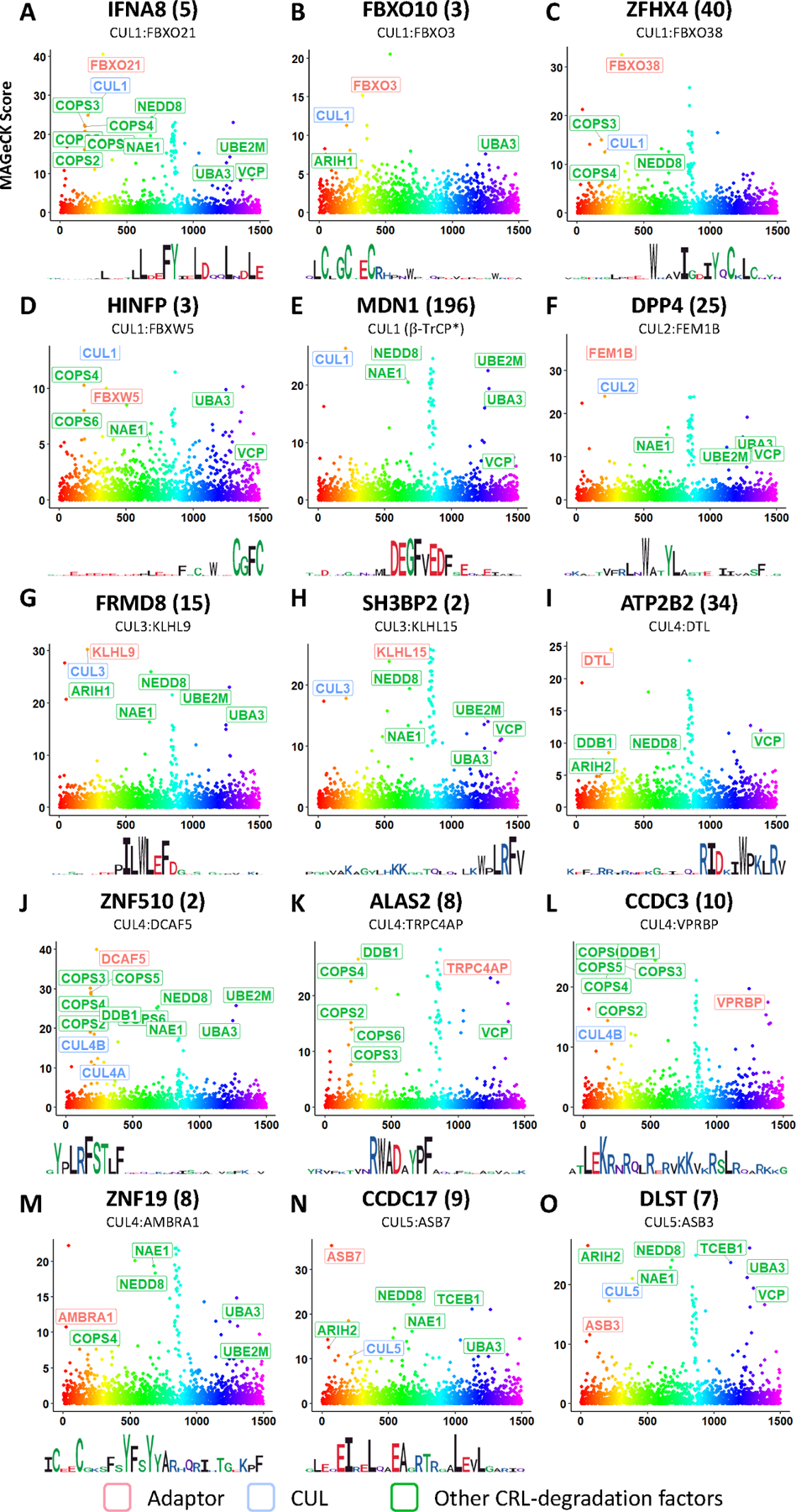
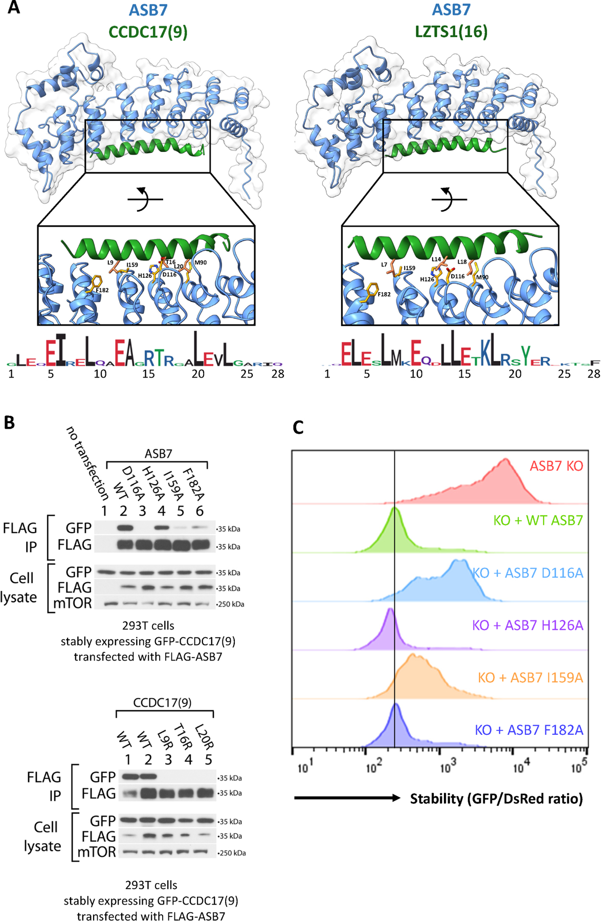
The ubiquitin-proteasome system plays a critical role in biology by regulating protein degradation. Despite their importance, precise recognition specificity is known for a few of the 600 E3s. Here, we establish a two-pronged strategy for identifying and mapping critical residues of internal degrons on a proteome-scale in HEK-293T cells. We employ global protein stability profiling combined with machine learning to identify 15,800 peptides likely to contain sequence-dependent degrons. We combine this with scanning mutagenesis to define critical residues for over 5,000 predicted degrons. Focusing on Cullin-RING ligase degrons, we generated mutational fingerprints for 219 degrons and developed DegronID, a computational algorithm enabling the clustering of degron peptides with similar motifs. CRISPR analysis enabled the discovery of E3-degron pairs, of which we uncovered 16 pairs that revealed extensive degron variability and structural determinants. We provide the visualization of these data on the public DegronID data browser as a resource for future exploration.

Keywords: Alphafold2; CRL; Cullin-RING ligase; DegronID; E3 ubiquitin ligase; GPS; degron; global protein stability; protein degradation; ubiquitination.


Conflict of interest statement

Declaration of interests S.J.E. is a founder of TSCAN Therapeutics, MAZE Therapeutics, ImmuneID and Mirimus; serves on the scientific advisory boards of Homology Medicines, ImmuneID, MAZE Therapeutics, and TSCAN Therapeutics; is an advisor for MPM Capital; and is on the Editorial Board for Molecular Cell. None of this affects this work.

Figures

Figure 1.
Figure 1.. Schematic diagram illustrating the workflow of mapping E3-degron pairs.
Overview of study workflow.
Figure 2.
Figure 2.. SVM machine learning-aided identification of a BAG6 degron motif.
(A) Scanning mutagenesis of non-CRL degron peptides. Peptides in the top group were subjected to CRISPR screens and BAG6 was identified as a destabilizing gene. BAG6-like motifs were found not only in terminal peptides (middle group) deriving from N-terminal signal peptide sequences, but also in internal peptides (bottom group). (B) Schematic diagram illustrating the workflow of the CRISPR screen designed to identify the genes required for the degradation of each degron peptide. (C) CRISPR screens of 4 representative degron peptides with motifs described in (A) using a sgRNA lentiviral library of UPS related genes identified BAG6 as a gene required for the degron activity.
Figure 3.
Figure 3.. The GPS-peptidome screen using MLN4924 identified CRL-dependent degron peptides.
(A) Schematic diagram illustrating the workflow of the GPS-peptidome screen in the presence of MLN4924. (B) Representative peptides identified in the screen in (A) as responsive to MLN4924 are shown. For each peptide, their relative distribution across the 6 bins in the control condition was compared to that of the MLN4924-treated condition. See also Figure S1. (C) Representative GPS measurements for peptide stability with dominant-negative (DN) CUL expression. Two peptides selectively stabilized by DN CUL1, DN CUL2/5, DN CUL3, DN CUL4A, DN CUL5 are shown.
Figure 4:
Figure 4:. Degron ID classified saturation mutagenesis motifs into clusters based on their sequence similarities.
(A) Distribution of peptides in terms of PSI observed in GPS screen (x) and composition-based PSI prediction (y) from the 260k library (black) and the top 200 scoring hits by DegronID (color). (B) Summary of top 200 hits by DegronID by MLN ΔPSI for (top) DegronID predictions for 198 CRL peptides; (bottom) iterations of random selections of 200 peptides from our 260k library. The bracket and asterisk indicate the instances for which the value of the green bar is greater than or equal to that which would be expected with an FDR of 0.1. (C) Hierarchical clustering of saturation mutagenesis degron footprints. Alpha helical structure predictions and paired ligases from validation experiments are indicated below the clustergram. Meta-clusters that correspond to groups of degrons with multiple members sharing the same CRL are boxed and labelled (see Figure S3). See also Figure S2.
Figure 5.
Figure 5.. Saturation mutagenesis identified degron motifs in peptides stabilized by MLN4924.
Saturation mutagenesis degron footprints for selected CRL degron peptides. The cognate E3 identified from subsequent CRISPR screening is also indicated. See also Figure 6 and Figure S3.
Figure 6.
Figure 6.. CRISPR screens identified the cognate E3 ligases for CRL-degron peptides.
MAGeCK scores from CRISPR screens to identify cognate E3 ligase of each shown peptide. (STAR Methods). Logo plots derived from the each peptide’s mutagenesis footprint are also shown. See also Table S7, Figure S4 and Figure S5.
Figure 7.
Figure 7.. E3-degron docking by AlphaFold2 identified critical degron residues consistent with that revealed by saturation mutagenesis, co-immunoprecipitation, and GPS.
(A) Alphafold2 multimer docking of ASB7 degron peptides onto ASB7. Selected residues at the interaction interface are shown and labelled. (B) Immunoblots for ASB7:CCDC17(9) binding in 293T ASB7 KO cells. (top) Co-immunoprecipitation of FLAG-ASB7 (WT or the indicated mutant) in cells stably expressing GFP-CCDC17(9) (WT). (bottom) Co-immunoprecipitation of FLAG-ASB7 (WT) in cells stably expressing GFP-CCDC17(9) (WT or the indicated mutant). Cells from lanes 1, 3, 4, and 5 stably express the specified GFP-degrons fusions flanked with C end sequence A (QGRARPNQEVQIGEMENQLS); while cells from lane 2 stably express GFP-CCDC17(9) flanked with C end sequence B (QGRARPNQEVQIGEMENQLD). (C) Flow stability data for CCDC17(9) GPS reporter peptide with KO, stably expressed WT, or stably expressed mutant ASB7. See also Figure S6 and Figure S7.

References

    1. Li W, Bengtson MH, Ulbrich A, Matsuda A, Reddy VA, Orth A, Chanda SK, Batalov S, and Joazeiro CAP (2008). Genome-Wide and Functional Annotation of Human E3 Ubiquitin Ligases Identifies MULAN, a Mitochondrial E3 that Regulates the Organelle’s Dynamics and Signaling. PLoS One 3, e1487. 10.1371/JOURNAL.PONE.0001487. - DOI - PMC - PubMed
    1. Collins GA, and Goldberg AL (2017). The Logic of the 26S Proteasome. Cell 169, 792–806. 10.1016/j.cell.2017.04.023. - DOI - PMC - PubMed
    1. Popovic D, Vucic D, and Dikic I (2014). Ubiquitination in disease pathogenesis and treatment. Nat Med 20, 1242–1253. 10.1038/nm.3739. - DOI - PubMed
    1. Juszkiewicz S, and Hegde RS (2018). Quality Control of Orphaned Proteins. Mol Cell 71, 443–457. 10.1016/J.MOLCEL.2018.07.001. - DOI - PMC - PubMed
    1. Yanagitani K, Juszkiewicz S, and Hegde RS (2017). UBE2O is a quality control factor for orphans of multiprotein complexes. Science (1979) 357, 472–475. 10.1126/science.aan0178. - DOI - PMC - PubMed

Publication types