. 2023 Sep 22;14(1):5922.
doi: 10.1038/s41467-023-41545-7.

Altered ubiquitin signaling induces Alzheimer's disease-like hallmarks in a three-dimensional human neural cell culture model

Affiliations

Altered ubiquitin signaling induces Alzheimer's disease-like hallmarks in a three-dimensional human neural cell culture model

Inbal Maniv et al. Nat Commun. .

Abstract

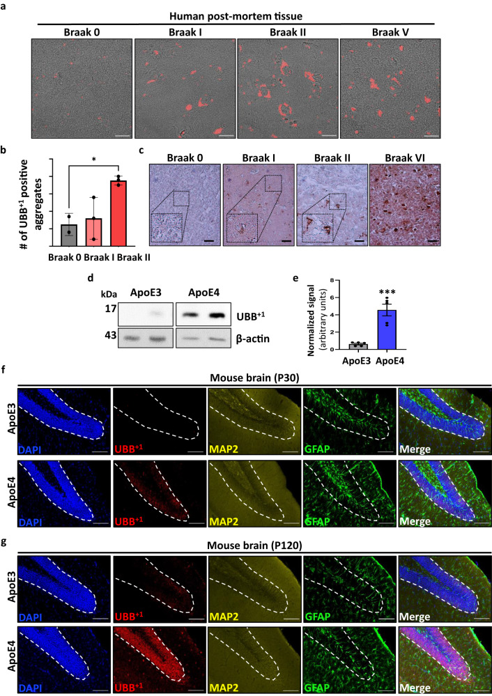
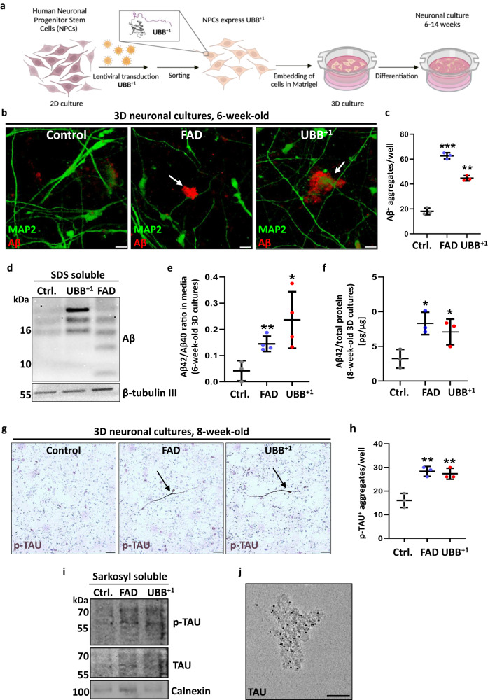
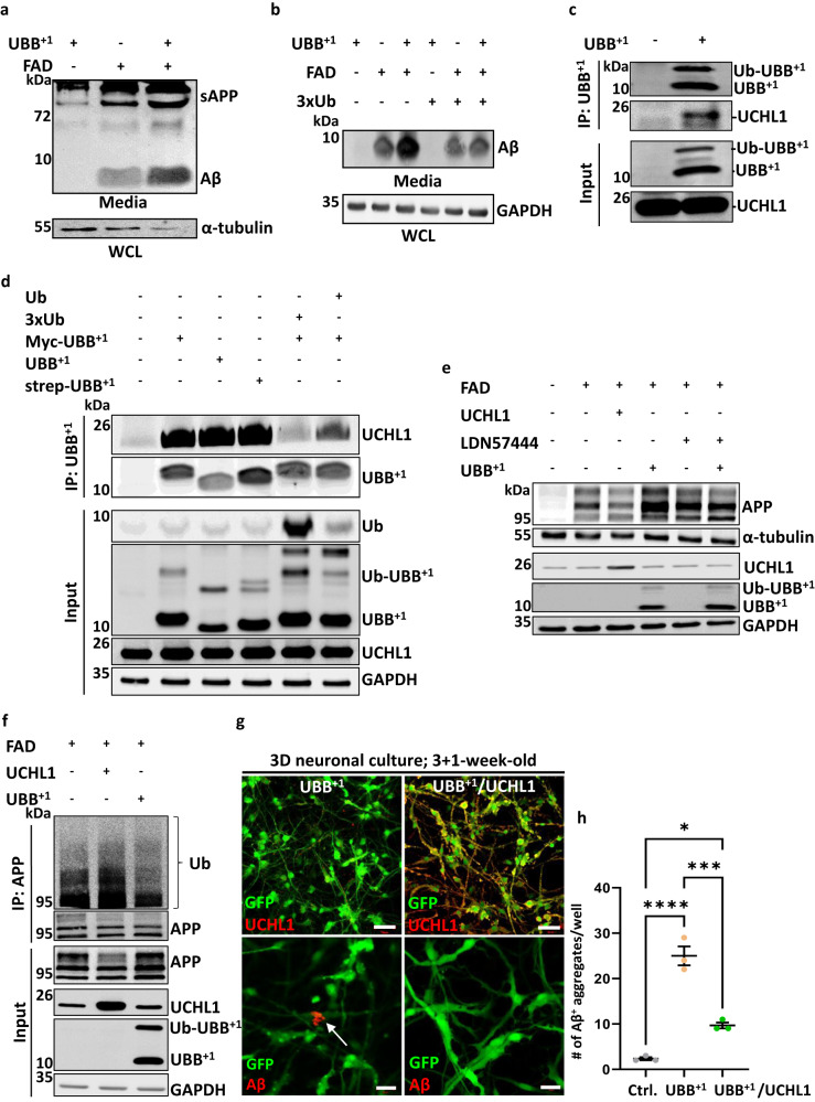
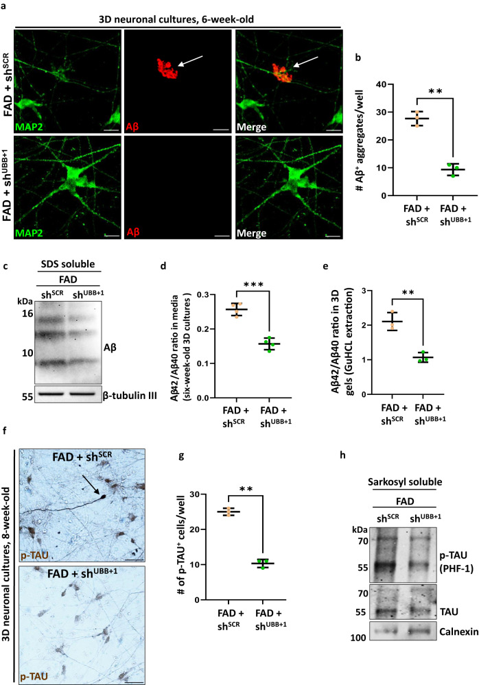
Alzheimer's disease (AD) is characterized by toxic protein accumulation in the brain. Ubiquitination is essential for protein clearance in cells, making altered ubiquitin signaling crucial in AD development. A defective variant, ubiquitin B + 1 (UBB+1), created by a non-hereditary RNA frameshift mutation, is found in all AD patient brains post-mortem. We now detect UBB+1 in human brains during early AD stages. Our study employs a 3D neural culture platform derived from human neural progenitors, demonstrating that UBB+1 alone induces extracellular amyloid-β (Aβ) deposits and insoluble hyperphosphorylated tau aggregates. UBB+1 competes with ubiquitin for binding to the deubiquitinating enzyme UCHL1, leading to elevated levels of amyloid precursor protein (APP), secreted Aβ peptides, and Aβ build-up. Crucially, silencing UBB+1 expression impedes the emergence of AD hallmarks in this model system. Our findings highlight the significance of ubiquitin signalling as a variable contributing to AD pathology and present a nonclinical platform for testing potential therapeutics.


Conflict of interest statement

The authors declare no competing interests.

Figures

Fig. 1
Fig. 1. UBB+1 accumulates in the early stages of AD postmortem tissue.
a Immunofluorescence staining of human tissue (ER = hippocampal complex, including entorhinal cortex) with an antibody against UBB+1 (red). b Quantification of UBB+1-positive aggregates counted in different biological samples taken from dentate gyrus (DG) of patients diagnosed at different Braak stages [n = 3 biologically independent samples] (Supplementary information Table 1) (p = 0.038). c Immunohistochemistry staining of human postmortem tissue (ER) with anti-UBB+1. d Representative immunoblots of proteins isolated from the hippocampus of Apolipoprotein E (ApoE3/4)-TR mice using an anti-UBB+1 antibody. e Quantification of (d) by densitometry and normalized to β-actin [n = 5 mice] (p = 0.00042) f, g Immunofluorescence staining of hippocampal sections of one (f) and 4-month-old (g) ApoE (ApoE3/4)-TR mice, showing UBB+1 is specifically expressed in neurons (MAP2) and not glial cells (GFAP). P-values were determined by unpaired two-tailed Student’s t-test. Error bars represent ± s.d. Images are representative of three independent wells. All experiments were repeated at least twice.Scale bars: 20 µm (a), 50 μm (c), 100 μm (f, g).
Fig. 2
Fig. 2. Expression of UBB+1 induces Amyloid beta (Aβ) and phosphorylated tau (p-tau) aggregates in a 3D human neural culture system.
a Schematic of UBB+1 expressing 3D system derived from ReN VM cells. Image was created using BioRender software. b Representative immunofluorescence images of 6-week-old differentiated control, APPSL-PSNE1ΔE9 (FAD) and UBB+1 expressing 3D cultures, showing Aβ-positive-aggregates in white arrows, indicating that they are extracellular deposits. c Quantification of the mean total number of extracellular Aβ deposits in 6-week-old control, FAD, and UBB+1 cultures [n = 3 biologically independent samples, the whole well was counted] (FAD/ Ctrl. p = 0.000029; UBB+1 /Ctrl. p = 0.00016). d Immunoblot analysis of SDS-solubilized aggregates isolated from 6-week-old -differentiated control, FAD, and UBB+1 cultures, showing Aβ oligomers. β-tubulin III levels were used as loading control. e ELISA of Aβ42 and Aβ40 in conditioned media collected from 3D 6-week-old control, FAD, and UBB+1 cultures. Aβ42/Aβ40 ratio is displayed [n = 3 biologically independent samples] (FAD/Ctrl. p = 0.007; UBB+1 /Ctrl. p = 0.04). f ELISA of Aβ42 in whole lysates of 3D 8-week-old control, FAD, and UBB+1 cultures. Results are displayed as Aβ42 normalized to total protein [n = 3 biologically independent samples] (FAD/ Ctrl. p = 0.014; UBB+1 /Ctrl. p = 0.043). g Representative immunohistochemistry images of 3D 8-week-old FAD and UBB+1 cultures stained with anti- p-tau. Cells displaying increased p-tau staining are marked by black arrowheads. h Quantification of the mean number of p-tau positive-stained cells [n = 3 biologically independent samples, whole well was counted] (FAD/ Ctrl. p = 0.004; UBB+1 /Ctrl. p = 0.007). i Sarkosyl-soluble fractions extracted from 3D FAD and UBB+1 -8-week-old thick cultures immunoblotted for tau or p-tau. Calnexin levels were used as loading control. j Representative image of Cryo-TEM of 14-week-old UBB+1 Sarkosyl-insoluble fraction showing specific binding of tau antibody to aggregates, visualized with 5 nm nano-gold particles. P-values were determined by unpaired two-tailed Student’s t-test. Error bars represent ± s.d. Images are representative of three independent samples. All experiments were repeated at least twice. Scale bars: 20 µm (b), 50 µm (g), and 100 nm (j).
Fig. 3
Fig. 3. UBB+1 binds UCH-L1 and increases APP protein levels.
a Representative immunoblot of Aβ from conditioned media collected from HEK293FT cells transfected with either APPSL-PSNE1ΔE9 (FAD), UBB+1, or both (top panel). α-tubulin immunoblot in whole cell lysate (WCL) from the same culture (bottom panel). b Representative immunoblot of Aβ from conditioned media collected from HEK293FT cells expressing FAD or UBB+1 or both, with or without overexpression of ubiquitin (3xUb). GAPDH immunoblot in WCL from the same culture (bottom panel). c Co-IP was performed with an anti-UBB+1 antibody from HEK293FT WCL expressing UBB+1 or an empty vector, immunoblotted for UCHL-1 and UBB+1. Note the higher migrating form of UBB+1 that has been confirmed to be a ubiquitinated form of UBB+1. d Co-immunoprecipitation of UBB+1 from HEK293FT cells expressing one of three UBB+1 constructs - UBB+1, STREP-UBB+1, or Myc-UBB+1 - and co-transfected with either one copy of ubiquitin or 3 copies of ubiquitin (3xUb). e Immunoblot of APP in HEK293FT cells transfected with FAD and UCHL1 or UBB+1 as noted. Some samples were treated with the UCHL1-specific chemical inhibitor LDN57444. f Ubiquitin immunoblot of APP IP from HEK293FT cells transfected with FAD and either UBB+1 or UCHL1 as noted. WCL was immunoblotted as indicated. GAPDH was used as a loading control. All blots in af were repeated at least twice. g Representative immunofluorescence images of UCH-L1 expression (top panel) and Aβ staining (3D6; white arrows, bottom panel) in 4-week-old 3D UBB+1 and UBB+1/UCH-L1 cultures. At 3 weeks, half of the UBB+1 cultures were transduced with UCHL1 lentiviral particles (right) and the other half with mock particles (left). h Quantification of the mean total number of extracellular Aβ+ deposits per well in UBB+1 and UBB+1/UCH-L1 cultures [n = 3 independent wells, whole well counted] (Ctrl./UBB+1 + UCHL1 p = 0.016; UBB+1 /UBB+1 + UCHL1 p = 0.0004 Ctrl./UBB+1 p = 0.0001). P-values were determined by one way ANOVA test. Error bars represent ± s.d. Scale bars: 50 µm (g, top panel), 20 µm (g, bottom panel).
Fig. 4
Fig. 4. Silencing UBB+1 expression decreases Aβ and p-tau aggregates in a 3D human neuronal culture.
a Immunofluorescence of Aβ deposits in 6-week- old FAD+shSCR or FAD+shshUBB+1 (green, MAP2; red, 3D6; arrowheads, extracellular Aβ deposits). b Quantification of extracellular Aβ deposits in FAD+shSCR or FAD+shshUBB+1 cultures [n = 3 biologically independent samples, the whole well was counted] (p = 0.0006)c Immunoblot of Aβ aggregates from 6-week-old FAD+shSCR or FAD+shshUBB+1 cultures. d Quantification of Aβ42/Aβ40 by ELISA of conditioned media from 6-week-old FAD+shSCR or FAD+shshUBB+1 cultures [n = 4 biologically independent samples] (p = 0.00018). e Quantification of Aβ42/Aβ40 by ELISA of lysates from 6-week-old FAD+shSCR or FAD+shshUBB+1 cultures [n = 4 biologically independent samples] (p = 0.0373). f Immunohistochemistry of 8-week-old FAD+shSCR or FAD+shshUBB+1 cultures showing p-tau staining (brown, p-tau; arrows indicate cells with high levels of p-tau). g Quantification of p-tau deposits in 8-week-old FADSCR or FADshUBB+1 cultures. [n = 3 biologically independent samples, the whole well was counted] (p = 0.00007). h Immunoblot of 8-week-old FAD+shSCR or FAD+shshUBB+1 cultures using anti-p-tau, and anti-tau, Calnexin was used as housekeeping gene. P-values were determined by unpaired two-tailed Student’s t-test where. Error bars represent ± s.d. Images are representative of three independent wells. All experiments were repeated at least twice. Scale bars: 20 µm (a), 50 µm (f).

References

    1. Perneczky R, et al. Anti-amyloid antibody therapies in Alzheimer’s disease. Brain: J. Neurol. 2023;146:842–849. - PubMed
    1. De Strooper B, Karran E. The cellular phase of Alzheimer’s disease. Cell. 2016;164:603–615. - PubMed
    1. Knopman DS, et al. Alzheimer disease. Nat. Rev. Dis. Prim. 2021;7:33. - PMC - PubMed
    1. Yarns BC, Holiday KA, Carlson DM, Cosgrove CK, Melrose RJ. Pathophysiology of Alzheimer’s disease. Psychiatr. Clin. North Am. 2022;45:663–676. - PubMed
    1. Tramutola A, Di Domenico F, Barone E, Perluigi M, Butterfield DA. It is all about (U)biquitin: role of altered ubiquitin-proteasome system and UCHL1 in Alzheimer disease. Oxid. Med. Cell Longev. 2016;2016:2756068. - PMC - PubMed

Publication types

MeSH terms

Substances