. 2023 Nov;43(8):2076-2089.
doi: 10.1007/s10875-023-01582-9. Epub 2023 Sep 23.

Functional Relevance of CTLA4 Variants: an Upgraded Approach to Assess CTLA4-Dependent Transendocytosis by Flow Cytometry

Affiliations

Functional Relevance of CTLA4 Variants: an Upgraded Approach to Assess CTLA4-Dependent Transendocytosis by Flow Cytometry

Jessica Rojas-Restrepo et al. J Clin Immunol. 2023 Nov.

Erratum in

Abstract

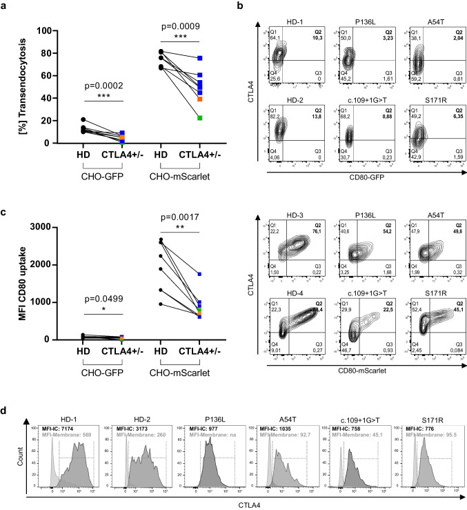
Variants of uncertain significance (VUS) in CTLA4 are frequently identified in patients with antibody deficiency or immune dysregulation syndromes including, but not limited to, patients with multi-organ autoimmunity and autoinflammation. However, to ascertain the diagnosis of CTLA4 insufficiency, the functional relevance of each variant needs to be determined. Currently, various assays have been proposed to assess the functionality of CTLA4 VUS, including the analysis of transendocytosis, the biological function of CTLA4 to capture CD80 molecules from antigen presenting cells. Challenges of this assay include weak fluorescence intensity of the internalized ligand, poor reproducibility, and poor performance upon analyzing thawed cells. In addition, the distinction of pathogenic from non-pathogenic variants and from wild-type CTLA4, and the classification of the different VUS according to its level of CTLA4 dysfunction, would be desirable. We developed a novel CD80-expressing cell line for the evaluation of CD80-transendocytosis and compared it to the published transendocytosis assay. Our approach showed lower inter-assay variability and better robustness regardless the type of starting material (fresh or thawed peripheral mononuclear cells). In addition, receiver operating characteristic analysis showed 100% specificity, avoiding false positive results and allowing for a clear distinction between pathogenic and non-pathogenic variants in CTLA4-variant carriers. With our transendocytosis assay, we assessed the pathogenicity of 24 distinct CTLA4 variants from patients submitted to our diagnostic unit. Significantly impaired transendocytosis was demonstrated for 17 CTLA4 variants, whereas seven variants tested normal. In conclusion, our upgraded transendocytosis assay allows a reliable assessment of newly identified variants in CTLA4.

Keywords: CTLA4; LRBA; diagnostics; inborn errors of immunity; transendocytosis.


Conflict of interest statement

The authors declare no competing interests.

Figures

Fig. 1
Fig. 1
The CTLA4 transendocytosis assay shows higher robustness when using CD80-mScarlet CHO cells. a Schematic illustration of the transendocytosis process. Human CD4+ T cells are co-cultured for 16h with CHO cells expressing CD80 tagged with either GFP (top panel) or mScarlet (bottom panel). After CTLA4-CD80 engagement, CTLA4 removes and internalizes the fluorescent tagged CD80- protein. Fluorescent CD80 is subsequently detected in CD4+CD45RO+FOXP3+ T cells by flow cytometry. bc Percentage of transendocytosis in activated CD4+FOXP3+ regulatory T cells from healthy donors (HD) (GFP, n=10; mScarlet, n=12) showing the inter-assay variability percentage; in b total number of samples and c fresh and thawed PBMCs. CD80-GFP/fresh PBMCS (n=7), CD80-GFP/thawed PMBCS (n=9); CD80-mScarlet/fresh PBMCS (n=8) and CD80-mScarlet/thawed PMBCS (n=10). All data shown in this figure are from ≥3 independent experiments with 2–4 biological replicates. Color-coded circles in CD80-GFP and -mScarlet represent replicates per individual. p-values were calculated using the Mann–Whitney test; the horizontal lines in b and c represents the median values. The coefficient of variation (CV) values for the transendocytosis assay were calculated as follows: CV= (standard deviation/mean) × 100%
Fig. 2
Fig. 2
CTLA4 transendocytosis using CD80-mScarlet CHO cells facilitates the identification of patients with functional CTLA4 defects. a Percentage of transendocytosis in stimulated CD4+FOXP3+ regulatory T cells from HD (n=8) and CTLA4-variant carriers (n=8), co-cultured with CHO cells expressing CD80-GFP or CD80-mScarlet. b Representative flow cytometry plots depicting the percentage of transendocytosis of CD80-GFP (top panel) or CD80-mScarlet (bottom panel) of HD versus patients carrying unique heterozygous mutations in CTLA4. c Mean fluorescence intensity (MFI) of internalized CD80-GFP or -mScarlet in regulatory T cells from HD versus patients. Type of mutations are color-coded: blue indicates missense; orange indicates frameshift, and green indicates splice-site variants. d Histograms show overlay of surface CTLA4 expression (light gray) and total intracellular CTLA4 (dark gray) in activated CD4+FOXP3+ regulatory T cells. p-values were calculated using the Mann–Whitney test
Fig. 3
Fig. 3
Both CD80-CHO cell lines show comparable receiver operating characteristics (ROC). ROC characteristics curve plot of performance of a percent of transendocytosis and b MFI for the assessment of CTLA4 variants with uncertain significance using CD80-GFP and CD80-mScarlet CHO cell lines. Cutoff points, areas under the curve (AUC), percentages of sensitivity, and percentages of specificity are depicted in the tables. Green circles represent CD80-GFP CHO cells and dark red triangles represent CD80-mScarlet CHO cells
Fig. 4
Fig. 4
CTLA4-variant carriers can be distinguished from healthy donors using both CD80-expressing CHO cells. Dot plots show a percentage and b MFI of CD80-GFP (n=15 CTLA4-variant carriers; n=16 healthy donors [HD]) and CD80-mScarlet (n=19 CTLA4-variant carriers; n=16 HD) transendocytosis in CD4+FOXP3+ regulatory T cells from HD and CTLA4-variant carriers. Filled circles represent data from HD, filled squares represent data from CTLA4-variant carriers. Type of mutations are color-coded: blue indicates missense mutations; orange, frameshift mutations; and green, splice-site mutations. Dot plots show the fold changes of percent of transendocytosis (c) and MFI (d). Fold change was calculated as a ratio between percent of transendocytosis or MFI values of each CTLA4-variant carrier and their matched HD. Green circles represent CD80-GFP CHO cells and dark red triangles represent CD80-mScarlet CHO cells (e) Percent of transendocytosis using CD80-GFP (n=18) or CD80-mScarlet (n=23) CHO cells in regulatory T cells from HD (filled circles), CTLA4-variant carriers (CD80-GFP n=17; CD80-mScarlet n=19; filled squares) and patients with LRBA deficiency (CD80-GFP n=2; CD80-mScarlet n=8; filled triangles). p-values were calculated using the Mann–Whitney test
Fig. 5
Fig. 5
The degree of impaired CD80 transendocytosis in CTLA4-variant carriers is not dependent on the affected amino acid position. Functional transendocytosis analysis was performed for 24 CTLA4 variants, including two splice-site, 18 missense, and four frameshift variants distributed throughout all four exons. Dark red bars represent percent of transendocytosis of CD80-mScarlet and green bars percent of transendocytosis of CD80-GFP. Dotted line indicates the cutoff for the percent of transendocytosis of CD80-GFP and for CD80-mScarlet, respectively
Fig. 6
Fig. 6
CTLA4-patients with a severe clinical phenotype according to the CHAI score have similarly reduced transendocytosis as mildly affected individuals with the same mutation in CTLA4. Bar graphs indicating the percent of transendocytosis in CTLA4-variant carriers grouped according to their disease severity. Disease state of CTLA4-variant carriers was calculated based on CHAI morbidity score and classified accordingly: ≥20% (severely affected); ≥10%<20% (mildly affected). Green squares represent percent of transendocytosis assay using CD80-GFP CHO cells and dark red triangles represent percent of transendocytosis using CD80-mScarlet CHO cell lines. p-values were calculated using the Mann–Whitney test

Similar articles

Cited by

References

    1. Mitsuiki N, Schwab C, Grimbacher B. What did we learn from CTLA-4 insufficiency on the human immune system? Immunol Rev. 2019;287(1):33–49. doi: 10.1111/imr.12721. - DOI - PubMed
    1. Qureshi OS, et al. Trans-endocytosis of CD80 and CD86: a molecular basis for the cell-extrinsic function of CTLA-4. Science. 2011;332(6029):600–603. doi: 10.1126/science.1202947. - DOI - PMC - PubMed
    1. Kuehn HS, et al. Immune dysregulation in human subjects with heterozygous germline mutations in CTLA4. Science. 2014;345(6204):1623–1627. doi: 10.1126/science.1255904. - DOI - PMC - PubMed
    1. Schubert D, et al. Autosomal dominant immune dysregulation syndrome in humans with CTLA4 mutations. Nat Med. 2014;20(12):1410–1416. doi: 10.1038/nm.3746. - DOI - PMC - PubMed
    1. Schwab C, et al. Phenotype, penetrance, and treatment of 133 cytotoxic T-lymphocyte antigen 4-insufficient subjects. J Allergy Clin Immunol. 2018;142(6):1932–1946. doi: 10.1016/j.jaci.2018.02.055. - DOI - PMC - PubMed

Publication types

LinkOut - more resources