. 2023 Oct:587:109866.
doi: 10.1016/j.virol.2023.109866. Epub 2023 Aug 19.

Human neurotropic polyomavirus, JC virus, late coding region encodes a novel nuclear protein, ORF4, which targets the promyelocytic leukemia nuclear bodies (PML-NBs) and modulates their reorganization

Affiliations

Human neurotropic polyomavirus, JC virus, late coding region encodes a novel nuclear protein, ORF4, which targets the promyelocytic leukemia nuclear bodies (PML-NBs) and modulates their reorganization

A Sami Saribas et al. Virology. 2023 Oct.

Abstract

We previously reported the discovery and characterization of two novel proteins (ORF1 and ORF2) generated by the alternative splicing of the JC virus (JCV) late coding region. Here, we report the discovery and partial characterization of three additional novel ORFs from the same coding region, ORF3, ORF4 and ORF5, which potentially encode 70, 173 and 265 amino acid long proteins respectively. While ORF3 protein exhibits a uniform distribution pattern throughout the cells, we were unable to detect ORF5 expression. Surprisingly, ORF4 protein was determined to be the only JCV protein specifically targeting the promyelocytic leukemia nuclear bodies (PML-NBs) and inducing their reorganization in nucleus. Although ORF4 protein has a modest effect on JCV replication, it is implicated to play major roles during the JCV life cycle, perhaps by regulating the antiviral response of PML-NBs against JCV infections and thus facilitating the progression of the JCV-induced disease in infected individuals.

Keywords: BKV; DNA replication; Interferon; JCV, SV40; Merkel cell carcinoma virus; ORF, splicing; PML-NBs; Papillomavirus; Polyomavirus; Progressive multifocal leukoencephalopathy; Promyelocytic leukemia nuclear bodies; RNA splicing; Transcription.


Conflict of interest statement

Declaration of competing interest The authors declare that they have no known competing financial interests or personal relationships that could have appeared to influence the work reported in this paper.

Figures

Fig 1.
Fig 1.. Analysis of the splicing patterns of the JCV late coding region by RT-PCR.
(A) A schematic representation of the splicing pattern of the JCV late coding region. The JCV late coding region primarily generates two major splice products, M1 and M2, by removing the intron 1 and intron 2. Both M1 and M2 splice products are produced by alternative cis-splicing. The JCV late coding region was also shown to produce two additional novel open reading frames (ORF1 and ORF2) by trans-splicing (Saribas et al., 2018). (B) Discovery of additional splice products from the JCV late coding region. Total RNA was isolated from PHFA infected with JCV Mad-1 strain [uninfected (lane 2) or infected (lane 3)] at 15th day post-infection and was subjected to RT-PCR using 5’-primer (JCV Mad-1 nt 277–294) and 3’-primer (JCV Mad-1 nt 2533–2511) as described in materials and methods. The RT-PCR products were then resolved on a 2% agarose gel. The band labeled with a star or an arrowhead or 3 dashed lines were subcloned into the pcDNA 3.1 vector at HindIII/Kpn1 sites, sequenced (clone 1, 2, 3 and 4) and analyzed by bioinformatics to determine the presence of the splice donor/acceptor sites and any open reading frame. The inserts in lane 7 and lane 9 represent the spliced and unspliced form of the M2 transcript respectively (panel A). In lane 10, DNA from the late coding region of the JCV Mad-1 strain was PCR-amplified using the same primers that were used for RT-PCR reaction above and loaded on the agarose gel as a positive control (+ control). Analysis of the sequencing data revealed that Clones 1, 2 and 3 contain open reading frames, designated as ORF3, ORF4 and ORF5 respectively. In addition, Clone 4, contains combinations of several open reading frames, including for the full-length of agnoprotein, ORF3, and a short coding sequence for the N-terminus of VP1. (C) Analysis of the total RNA obtained from a non-progressive multifocal leukoencephalopathy and progressive multifocal leukoencephalopathy brain tissue samples by RT-PCR to determine the splicing pattern of the JCV late coding region. The total RNA was isolated from a non-progressive multifocal leukoencephalopathy and progressive multifocal leukoencephalopathy brain in vivo tissue samples (Saribas et al., 2018), subjected to RT-PCR using the same PCR primer set that is used for panel B and RT-PCR products were resolved on a 2% agarose gel. The bands indicated by an asterisk, a yellow-colored arrow and an arrowhead correspond to the same bands indicated on panel B. The bands indicated by the labeled arrows on panel C were determined to be corresponding to the ORF3- and ORF4-containing RT-PCR products obtained from in vitro infected cells (panel B) after DNA sequencing.
Fig. 2.
Fig. 2.. The splicing products of the JCV late transcripts revealed novel open reading frames.
(A) Graphical representation of two major splicing patterns of the JCV late transcripts (M1 and M2). (B) The splicing pattern in Clone 1 produces a novel open reading frame, designated as ORF3, which is created by the removal of an internal intron and rejoining the exons within the VP1 coding region followed by a frame shift occurring at the splice junction. The N-terminus 41 aa region is identical to the VP1 coding sequences, but its C-terminus 29 aa region is unique. (C) The splicing pattern in Clone 2 also produces a unique ORF called ORF4. ORF4 is a truncated version of VP1 and encodes a 173 aa long protein (Table 2). (D) The splicing pattern in Clone 3 created a fusion protein (ORF5) after joining the N-terminus of agnoprotein (14 aa long) with the C-terminus coding region of VP1 (251 aa long). ORF5 can encode a 265 aa long fusion protein (Table 2). (E) The splicing pattern in Clone 4 creates multiple ORFs, where a short N-terminus of VP1 is duplicated as indicated. The C-terminus of agnoprotein is spliced into between the duplicated short N-terminus of VP1. Resulting ORFs are as follows: (i) Full-length agnoprotein coding region. (ii) An ORF designated as “VP1 N-terminus” which encodes a 45 aa long protein, 42 aa region of which is identical with VP1 and 3 aa are unique. (iii) This splicing pattern also creates an ORF identical to the ORF3 observed for the Clone 1. Note that partially duplicated agnoprotein coding region does not create any ORF.
Fig. 3.
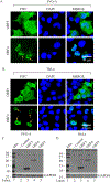
Fig. 3.. Analysis of the expression profiles of ORFs in SVG-A and HeLa cells by immunocytochemistry (ICC) and Western blotting.
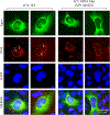
(A) Analysis of the subcellular distribution patterns of ORF3 and ORF4 in SVG-A cells. pCGT7-ORF3 and pCGT7-ORF4 expression plasmids were separately transfected into SVG-A cells by lipofectamine 3000 as described in materials and methods; and at 24h post-transfection, cells were processed for ICC using a primary mouse α-T7 monoclonal antibody and the secondary FITC-conjugated goat α-mouse antibody. Samples were then stained with DAPI and examined under a fluorescence microscope. Arrows point to a distinct punctate nuclear localization of ORF4 in the nucleus. (B) Analysis of the subcellular distribution patterns of ORF3 and ORF4 in HeLa cells. In parallel to the ICC experiments described for panel A, HeLa cells were also transfected separately with the same expression plasmids and processed for ICC as described for panel A. Scale bar: 40 µm in both panels. Immunocytochemistry experiments were repeated more than three times in different cell lines. Without exception, all ORF4-transfected cells showed a similar punctate distribution of ORF4 protein in the nucleus. (C and D) Analysis of the ORF3, ORF4 and ORF5 expression by Western blotting. In parallel to the ICC experiments described for panel A and B, whole-cell extracts were also prepared from SVG-A (C) and HeLa (D) cells and analyzed by Western blotting using α-T7 monoclonal antibody. GAPDH was used as a gel loading control.
Fig. 4.
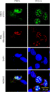
Fig. 4.. ORF4 co-localizes with PML-NBs in PHFA and SVG-A cells.
Co-localization of ORF4 protein with PML-NBs is demonstrated by ICC. pCGT7-ORF4 plasmid was transfected into PHFG and SVG-A cells and at 24h post-transfection, cells were fixed with cold acetone and processed for ICC as follows: The sample slides were incubated with the primary (α-T7 polyclonal and α-PML monoclonal) and the secondary (FITC-conjugated goat α-rabbit and Rhodamine-conjugated goat α-mouse) antibodies. Finally, the cells were stained with DAPI and examined under a florescence microscope as described under materials and methods. The cells with a punctate nuclear expression of ORF4 is demarcated by the dashed circles. These ICC experiments were repeated more than three times with the same consistent results. A representative group of cells is shown. Scale bar: 20 µm
Fig. 5.
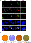
Fig. 5.. Analysis of the effect of ORF4 on the PML-NB members.
(A) SVG-A cells were transfected with a T7-tagged ORF4 expression plasmid (pCGT7-ORF4) and the next day, samples were processed for ICC. T7-ORF4, PML, hDaxx, ATRX proteins were detected by using α-T7 (polyclonal, catalog no. AB3790, Millipore), α-PML (monoclonal, catalog no. sc-966, Santa Cruz), α-hDaxx (monoclonal, catalog no. sc-8043, Santa Cruz), α-ATRX (monoclonal, catalog no. sc-55584, Santa Cruz) antibodies. In Sp100 column, T7-ORF4 and Sp100 were detected using α-T7 (monoclonal, catalog no. 69522, Novagen) and α-Sp100 (polyclonal, catalog no. NBP1–89457, Novus) antibodies. The white and red color-labeled circles in PML, hDaxx and ATRX columns indicate the ORF4-negative and -positive cells respectively. The red and green color-labeled circles in the Sp100 column indicate ORF4-positive and –negative cells respectively. These ICC experiments were repeated more than three times. A representative group of cells is shown here. Scale bar: 20 µm. (B) Presentation of the quantification of the results in the form of Pie graph.
Fig. 6.
Fig. 6.. JCV ORF4 protein contains a novel nuclear localization signal (NLS)-like motif.
(A) Sequence alignment of JCV, BKV and SV40 VP1 proteins. VP1 protein sequence of JCV, BKV and SV40 were aligned with each other using the “Clustal Omega” program (https://www.ebi.ac.uk/Tools/msa/clustalo). Each protein contains two putative NLS designated as NLS-1 and NLS-2 as indicated in yellow color. Methionine residues in each VP1 protein sequence are highlighted in red color. ORF4 coding region is highlighted in light blue. The putative ORF4 protein of JCV, BKV and SV40 contains a predicted NLS sequence (NLS-2) of their own, which was predicted by using the following website: http://nls-mapper.iab.keio.ac.jp/cgi-bin/NLS_Mapper_form.cgi. CLUSTAL O (1.2.4) multiple sequence alignment for JCV, BKV and SV40 VP1 proteins
Fig. 7.
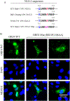
Fig. 7.. ORF4 lost its predominant nuclear localization feature when its NLS is altered (RR105–106AA).
(A) Comparison of the NLS-2 sequences of JCV, BKV and SV40. (B) ORF4 protein containing the mutant NLS-2 sequence lost its predominant nuclear localization feature. The ORF4 (pCGT7-ORF4) and its NLS-2 mutant (pCGT7-ORF4-NLS-2 mut) expression plasmids were separately transfected into SVG-A cell and at 24h post-transfection, cells were processed for ICC using a primary α-T7 polyclonal and a secondary α-rabbit FITC conjugated antibodies and examined under a fluorescence microscope. Scale bar: 40 µm.
Fig. 8.
Fig. 8.. Analysis of the ORF4 mutant of JCV in reorganization of PML-NBs.
The JCV WT (Bluescript KS+JCV Mad-1) and its mutant [Bluescript KS+JCV Mad-1 VP1 Mut (M182I)] plasmids were digested with the BamHI restriction enzyme to release the viral DNA from the vector and transfected into SVG-A cells. On the 5th day post-transfection/infection, cells were processed for ICC to detect the expression of PML, using the primary (α-PML, monoclonal and anti-Agno, polyclonal) and secondary (rhodamine-conjugated goat α-mouse and FITC-conjugated goat α-rabbit) antibodies. Note that agnoprotein detection was used as a positive control to pinpoint the infected cells. Cells were then examined under a florescence microscope after staining the nucleus with DAPI. Note that the nucleus of the cells infected with JCV WT and those infected with JCV harboring ORF4 mutant (M182I) sequences are demarcated with yellow dashed-line circles to demonstrate the differential distribution pattern of PML protein in WT versus mutant virus-infected cells. The nuclei of the several uninfected cells are demarcated with red dashed-line circles. Arrows point to the reorganized PML-NBs. Scale bar: 30 µm.
Fig. 9.
Fig. 9.. The newly raised anti-ORF4 antibody immunoprecipitates and detects ORF4 protein.
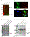
(A) Detection of JCV Agnoprotein and VP1 by Western blotting in extracts prepared from the JCV-infected SVG-A cells using anti-Agno (rabbit polyclonal (Del Valle et al., 2005)) and anti-VP1 (monoclonal, PAB597, a gift from Dr. Walter Atwood, Brown University, USA) antibodies. (B) Detection of Agnoprotein and JCV VP1 protein by immunocytochemistry using the same anti-Agno and anti-VP1 antibodies mentioned for panel A in cells infected with JCV. (C) Detection of ORF4 and VP1 proteins by the newly raised anti-ORF4 antibody. Whole-cell extracts prepared from the uninfected, ORF4-transfected, and JCV-infected SVG-A cells were analyzed by Western blotting using the newly raised anti-ORF4 antibody (rabbit polyclonal, #31 antibody). Anti-ORF4 antibody detects ORF4 and VP1 proteins. (D) Anti-ORF4 antibody immunoprecipitates ORF4 and VP1. Whole cell extracts (200 µg) prepared from the ORF4-transfected, untransfected and JCV-infected cells were subjected to immunoprecipitation by anti-ORF4 antibody (10 µl/sample) and immunoprecipitants were analyzed by immunoblotting using the anti-ORF4 antibody (1:200 dilution).
Fig. 10.
Fig. 10.. Proteomics studies confirm the expression of ORF4 protein in JCV-infected cells.
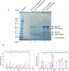
(A) Analysis of the immunoprecipitated ORF4 protein on an SDS-PAGE gradient gel (4–20%). Whole-cell extracts (1 mg) prepared from the SVG-A cells (untransfected or JCV-infected) were subjected to immunoprecipitation in duplicate with the newly raised anti-ORF4 antibody (10 µl/sample) using protein G magnetic beads (catalog no. 101945, Active Motive) and analyzed on a gradient gel (catalog no. 5671094, Bio-Rad). Gel was stained with “gel code blue safe protein stain” (catalog no. 1860957, ThermoScientific) to reveal the immunoprepitated proteins; and antibody heavy (HC) and light (LC) chains. The labeled bands on the gel were marked with the dashed rectangles (bands 1 and 2 for uninfected cells, bands 3 and 4 for infected cells where ORF4 is supposed to migrate, bands 5 and 6 for infected cells, where VP1 is supposed to migrate). These labeled bands were individually cut out, subjected to in-gel digestion with trypsin, and analyzed by proteomics (LC-MS/MS) to detect the ORF4 and VP1 proteins.
Fig. 11.
Fig. 11.. Analysis of the replication efficiency of a ORF4 mutant by Southern blotting.
(A and B) Western blot analysis of JCV VP1 expression. The plasmid constructs [Bluescript KS-JCV Mad-1 WT and its mutant (Bluescript KS-JCV Mad-1 VP1 M182I) were separately transfected/infected into SVG-A cells. At the indicated time points, whole-cell extracts were prepared from SVG-A cells (uninfected and transfected/infected), immunoprecipitated (150 µg protein/sample) using an α-VP1 (PAB597) antibody (2 µl/sample) and analyzed by Western blotting by using the same antibody. (C) In parallel, the low-molecular-weight DNA containing both input and replicated viral DNA was isolated from the uninfected and transfected/infected cells, digested with BamHI and DpnI restriction enzymes. Digested DNA was resolved on a 1% agarose gel, transferred onto a nitrocellulose membrane (Bio-Rad, catalog no, 1620094) and probed for detection of the newly replicated DNA using a probe prepared from the JCV Mad-1 WT as a template. In lane 1, 2 ng of JCV Mad-1 WT linearized by BamHI digestion was loaded as positive control (+ Cont.). Replication assays were repeated twice in duplicate, and a representative data is shown here. (D) Quantification analysis of Southern blots by a semi-quantitative densitometry method (using the NIH Image J program) and presentation of the results in arbitrary units. Results were statistically analyzed by the GraphPad program using One-way ANOVA and data columns were compared by Tukey’s multiple comparison test.

Similar articles

Cited by

References

    1. Ascoli CA, Maul GG, 1991. Identification of a Novel Nuclear Domain. Journal of Cell Biology 112, 785–795. - PMC - PubMed
    1. Becker KA, Florin L, Sapp C, Sapp M, 2003. Dissection of human papillomavirus type 33 L2 domains involved in nuclear domains (ND) 10 homing and reorganization. Virology 314, 161–167. - PubMed
    1. Berger JR, 1992. PML in AIDS. Neurology 42, 1845–1846. - PubMed
    1. Berger JR, 2000. Progressive Multifocal Leukoencephalopathy. Curr Treat Options Neurol 2, 361–368. - PubMed
    1. Berger JR, 2007. Progressive multifocal leukoencephalopathy. Curr Neurol Neurosci Rep 7, 461–469. - PubMed

Publication types

Substances