. 2023 Oct 20;82(11):966-969.
doi: 10.1093/jnen/nlad074.

A novel ARIH1::BRAF fusion in a glioma

Affiliations

A novel ARIH1::BRAF fusion in a glioma

Emily Xu et al. J Neuropathol Exp Neurol. .
No abstract available


Conflict of interest statement

The authors have no duality or conflicts of interest to declare.

Figures

Figure 1
Figure 1
Interval growth and transformation of a right mesial temporal lesion. Coronal T2 (A, D), axial FLAIR (B, E), and axial T1 with contrast MRI images (C, F) demonstrated interval growth in size and transformation of the mesial temporal lesion (white arrow) from 2015 to 2022 (D–F). This was associated with a 3-mm nodular contrast-enhancing lesion (F, open arrow) next to the cavernous sinus, which was not present in 2015. No mass effect or abnormal diffusion restriction was observed.
Figure 2
Figure 2
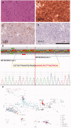
(A) H&E stain of the lesion tissue shows a few atypical cells. Immunohistochemistry staining demonstrates strong GFAP expression (B) and scattered P53 (C) and Ki-67 (D) stained nuclei. Scale bar: 200 microns, lower right. (E) Schematic representation of the ARIH1::BRAF fusion. RNA-seq and confirmatory Sanger sequencing demonstrate a fusion of exon 8 of ARIH1 (NM_005744.5) to exon 11 of BRAF (NM_004333.6). The red double arrow indicates the junction of the fusion. (F) UMAP visualizing our case (arrow) in proximity to cases called low-grade glial tumors and control brain tissue by the Heidelberg classifier. DLGNT: diffuse leptomeningeal glioneuronal tumor, GG: ganglioglioma, HGG_F: Adult-type diffuse high-grade glioma, IDH-wildtype, subtype F, PA-CORT: hemispheric pilocytic astrocytoma, PLNTY: polymorphous low-grade neuroepithelial tumor of the young, PXA: pleomorphic xanthoastrocytoma, RGNT: rosette-forming glioneuronal tumor, CTRL_CORPCAL: control, corpus callosum, CTRL_HEMI: control, hemisphere; CTRL_REACTIVE: control, reactive brain tissue.

References

    1. Louis DN, Wesseling P, Paulus W, et al. cIMPACT-NOW update 1: Not otherwise specified (NOS) and not elsewhere classified (NEC). Acta Neuropathol 2018;135:481–4 - PubMed
    1. Chang F, Lin F, Cao K, et al. Development and clinical validation of a large fusion gene panel for pediatric cancers. J Mol Diagn 2019;21:873–83 - PMC - PubMed
    1. Capper D, Jones DTW, Sill M, et al. DNA methylation-based classification of central nervous system tumours. Nature 2018;555:469–74 - PMC - PubMed
    1. Wu Z, Abdullaev Z, Pratt D, et al. Impact of the methylation classifier and ancillary methods on CNS tumor diagnostics. Neuro Oncol 2022;24:571–81 - PMC - PubMed
    1. Maraka S, Janku F. BRAF alterations in primary brain tumors. Discov Med 2018;26:51–60 - PubMed