. 2023 Nov;299(11):105278.
doi: 10.1016/j.jbc.2023.105278. Epub 2023 Sep 22.

Sequence tolerance of immunoglobulin variable domain framework regions to noncanonical intradomain disulfide linkages

Affiliations

Sequence tolerance of immunoglobulin variable domain framework regions to noncanonical intradomain disulfide linkages

Dae Young Kim et al. J Biol Chem. 2023 Nov.

Abstract

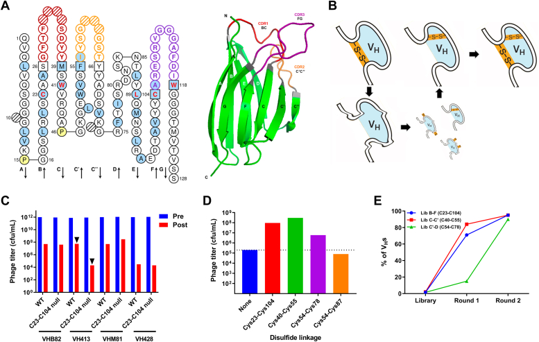
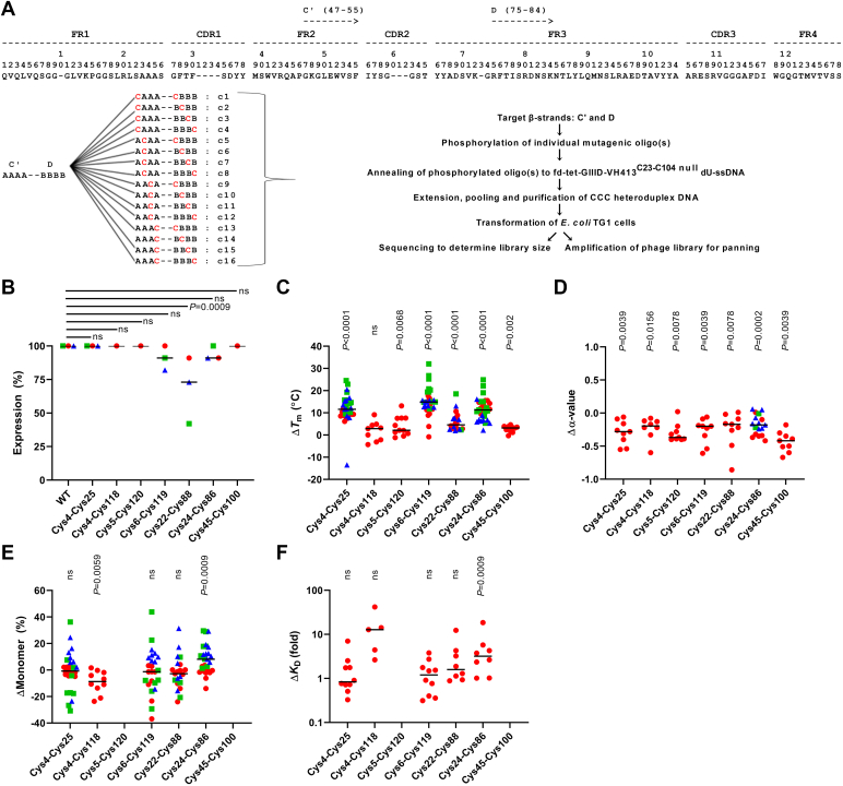
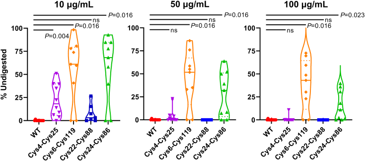
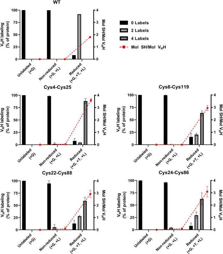
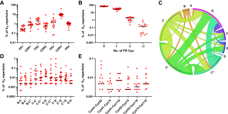
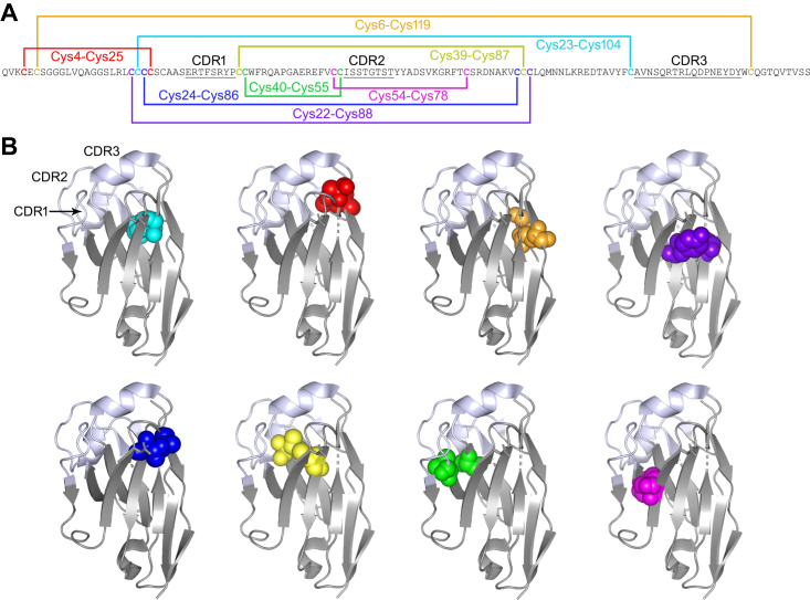
Most immunoglobulin (Ig) domains bear only a single highly conserved canonical intradomain, inter-β-sheet disulfide linkage formed between Cys23-Cys104, and incorporation of rare noncanonical disulfide linkages at other locations can enhance Ig domain stability. Here, we exhaustively surveyed the sequence tolerance of Ig variable (V) domain framework regions (FRs) to noncanonical disulfide linkages. Starting from a destabilized VH domain lacking a Cys23-Cys104 disulfide linkage, we generated and screened phage-displayed libraries of engineered VHs, bearing all possible pairwise combinations of Cys residues in neighboring β-strands of the Ig fold FRs. This approach identified seven novel Cys pairs in VH FRs (Cys4-Cys25, Cys4-Cys118, Cys5-Cys120, Cys6-Cys119, Cys22-Cys88, Cys24-Cys86, and Cys45-Cys100; the international ImMunoGeneTics information system numbering), whose presence rescued domain folding and stability. Introduction of a subset of these noncanonical disulfide linkages (three intra-β-sheet: Cys4-Cys25, Cys22-Cys88, and Cys24-Cys86, and one inter-β-sheet: Cys6-Cys119) into a diverse panel of VH, VL, and VHH domains enhanced their thermostability and protease resistance without significantly impacting expression, solubility, or binding to cognate antigens. None of the noncanonical disulfide linkages identified were present in the natural human VH repertoire. These data reveal an unexpected permissiveness of Ig V domains to noncanonical disulfide linkages at diverse locations in FRs, absent in the human repertoire, whose presence is compatible with antigen recognition and improves domain stability. Our work represents the most complete assessment to date of the role of engineered noncanonical disulfide bonding within FRs in Ig V domain structure and function.

Keywords: disulfide; immunoglobulin variable domain; phage display; protein engineering; protein stability; single-domain antibody (sdAb, nanobody).


Conflict of interest statement

Conflict of interest The authors declare that they have no conflicts of interest with the contents of this article.

Figures

Figure 1
Figure 1
Development of a model system for identification of stabilizing noncanonical disulfide linkages in Ig V domains.A, secondary and tertiary structure of immunoglobulin V domains. Blue shading, hydrophobic residue in ≥50% of V domains; red bold, highly conserved residues in V domains; square, complementarity-determining region (CDR) anchor position; hashed, gaps according to IMGT numbering; yellow, proline. CDR1, CDR2, and CDR3 are shown in red, orange, and purple, respectively. β-strands of the two opposing β-sheets are labeled in the tertiary structure in black and white. Left, figure produced using the sequence of VH413 with IMGT/Collier-de-Perles. Right, VH domain of murine anti-lipoprotein A antibody 2E8 (PDB ID: 12E8). B, cartoon representation showing rescue of a destabilized Cys23-Cys104 null immunoglobulin VH domain by Cys pair scanning of FRs, followed by enrichment of phage-displayed VHs bearing novel stabilizing intradomain noncanonical disulfide linkages from phage libraries using protein A selection. C, panning of fd phage displaying VHs (WT) or their Cys23-Cys104 null derivatives against protein A. Only VH413C23-C104 null phage showed a significant difference in postselection output phage titer compared with the WT VH–displaying phage (arrow heads). Pre, preselection; Post, postselection. D, effect of introducing known canonical (Cys23-Cys104) and noncanonical (Cys40-Cys55 and Cys54-Cys78) disulfide linkages into VH413C23-C104 null fd phage on output phage titer following protein A selection. “None” refers to unmodified VH413C23-C104 null displaying fd phage (blue bar and dotted line). The effects of a negative control Cys pair (Cys54-Cys87) were also assessed. E, panning of test VH413C23-C104 null Cys pair scan libraries (β-strands B–F, C–C', and C'–D) on protein A. The percent frequencies of known noncanonical stabilizing disulfide linkages formed between these β-strands is indicated. Four rounds of panning were performed. The pannings in panels C, D, and E were performed at room temperature with no heating step. “Library” denotes the unpanned libraries. cfu, colony-forming units; FR, framework region; VH, variable heavy chain; WT, wild-type.
Figure 2
Figure 2
Strategy for generation of Cys pair scan VH libraries and effects of novel noncanonical disulfide linkages on Ig V domain biophysical properties.A, schematic of phage-displayed VH413C23-C104 null Cys pair scan library construction. The example illustrates the construction of the C'–D library (only 16 representative Cys pair combinations, c1-c16, are pictured from the full set of 90 theoretical pairs). The letter A denotes amino acid positions in β-strand C', and the letter B denotes amino acid positions in β-strand D', while red letter C denotes positions mutated to Cys. Mutagenic oligonucleotides targeting two positions on two different β-strands were phosphorylated, annealed (either two oligonucleotides targeting separate β-strands or a single oligonucleotide spanning two contiguous β-strands in the primary amino acid sequence) to fd-tet GIIID-VH413C23-C104 null dU-ssDNA, and extended. Heteroduplex DNA for all mutagenesis reactions targeting β-strands C'–D was pooled and used to transform Escherichia coli TG1 cells. The same steps were used for the construction of the remaining 16 Cys pair scan libraries, except that different mutagenic oligonucleotide pairs were used. BF, effect of introducing seven Cys pairs forming putative noncanonical FR disulfide linkages on Ig V domain thermostability, other biophysical properties and antigen recognition. VHHs are shown in red circles, VHs in green squares, and VLs in blue triangles. B, expression yields of Ig V domains bearing Cys pairs forming putative noncanonical FR disulfide linkages. Expression (%) refers to the percentage of sdAbs tested that expressed in adequate yields for subsequent experiments. Changes in (C) sdAb Tm and (D) fraction refolded (α-value) following introduction of FR Cys pairs were measured by CD. E, changes in sdAb monomericity following introduction of FR Cys pairs were measured by SEC-MALS. F, changes in the KDs of antigen-specific VHHs following introduction of FR Cys pairs were assessed by SPR. For changes (Δ) in biophysical properties, values above the null (ΔMonomer 0%, ΔTm 0 °C, and Δα-value 0) indicate improvement, while for changes in binding affinity, values above the null (ΔKD 1) reflect weaker binding. Median values are indicated by black lines. p-values are from Fisher’s exact test (B) or Wilcoxon matched–pairs signed-rank test (CF) comparing WT to Cys-engineered sdAbs. CD, circular dichroism; dU-ssdDNA, dUTP-containing fd-tetGIIID-VH413C23-C104 null ssDNA; FR, framework region; Ig, immunoglobulin; ns, not significant; sdAb, single-domain antibody; SEC-MALS, size-exclusion chromatography-multiangle light scattering; SPR, surface plasmon resonance; V, variable; VH, variable heavy chain; VHH, variable heavy chain of camelid heavy chain–only antibody; VL, variable light chain; WT, wild-type.
Figure 3
Figure 3
Effect of introducing Cys pairs forming putative noncanonical FR disulfide linkages on VHH pepsin resistance. The three Cys pairs conferring the highest increase in sdAb Tm (Cys4-Cys25, Cys6-Cys119, and Cys24-Cys86) were assessed as well as one Cys pair (Cys22-Cys88) that had an intermediate effect on Tm. The VHHs were digested with three concentrations of pepsin (10, 50 and 100 µg/ml) for 1 h at 37 °C, and then the remaining undigested VHH was quantitated by SDS-PAGE and band densitometry. Solid lines indicate medians and dashed lines indicate quartiles. The contours of the violin plots show probability density. p-values are from Wilcoxon matched–pairs signed-rank test comparing WT to Cys-engineered VHHs. FR, framework region; ns, not significant; sdAb, single-domain antibody; VHH, variable heavy chain of camelid heavy chain–only antibody; WT, wild-type.
Figure 4
Figure 4
LC-MS analysis of free sulfhydryl abundance in WT and Cys-engineered VHHs. For each category of VHH (WT VHHs or VHHs bearing the indicated engineered Cys pair), the percentage of unlabeled, reduced, and nonreduced protein analyzed bearing 0, 2, and 4 maleimide-PEG2-biotin labels is indicated (left y-axis; sum: 100%). The molar ratio of free sulfhydryl (SH) groups to total VHH protein is shown in red (right y-axis). Bars represent means and error bars represent SEMs. G, GdnHCl, guanidine hydrochloride; L, label (maleimide-PEG2-biotin); LC-MS, liquid chromatography-mass spectrometry; T, TCEP, tris(2-carboxyethyl)phosphine; VHH, variable heavy chain of camelid heavy chain–only antibody; WT, wild-type.
Figure 5
Figure 5
Presence of noncanonical Cys residues in the human expressed VHrepertoires of 17 individuals. The canonical intradomain disulfide linkage formed by Cys residues at IMGT positions 23 and 104 was disregarded in all analyses presented. A, frequency of noncanonical Cys residues in VH FRs and CDRs. B, frequency of VHs in the expressed repertoire bearing the indicated number of Cys residues. C, Circos plot showing the relative frequencies and β-strand locations of noncanonical Cys pairs in the subset of expressed human VHs bearing ≧4 FR Cys residues. The sizes of β-strands along the circumference of the circle reflect the frequency of noncanonical Cys, and links show the relative frequencies of the locations of the second Cys in the pair. D, most frequent β-strands bearing noncanonical Cys pairs among human VHs bearing ≧4 FR Cys residues. E, most frequent noncanonical Cys pairs among human VHs bearing ≧4 FR Cys residues. Black lines represent median values across 17 VH repertoires. Note that y-axes are log-scaled and zero values are not plotted. CDR, complementarity-determining region; FR, framework region; VH, variable heavy chain.
Figure 6
Figure 6
Locations of known noncanonical intradomain disulfide linkages in Ig V domain FRs in a representative VHH.A, amino acid sequence of the Clostridioides difficile toxin A–specific VHH A26.8 (21) with the locations of the canonical Cys23-Cys104 disulfide linkage (cyan) and noncanonical disulfide linkages in Ig V domain FRs: Cys4-Cys25 (red), Cys6-Cys119 (orange), Cys22-Cys88 (indigo), Cys24-Cys86 (blue), Cys39-Cys87 (yellow), Cys40-Cys55 (green), and Cys54-Cys78 (magenta). The first four noncanonical disulfide linkages were identified in this study. CDRs are underlined. Note that while the positions of the noncanonical disulfide linkages are shown using IMGT numbering, IMGT gaps are not shown in the sequence of A26.8. Refer to Figure 1A for an example of IMGT numbering including gaps. B, crystal structure of A26.8 VHH (PDB ID: 4NBZ) with the locations of the canonical and noncanonical disulfide linkages colored as in A: canonical Cys23-Cys104 (cyan) and noncanonical Cys4-Cys25 (red), Cys6-Cys119 (orange), Cys22-Cys88 (indigo), Cys24-Cys86 (blue), Cys39-Cys87 (yellow), Cys40-Cys55 (green), and Cys54-Cys78 (magenta). CDRs are shown in white and FRs in gray. The first four noncanonical disulfide linkages were identified in this study. CDR, complementarity-determining region; FR, framework region; Ig, immunoglobulin; VHH, variable heavy chain of camelid heavy chain–only antibody.

References

    1. Bechtel T.J., Weerapana E. From structure to redox: the diverse functional roles of disulfides and implications in disease. Proteomics. 2017;17 doi: 10.1002/pmic.201600391. - DOI - PMC - PubMed
    1. Liu H., May K. Disulfide bond structures of IgG molecules: structural variations, chemical modifications and possible impacts to stability and biological function. mAbs. 2012;4:17–23. - PMC - PubMed
    1. Dombkowski A.A., Sultana K.Z., Craig D.B. Protein disulfide engineering. FEBS Lett. 2013;588:206–212. - PubMed
    1. Willuda J., Honegger A., Waibel R., Schubiger P.A., Stahel R., Zangemeister-Wittke U., et al. Thermal stability is essential for tumor targeting of antibody fragments: engineering of a humanized anti-epithelial glycoprotein-2 (epithelial cell adhesion molecule) single-chain Fv fragment. Cancer Res. 1999;59:5758–5767. - PubMed
    1. Gong R., Vu B.K., Feng Y., Prieto D.A., Dyba M.A., Walsh J.D., et al. Engineered human antibody constant domains with increased stability. J. Biol. Chem. 2009;284:14203–14210. - PMC - PubMed

Publication types

MeSH terms

Substances

LinkOut - more resources