Prenatal allergic inflammation in rats confers sex-specific alterations to oxytocin and vasopressin innervation in social brain regions
- PMID: 37743114
- PMCID: PMC10842952
- DOI: 10.1016/j.yhbeh.2023.105427
Prenatal allergic inflammation in rats confers sex-specific alterations to oxytocin and vasopressin innervation in social brain regions
Abstract
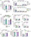
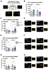
Prenatal exposure to inflammation via maternal infection, allergy, or autoimmunity increases one's risk for developing neurodevelopmental and psychiatric disorders. Many of these disorders are associated with altered social behavior, yet the mechanisms underlying inflammation-induced social impairment remain unknown. We previously found that a rat model of acute allergic maternal immune activation (MIA) produced deficits like those found in MIA-linked disorders, including impairments in juvenile social play behavior. The neuropeptides oxytocin (OT) and arginine vasopressin (AVP) regulate social behavior, including juvenile social play, across mammalian species. OT and AVP are also implicated in neuropsychiatric disorders characterized by social impairment, making them good candidate regulators of social deficits after MIA. We profiled how acute prenatal exposure to allergic MIA changed OT and AVP innervation in several brain regions important for social behavior in juvenile male and female rat offspring. We also assessed whether MIA altered additional behavioral phenotypes related to sociality and anxiety. We found that allergic MIA increased OT and AVP fiber immunoreactivity in the medial amygdala and had sex-specific effects in the nucleus accumbens, bed nucleus of the stria terminalis, and lateral hypothalamic area. We also found that MIA reduced ultrasonic vocalizations in neonates and increased the stereotypical nature of self-grooming behavior. Overall, these findings suggest that there may be sex-specific mechanisms underlying MIA-induced behavioral impairment and underscore OT and AVP as ideal candidates for future mechanistic studies.
Keywords: Allergy; Maternal immune activation; Oxytocin; Sex differences; Social behavior; Social play; Vasopressin.
Copyright © 2023 Elsevier Inc. All rights reserved.
Conflict of interest statement
Declaration of competing interest The authors declare no competing financial interests.
Figures


Similar articles
-
Quantitative mapping reveals age and sex differences in vasopressin, but not oxytocin, immunoreactivity in the rat social behavior neural network.J Comp Neurol. 2017 Aug 1;525(11):2549-2570. doi: 10.1002/cne.24216. Epub 2017 May 8. J Comp Neurol. 2017. PMID: 28340511 Free PMC article.
-
Prenatal allergic inflammation in rats programs the developmental trajectory of dendritic spine patterning in brain regions associated with cognitive and social behavior.Brain Behav Immun. 2022 May;102:279-291. doi: 10.1016/j.bbi.2022.02.026. Epub 2022 Mar 1. Brain Behav Immun. 2022. PMID: 35245680 Free PMC article.
-
Sex differences in oxytocin receptor binding in forebrain regions: correlations with social interest in brain region- and sex- specific ways.Horm Behav. 2013 Sep;64(4):693-701. doi: 10.1016/j.yhbeh.2013.08.012. Epub 2013 Sep 18. Horm Behav. 2013. PMID: 24055336
-
Vasopressin and oxytocin receptor systems in the brain: Sex differences and sex-specific regulation of social behavior.Front Neuroendocrinol. 2016 Jan;40:1-23. doi: 10.1016/j.yfrne.2015.04.003. Epub 2015 May 4. Front Neuroendocrinol. 2016. PMID: 25951955 Free PMC article. Review.
-
Neuroendocrine underpinning of social recognition in males and females.J Neuroendocrinol. 2022 Feb;34(2):e13070. doi: 10.1111/jne.13070. Epub 2021 Dec 19. J Neuroendocrinol. 2022. PMID: 34927288 Review.
Cited by
-
Peripartum buprenorphine and oxycodone exposure impair maternal behavior and increase neuroinflammation in new mother rats.Brain Behav Immun. 2025 Feb;124:264-279. doi: 10.1016/j.bbi.2024.11.027. Epub 2024 Nov 27. Brain Behav Immun. 2025. PMID: 39612963
References
-
- Argue KJ, Vanryzin JW, Falvo DJ, Whitaker AR, Yu SJ, & McCarthy MM (2017). Activation of Both CB1 and CB2 Endocannabinoid Receptors Is Critical for Masculinization of the Developing Medial Amygdala and Juvenile Social Play Behavior. eNeuro, 4(1), ENEURO.0344-0316. 10.1523/eneuro.0344-16.2017 - DOI - PMC - PubMed
-
- Baribeau DA, Dupuis A, Paton TA, Scherer SW, Schachar R, Arnold PD, Szatmari P, Nicolson R, Georgiades S, Crosbie J, Brian J, Laboni A, Lerch JP, & Anagnostou E (2017). Oxytocin Receptor Polymorphisms are Differentially Associated with Social Abilities across Neurodevelopmental Disorders. Sci Rep, 7(11618). 10.1038/s41598-017-10821-0 - DOI - PMC - PubMed
-
- Bauminger N, Shulman C, & Agam G (2003). Peer Interaction and Loneliness in High-Functioning Children with Autism. Journal of Autism and Developmental Disorders, 33, 489–507. - PubMed
Publication types
MeSH terms
Substances
Grants and funding
LinkOut - more resources
Full Text Sources
Miscellaneous

