. 2023 Sep 25;8(1):366.
doi: 10.1038/s41392-023-01603-4.

Non-small cell lung cancers (NSCLCs) oncolysis using coxsackievirus B5 and synergistic DNA-damage response inhibitors

Affiliations

Non-small cell lung cancers (NSCLCs) oncolysis using coxsackievirus B5 and synergistic DNA-damage response inhibitors

Bopei Cui et al. Signal Transduct Target Ther. .

Abstract

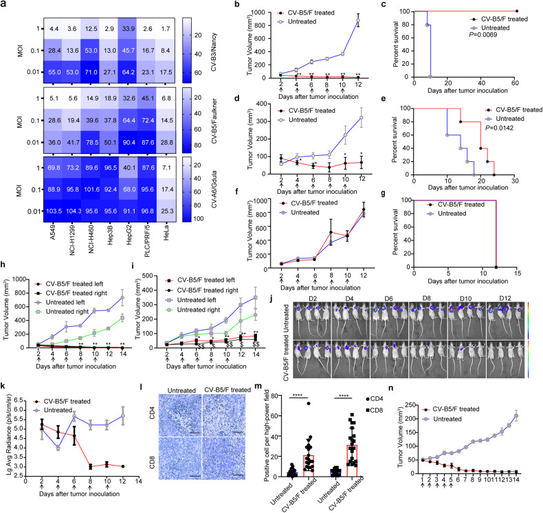
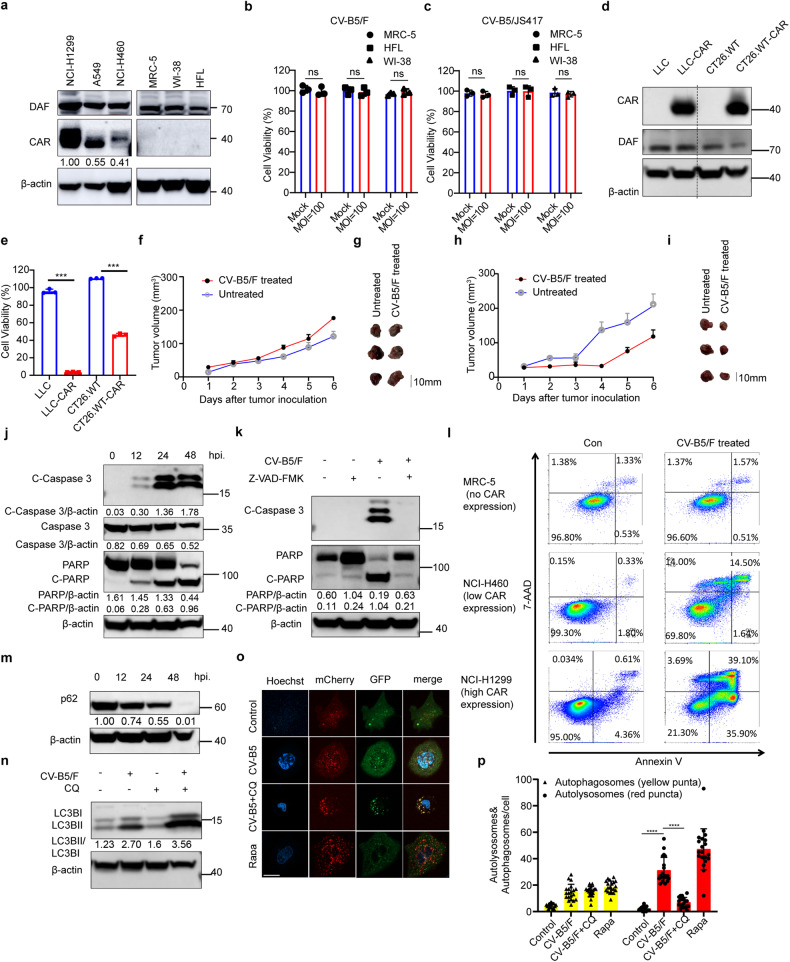
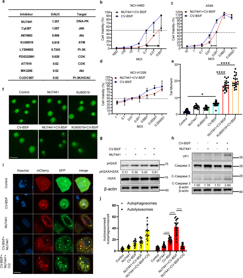
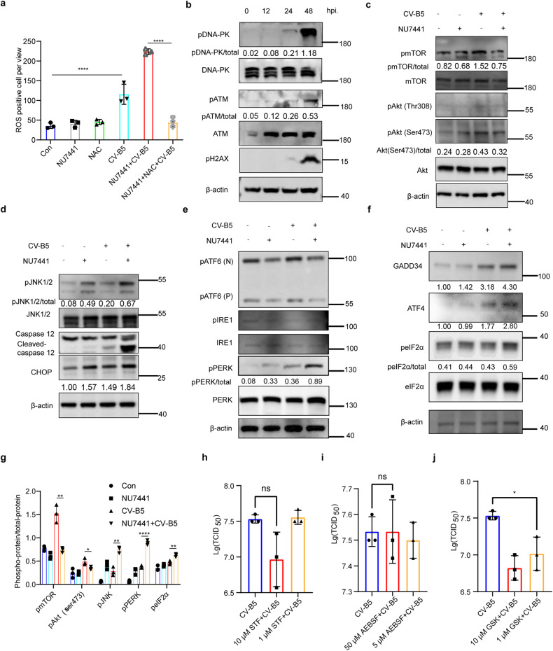
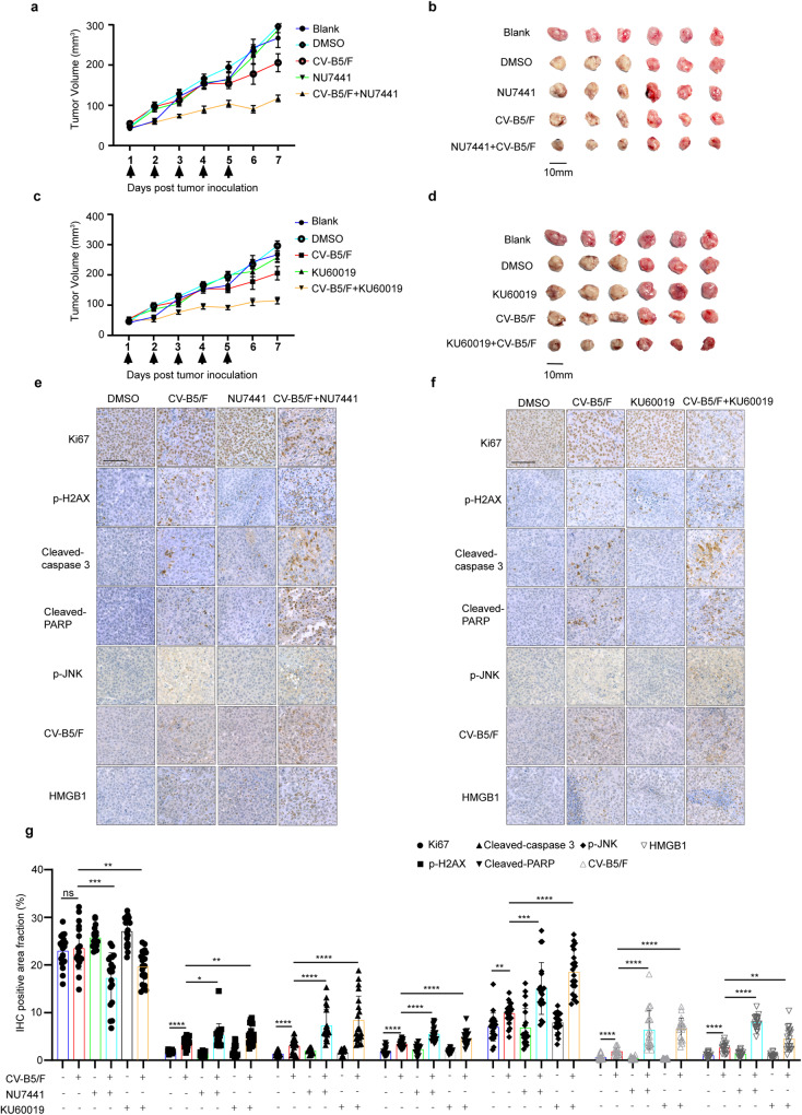
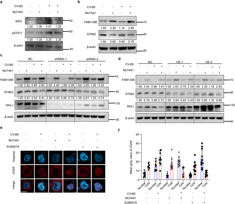
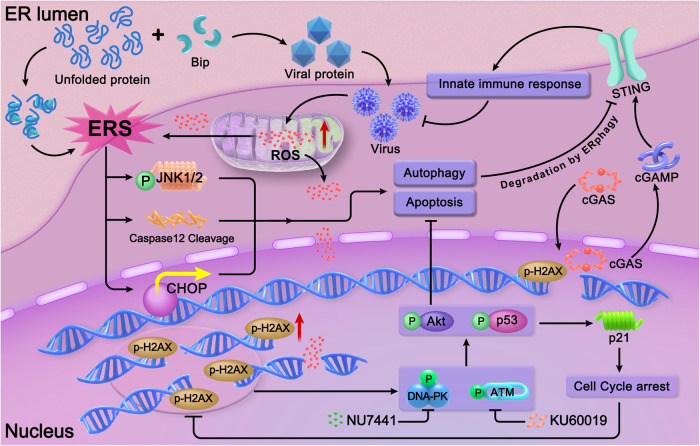
With the continuous in-depth study of the interaction mechanism between viruses and hosts, the virus has become a promising tool in cancer treatment. In fact, many oncolytic viruses with selectivity and effectiveness have been used in cancer therapy. Human enterovirus is one of the most convenient sources to generate oncolytic viruses, however, the high seroprevalence of some enteroviruses limits its application which urges to exploit more oncolytic enteroviruses. In this study, coxsackievirus B5/Faulkner (CV-B5/F) was screened for its potential oncolytic effect against non-small cell lung cancers (NSCLCs) through inducing apoptosis and autophagy. For refractory NSCLCs, DNA-dependent protein kinase (DNA-PK) or ataxia telangiectasia mutated protein (ATM) inhibitors can synergize with CV-B5/F to promote refractory cell death. Here, we showed that viral infection triggered endoplasmic reticulum (ER) stress-related pro-apoptosis and autophagy signals, whereas repair for double-stranded DNA breaks (DSBs) contributed to cell survival which can be antagonized by inhibitor-induced cell death, manifesting exacerbated DSBs, apoptosis, and autophagy. Mechanistically, PERK pathway was activated by the combination of CV-B5/F and inhibitor, and the irreversible ER stress-induced exacerbated cell death. Furthermore, the degradation of activated STING by ERphagy promoted viral replication. Meanwhile, no treatment-related deaths due to CV-B5/F and/or inhibitors occurred. Conclusively, our study identifies an oncolytic CV-B5/F and the synergistic effects of inhibitors of DNA-PK or ATM, which is a potential therapy for NSCLCs.


Conflict of interest statement

The authors declare no competing interests.

Figures

Fig. 1
Fig. 1
Screening of an oncolytic virus and its anti-tumor effects in multiple tumor models. a Non-small cell lung cancer cells (NSCLCs; A549, NCI-H1299, NCI-H460), hepatocarcinoma cells (Hep3B, HepG2, PLC/PRF/5), cervical carcinoma cells (HeLa) were infected with CV-B3/Nancy, CV-B5/Faulkner (CV-B5/F), CV-A6/Gdula at an MOI of 1, 0.1, 0.01 for 48 h. Cell viability was assessed by CCK8 assay. bg NCI-H1299 (n = 5, b), A549 (n = 5, d), and NCI-H460 (n = 5, f) cells were subcutaneously injected into the right flank of BALB/c nude mice. Each mouse received five doses of CV-B5/F intratumorally when the diameter of the tumor reached 4–5 mm. Tumor volumes are expressed as mean ± SEMs. *P < 0.05; **P < 0.01. Kaplan–Meier survival analyses were performed for CV-B5/F-treated mice with five doses (c: NCI-H1299; e: A549; g: NCI-H460). h, i Administration of CV-B5/F to the right tumor (h: NCI-H1299, n = 5; i: A549, n = 5) was performed in BALB/c nude mice with bilateral tumors. *, $P < 0.05; **, $$P < 0.01. Each symbol represents the statistical significance of the right and left lateral tumors between untreated and treated mice. j, k NCI-H1299 cells expressing firefly luciferase were subcutaneously injected into the axillia of BALB/c nude mice (n = 4). The bioluminescent images were measured using the IVIS-Lumina II imaging system on days 2, 4, 6, 8, 10, and 12. Relative bioluminescence intensity is shown in pseudocolor, with red representing the strongest and blue representing the weakest photon fluxes (j). The bioluminescence intensities of control and CV-B5/F-treated mice are shown as mean ± deviation (k). l, m NCI-H1299 was combined with Matrigel at a 1:1 ratio immediately before subcutaneous injection into the axillia of B-NDG mice. Seven days after treatment, PBMCs were injected intraperitoneally in PBS. On day 10, tumors were dissected for immunohistochemistry (IHC) using anti-CD4 and CD8 antibodies. Numbers of positive cell in high-power field (n = 20) were analyzed. One-way ANOVA was used to analyze the data. Scale bars, 100 μm. ****P < 0.0001. n Tumor volume curves of patient-derived xenografts (PDX) for NSCLC after treatment with five doses of CV-B5/F
Fig. 2
Fig. 2
Expression profile of CAR and DAF on normal lung and cancer cells and its correlation with CVB5-mediated cytotoxicity. a Expression of CAR and DAF on normal lung cells (MRC-5, WI-38, and HFL) and NSCLC cells analyzed by western blot. Gray value ratios to the first lane of CAR were shown. b, c Normal lung cells infected with CV-B5/F (b) or CV-B5/JS417 (c) at 100 MOI and analyzed at 48 h for cell viability by CCK8 assay (n = 3). Each value represents the mean ± deviation. d Expression of CAR and DAF on wild-type or CAR over-expressing mouse Lewis lung cancer (LLC; LLC-CAR) and colorectal carcinoma (CT26.WT; CT26.WT-CAR). e LLC, LLC-CAR, CT26.WT, CT26.WT-CAR infected with CV-B5/F at 10 MOI was analyzed at 48 h for cell viability by CCK8 assay (n = 3). Each value represents the mean ± standard deviation. fi LLC (f, g) or LLC-CAR (h, i) were subcutaneously injected into the axillia of C57BL/6 mice. Each mouse received 5 doses of CV-B5/F or with MEM intratumorally. Tumor were measured every day and anatomized ultimately (n = 3). j NCI-H1299 infected with CV-B5/F (MOI = 0.01) were analyzed at different time points. Each cellular lysate obtained was subjected to immunoblot analysis. Full-length PARP (116 kDa), cleaved-PARP (85 kDa), full-length caspase 3 (35 kDa) and cleaved-caspase 3 (17/19 kDa) were shown. hpi, hours post infection. k NCI-H1299 pretreated with 100 µM Z-VAD-FMK or MOCK and incubated with MEM or CV-B5/F at 0.01 MOI were subjected to immunoblot analysis. Full-length PARP (116 kDa), cleaved-PARP (85 kDa), and cleaved-caspase 3 (17/19 kDa) were shown. l NCI-1299, NCI-H460, and MRC-5 were infected with 1 MOI CV-B5/F for 24 h. Apoptotic population was represented as Annexin V +/7-AAD- or Annexin V+/7-AAD+ cells. m NCI-H1299 infected with CV-B5/F (MOI = 0.01) were analyzed at 0, 12, 24, and 48 h. p62 (62 kDa) was detected for the whole cell lysis (WCL). n NCI-H1299 was pretreated with 100 μM CQ for 2 h, and then treated with 0.01 MOI CV-B5/F for 24 h. LC3B (14/16 kDa) was detected for the WCL. o, p NCI-H1299 was transfected with mcherry-GFP-LC3B for 24 h, and then treated with 0.01 MOI CV-B5/F with or without 100 μM CQ (10 μM Rapamycin as positive control). Autophagosomes display both GFP and mCherry fluorescence (yellow-green), whereas autolysosomes display only mCherry fluorescence (red) because GFP is denatured by the acidity of the lysosome (o). Number of autophagosomes and autolysosomes were enumerated for at least 20 cells (n = 20, p). One-way ANOVA was used to analyze the data. ***P < 0.001, ****P < 0.0001, ns, not significant. Scale bars, 10 μm
Fig. 3
Fig. 3
Combinatorial drug screen identifies DNA-PKI NU7441 and ATMI KU60019 as synergistic sensitizers for CV-B5/F in NSCLC cells. a NCI-H460 cells were seeded in 96-well plates and treated with escalating doses of each of the 30 compounds in the drug screen, either singly or in combination with CV-B5/F (MOI = 0.01). Top 9 candidate drugs for NCI-H460 identified through this screening. DNA-PK DNA-dependent protein kinase, JAK Janus kinase, Akt serine/threonine kinase, ATM ataxia telangiectasia mutated protein, PI3K phosphatidylinositol 3-kinase, CDK cyclin-dependent kinase, HDAC histone deacetylase. bd NCI-H460, A549 and NCI-H1299 cells treated with escalating titers of CV-B5/F with or without 1 μM NU7441 for 48 h (n = 3). EC50 shifts were shown. e, f NCI-H460 cells were treated with NU7441 (1 μM), KU60019 (1 μM), CV-B5/F (MOI = 0.01), NU7441/CV-B5/F or KU60019/CV-B5/F for 24 h, and comet assay was used to assess double-strand breaks (DSBs). Quantification of tail moment was analyzed by OpenComet software (n = 20). Scale bar, 40 μm. g NCI-H460 cells were exposed to NU7441 (1 μM), CV-B5/F (MOI = 0.01) or a combination as indicated. p-H2AX (a marker of DNA damage response) was determined by western blot. h NCI-H460 cells were exposed to NU7441 (1 μM), CV-B5/F (MOI = 0.01) or a combination as indicated. Structural viral protein VP1 and cleaved-caspase 3 were determined by western blot. i, j NCI-H460 was transfected with mcherry-GFP-LC3B for 24 h, and then treated with 0.01 MOI CV-B5/F, 1 μM NU7441 or a combination with or without 100 μM CQ. The number of autophagosomes and autolysosomes were enumerated for 20 cells at least. One-way ANOVA was used to analyze data. *P < 0.05, ****P < 0.0001, Scale bar, 10 μm
Fig. 4
Fig. 4
DNA-PK inhibitors plus CV-B5/F virus-induced irresolvable ER stress-associated apoptosis in NCI-H460 cells. a NCI-H460 was treated with 1 μM NU7441, 10 μM NAC, 0.01 MOI CV-B5/F, or a combination of NU7441 and CV-B5/F with or without 10 μM NAC. Reactive oxygen species (ROS) of each were detected (n = 3). b Immunoblots of p-DNA-PK, p-ATM and p-H2AX after infection with CV-B5/F (MOI = 0.01) for 0, 12, 24, and 24 h. cg NCI-H460 was treated with 1 μM NU7441, 0.01 MOI CV-B5/F, or a combination for 24 h. Immunoblots of p-mTOR, p-Akt (Ser 473), and p-Akt (Thr 308) were detected (c). ER stress-associated apoptotic pathways, including caspase 12, CHOP (C/EBP-homologous protein), JNK1/2 (Jun N-terminal kinase 1/2) were analyzed by western blot (d). Three initiators of ERS, including ATF6, IRE1, and PERK were detected (e). Immunoblots of downstream of PERK were detected (f). Data of densitometry analysis were shown (g). hj NCI-H460 was treated for CV-B5/F (MOI = 1) with or without STF-083010 (an inhibitor for IRE1, h), AEBSF (an inhibitor for ATF6, i) or GSK2606414 (an inhibitor for PERK, j) for 24 h. Viral titers in supernatant were measured (n = 3). One-way ANOVA was used to analyze data. *P < 0.05, **P < 0.01, ****P < 0.0001, ns, not significant
Fig. 5
Fig. 5
Inhibiting DNA-PK or ATM enhances oncolytic therapy in BALB/c nude models of NCI-H460 xenografts. a, b NCI-H460 xenografts were treated with vehicle, CV-B5/F (5 × 106 TCID50, intratumorally injection), NU7441 (10 mg/kg/day, intraperitoneally) or a combination. Blank, n = 5; Vehicle, n = 5; CV-B5/F, n = 5; NU7441, n = 5; NU7441 + CV-B5/F, n = 5. Tumor growth presented as the mean tumor volume ± SEM (a). Images of tumors from each group in (a) at the experimental endpoints (b). c, d NCI-H460 xenografts were treated with vehicle, CV-B5/F (5 × 106 TCID50, intratumorally injection), KU60019 (10 mg/kg/day, intraperitoneally) or a combination. Blank, n = 5; Vehicle, n = 5; CV-B5/F, n = 5; KU60019, n = 5; KU60019 + CV-B5/F, n = 5. Tumor growth presented as the mean tumor volume ± SEM (c). Images of tumors from each group in (c) at the experimental endpoints (d). eg Tumor tissues from B (e) or D (f) were evaluated through IHC for Ki-67 (a marker of proliferation), p-H2AX, cleaved-caspase-3, cleaved-PARP, p-JNK, CV-B5/F, HMGB1. One-way ANOVA was used to analyze data (n = 20). *P < 0.05, **P < 0.01, ***P < 0.001, ****P < 0.0001, Scale bar, 100 μm
Fig. 6
Fig. 6
Exacerbated ERphagy induced by the combination of NU7441 and CV-B5/F degraded STING activated by nuclear cGAS recruited by DSBs. a NCI-H460 was treated with 1 μM NU7441, 0.01 MOI CV-B5/F, or a combination for 24 h. Immunoblots of anti-viral proteins including IRF9 and p-STAT1 were detected. b NCI-H460 cells were treated with 1 μM NU7441, 0.01 MOI CV-B5/F, or a combination for 24 h. FAM134B (a marker of ERphagy) and STING were determined by western blot. c NCI-H460 cells knocked down with shRNA for RIG-I were treated with 1 μM NU7441, 0.01 MOI CV-B5/F, or a combination for 24 h. RIG-I, FAM134B and STING were determined by western blot. Gray values of NC, shRNA-1 and shRNA-2 were compared separately. d NCI-H460 cells overexpressing RIG-I were treated with 1 μM NU7441, 0.01 MOI CV-B5/F, or a combination for 24 h. RIG-I, FAM134B and STING were determined by western blot. Gray values of NC, OE-1 and OE-2 were compared separately. e, f NCI-H460 cells were treated with 1 μM NU7441 or 1 μM KU60019, 0.01 MOI CV-B5/F, or a combination for 24 h. Immunofluorescence of cGAS of nuclear was analyzed by confocal microscopy. Mean gray value of cGAS in nuclear and cytoplasma was separately analyzed by Image J software (n = 20)
Fig. 7
Fig. 7
Graphical model of DNA-PK or ATM and CV-B5/F combination therapy. Normally, viral proteins are folded in the ER and assembled in the cytoplasm. Massive accumulation of unfolded viral proteins can induce ERS, which is initiated by the combination of bip and unfolded proteins. Irresolvable ERS can induce apoptosis, and autophagy which relies on the activation of p-JNK, caspase 12, and CHOP. In addition to inducing ERS, viral propagation can also induce the release of ROS from the mitochondria, which can cause DSBs. DSBs can further activate the DNA damage response, including the activation of Akt, which inhibits apoptosis and autophagy, and the activation of p53, which causes cell cycle arrest and diminishes DSBs. Thus, co-treatment with DNA-PKI or ATMI hinders DNA repair and promotes DNA damage induced by CV-B5/F. Furthermore, excess DSBs can recruit cGAS into the nucleus and activate the STING pathway for the innate immune response. However, ERphagy, a form of autophagy, is also initiated by inhibitors and can degrade STING to offset this response. Above all, DNA-PK or ATM inhibitors can synergize with CV-B5/F by attenuating the antiviral pathway and increasing DNA damage-mediated cell apoptosis and autophagy

Similar articles

Cited by

References

    1. GBD 2019 Cancer Risk Factors Collaborators. The global burden of cancer attributable to risk factors, 2010–19: a systematic analysis for the Global Burden of Disease Study 2019. Lancet. 2022;400:563–591. - PMC - PubMed
    1. Wang S, et al. The landscape of cell and gene therapies for solid tumors. Cancer Cell. 2021;39:7–8. - PubMed
    1. Kaufman HL, Kohlhapp FJ, Zloza A. Oncolytic viruses: a new class of immunotherapy drugs. Nat. Rev. Drug Discov. 2015;14:642–662. - PMC - PubMed
    1. Corrigan PA, Beaulieu C, Patel RB, Lowe DK. Talimogene Laherparepvec: an oncolytic virus therapy for melanoma. Ann. Pharmacother. 2017;51:675–681. - PubMed
    1. Su Y, Su C, Qin L. Current landscape and perspective of oncolytic viruses and their combination therapies. Transl. Oncol. 2022;25:101530. - PMC - PubMed

Publication types