

Blood-spinal cord barrier disruption in degenerative cervical myelopathy
- PMID: 37743487
- PMCID: PMC10519090
- DOI: 10.1186/s12987-023-00463-y
Blood-spinal cord barrier disruption in degenerative cervical myelopathy
Abstract
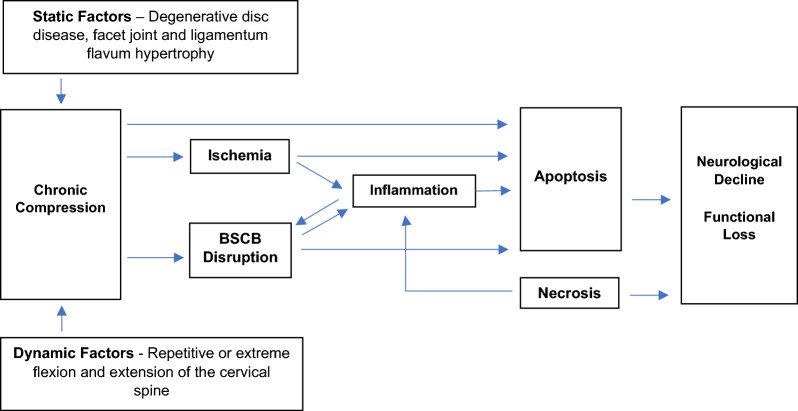
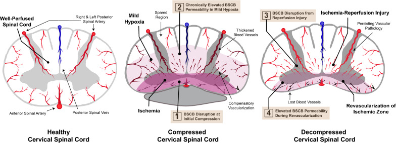
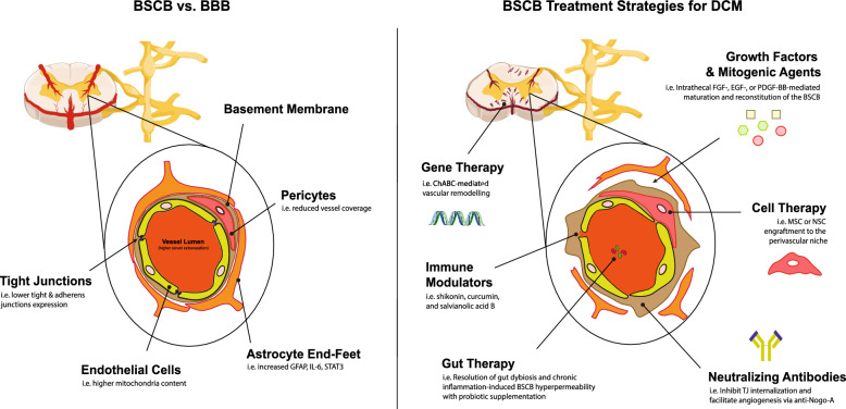
Degenerative cervical myelopathy (DCM) is the most prevalent cause of spinal cord dysfunction in the aging population. Significant neurological deficits may result from a delayed diagnosis as well as inadequate neurological recovery following surgical decompression. Here, we review the pathophysiology of DCM with an emphasis on how blood-spinal cord barrier (BSCB) disruption is a critical yet neglected pathological feature affecting prognosis. In patients suffering from DCM, compromise of the BSCB is evidenced by elevated cerebrospinal fluid (CSF) to serum protein ratios and abnormal contrast-enhancement upon magnetic resonance imaging (MRI). In animal model correlates, there is histological evidence of increased extravasation of tissue dyes and serum contents, and pathological changes to the neurovascular unit. BSCB dysfunction is the likely culprit for ischemia-reperfusion injury following surgical decompression, which can result in devastating neurological sequelae. As there are currently no therapeutic approaches specifically targeting BSCB reconstitution, we conclude the review by discussing potential interventions harnessed for this purpose.
Keywords: Blood-spinal cord barrier; Cell therapy; Cervical decompression; Degenerative cervical myelopathy; Gene therapy; Inflammation; Ischemia.
© 2023. The Author(s).
Conflict of interest statement
None to declare.
Figures
Similar articles
-
A preclinical study on cell therapy as an adjunct to surgical decompression in degenerative cervical myelopathy via accelerating blood spinal cord barrier reconstitution and neurological recovery.Stem Cell Res Ther. 2025 May 28;16(1):262. doi: 10.1186/s13287-025-04348-9. Stem Cell Res Ther. 2025. PMID: 40437637 Free PMC article.
-
Blood spinal cord barrier disruption recovers in patients with degenerative cervical myelopathy after surgical decompression: a prospective cohort study.Sci Rep. 2023 May 6;13(1):7389. doi: 10.1038/s41598-023-34004-2. Sci Rep. 2023. PMID: 37149638 Free PMC article.
-
Patients with degenerative cervical myelopathy have signs of blood spinal cord barrier disruption, and its magnitude correlates with myelopathy severity: a prospective comparative cohort study.Eur Spine J. 2020 May;29(5):986-993. doi: 10.1007/s00586-020-06298-7. Epub 2020 Jan 25. Eur Spine J. 2020. PMID: 31982957 Clinical Trial.
-
[Degenerative cervical myelopathy].Rev Med Chil. 2022 Mar;150(3):339-352. doi: 10.4067/S0034-98872022000300339. Rev Med Chil. 2022. PMID: 36156719 Review. Spanish.
-
Magnetic resonance imaging assessment of degenerative cervical myelopathy: a review of structural changes and measurement techniques.Neurosurg Focus. 2016 Jun;40(6):E5. doi: 10.3171/2016.3.FOCUS1667. Neurosurg Focus. 2016. PMID: 27246488 Review.
Cited by
-
Spinal Cord Injury Management Based on Microglia-Targeting Therapies.J Clin Med. 2024 May 8;13(10):2773. doi: 10.3390/jcm13102773. J Clin Med. 2024. PMID: 38792314 Free PMC article. Review.
-
Restoring brain barriers: an innovative approach for treating neurological disorders.Fluids Barriers CNS. 2025 Jul 10;22(1):72. doi: 10.1186/s12987-025-00688-z. Fluids Barriers CNS. 2025. PMID: 40640916 Free PMC article. Review.
-
A preclinical study on cell therapy as an adjunct to surgical decompression in degenerative cervical myelopathy via accelerating blood spinal cord barrier reconstitution and neurological recovery.Stem Cell Res Ther. 2025 May 28;16(1):262. doi: 10.1186/s13287-025-04348-9. Stem Cell Res Ther. 2025. PMID: 40437637 Free PMC article.
-
Regulation of dynamic spatiotemporal inflammation by nanomaterials in spinal cord injury.J Nanobiotechnology. 2024 Dec 19;22(1):767. doi: 10.1186/s12951-024-03037-8. J Nanobiotechnology. 2024. PMID: 39696584 Free PMC article. Review.
-
Biomaterials targeting the microenvironment for spinal cord injury repair: progression and perspectives.Front Cell Neurosci. 2024 May 9;18:1362494. doi: 10.3389/fncel.2024.1362494. eCollection 2024. Front Cell Neurosci. 2024. PMID: 38784712 Free PMC article. Review.
References
Publication types
MeSH terms
LinkOut - more resources
Full Text Sources
Medical
Research Materials
