. 2023 Sep;13(9):e1406.
doi: 10.1002/ctm2.1406.

Rnd3 suppresses endothelial cell pyroptosis in atherosclerosis through regulation of ubiquitination of TRAF6

Affiliations

Rnd3 suppresses endothelial cell pyroptosis in atherosclerosis through regulation of ubiquitination of TRAF6

Yan Zhang et al. Clin Transl Med. 2023 Sep.

Abstract

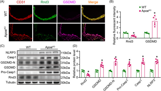
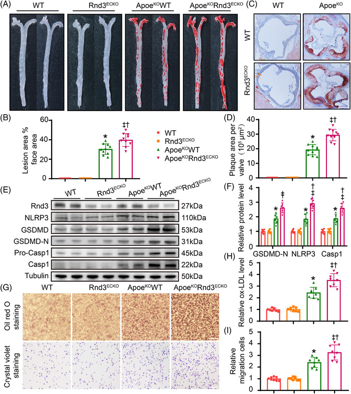
Background: As the main pathological basis for various cardiovascular and cerebrovascular diseases, atherosclerosis has become one of the leading causes of death and disability worldwide. Emerging evidence has suggested that Rho GTPase Rnd3 plays an indisputable role in cardiovascular diseases, although its function in atherosclerosis remains unclear. Here, we found a significant correlation between Rnd3 and pyroptosis of aortic endothelial cells (ECs).

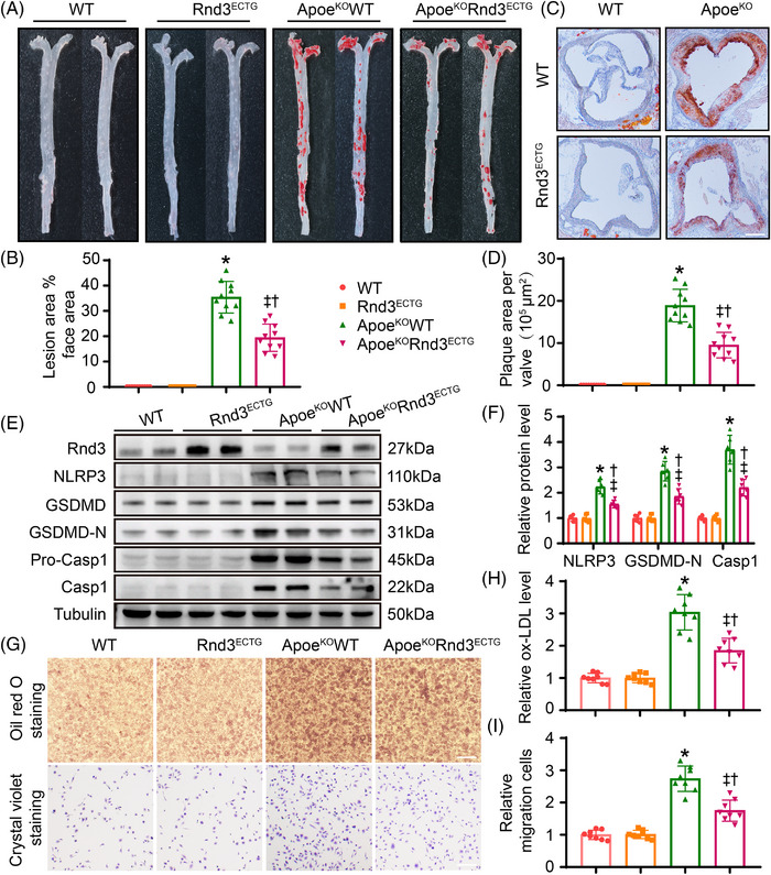
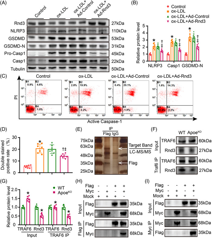
Methods: ApoeKO mice were utilized as a model for atherosclerosis. Endothelium-specific transgenic mice were employed to disrupt the expression level of Rnd3 in vivo. Mechanistic investigation of the impact of Rnd3 on endothelial cell pyroptosis was carried out using liquid chromatography tandem mass spectrometry (LC-MS/MS), co-immunoprecipitation (Co-IP) assays, and molecular docking.

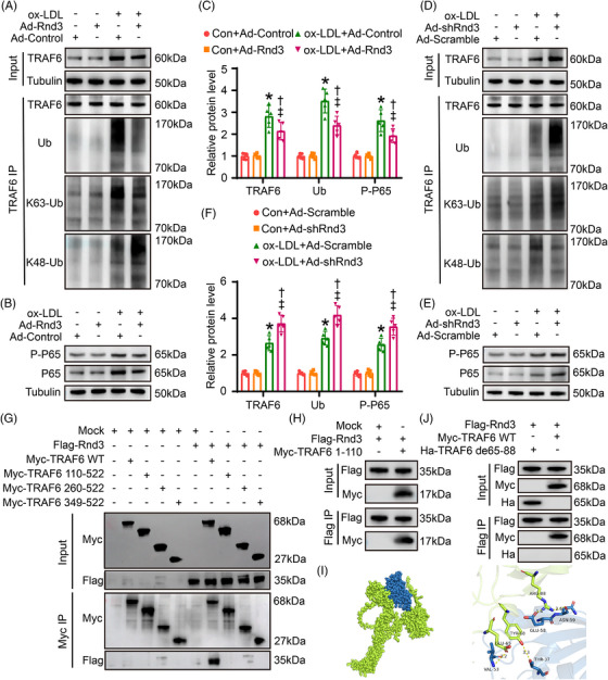
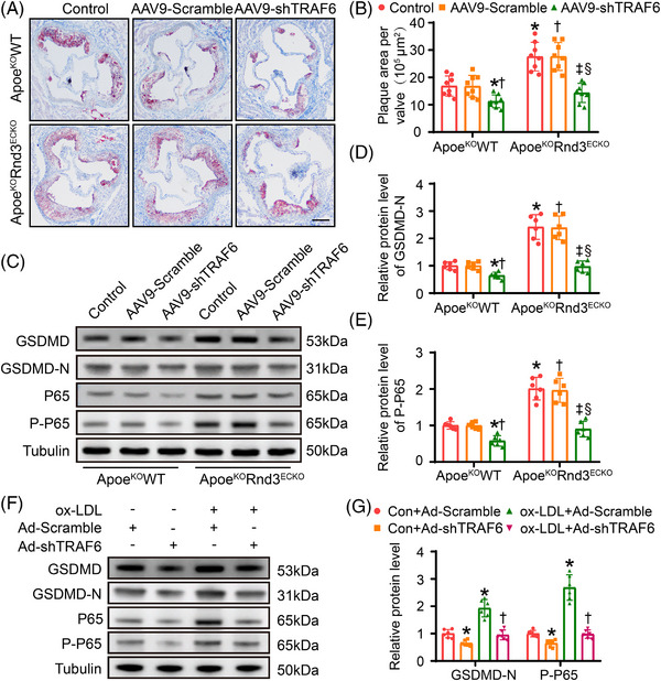
Results: Evidence from gain-of-function and loss-of-function studies denoted a protective role for Rnd3 against ECs pyroptosis. Downregulation of Rnd3 sensitized ECs to pyroptosis under oxidized low density lipoprotein (oxLDL) challenge and exacerbated atherosclerosis, while overexpression of Rnd3 effectively prevented these effects. LC-MS/MS, Co-IP assay, and molecular docking revealed that Rnd3 negatively regulated pyroptosis signaling by direct interaction with the ring finger domain of tumor necrosis factor receptor-associated factor 6 (TRAF6). This leads to the suppression of K63-linked TRAF6 ubiquitination and the promotion of K48-linked TRAF6 ubiquitination, inhibiting the activation of NF-κB and promoting the degradation of TRAF6. Moreover, TRAF6 knockdown countered Rnd3 knockout-evoked exacerbation of EC pyroptosis in vivo and vitro.

Conclusions: These findings establish a critical functional connection between Rnd3 and the TRAF6/NF-κB/NLRP3 signaling pathway in ECs, indicating the essential role of Rnd3 in preventing pyroptosis of ECs.

Keywords: Rho-GTPase; atherosclerosis; endothelial cell; pyroptosis; ubiquitination.


Conflict of interest statement

The authors declare no conflicts of interest.

Figures

FIGURE 1
FIGURE 1
ApoeKO mice present increased pyroptosis and reduced Rnd3 levels in endothelial cells (ECs) of atherosclerotic lesion. (A) The protein level of Rnd3 and gasdermin‐D (GSDMD) was evaluated using immunofluorescence staining in the aortic roots of wild‐type (WT) mice and ApoeKO mice (n = 6). Scale bars represent 200 μm. (B) Quantification of the fluorescent intensity of Rnd3 and GSDMD immunofluorescence staining in ECs of wild‐type (WT) mice and ApoeKO mice. (C) The expression of Rnd3, NLRP3, GSDMD, GSDMD‐N, Caspase1 and Pro‐Caspase 1 in different groups by western blot (n = 5). (D) Quantitative analysis of Rnd3, NLRP3, GSDMD, GSDMD‐N, Caspase 1, and Pro‐Caspase1 protein levels in different groups. *p < .05 versus WT.
FIGURE 2
FIGURE 2
Overexpression of Rnd3 inhibits endothelial pyroptosis and atherosclerosis in ApoeKO mice. (A) Representative Oil Red O staining of aorta in different groups (n = 10). (B) Quantification of the percentage of aortic lesion areas in different groups. (C) Representative Oil Red O staining of aortic roots in different groups (n = 10). Scale bars represent 500 μm. (D) Quantification of the percentage of lesion areas in aortic roots of different groups. (E and F) Representative and pooled western blot analysis of Rnd3 and pyroptosis‐associated proteins in different groups (n = 6). (G) Assessment of macrophage migration and phagocytosis by Oil Red O staining and crystal violet staining, which treated by condition medium of endothelial cells (ECs) from different groups (n = 8). Scale bars represent 200 μm. (H) Relative oxLDL levels in different groups were quantified. (I) Relative migration cells in different groups were quantified. *p < .05 versus WT; †p < .05 versus Rnd3ECTG; ‡p < .05 versus ApoEKOWT.
FIGURE 3
FIGURE 3
Rnd3 interacts with TRAF6 in endothelial cells (ECs). (A and B) Representative and pooled western blot analysis of Rnd3 and pyroptosis‐associated proteins in different groups (n = 6). (C and D) Flow cytometry and associated quantitative analysis of propidium iodide (PI) and Caspase‐1 double‐staining positive cells treated as indicated (n = 5). *p < .05 versus control; p < .05 versus oxLDL; p < .05 versus oxLDL + Ad‐Control. (E) Flag antibody or IgG (negative control antibody) was used for immunoprecipitation, electrophoresed and silver staining (n = 5). (F and G) Interaction between Rnd3 and TRAF6 was demonstrated by immunoprecipitation in ECs of wild‐type (WT) mice and ApoeKO mice (n = 6). *p < .05 versus WT. (H and I) Primary aortic ECs were transfected with mock, Ad‐Flag‐Rnd3, or Ad‐Myc‐TRAF6 as indicated and performed immunoprecipitation using anti‐Flag or anti‐Myc antibody.
FIGURE 4
FIGURE 4
Rnd3 inhibits endothelial pyroptosis by regulating ubiquitination of TRAF6. (A) Effects of Rnd3 overexpression on TRAF6 ubiquitination. The protein level of TRAF6 in input, and Ub, K63‐Ub, and K48‐Ub after TRAF6 immunoprecipitation (IP) was evaluated by western blot (n = 5). (B) Effects of Rnd3 overexpression on NF‐κB activation. The protein level of P‐P65 and P65 was evaluated by western blot (n = 5). (C) Quantitative analysis of TRAF6, Ub, and P‐P65 protein levels in different groups. *p < .05 versus Con+Ad‐Control; p < .05 versus Con+Ad‐Rnd3; p < .05 versus oxLDL+Ad‐Control. (D) Western blotting was used to evaluate the effect of Rnd3 knockdown on TRAF6 ubiquitination (n = 5). (E) Western blotting was used to evaluate the effect of Rnd3 knockdown on NF‐κB activation (n = 5). (F) Quantitative analysis of TRAF6, Ub, and P‐P65 protein levels in different groups. *p < .05 versus Con+Ad‐Scramble; p < .05 versus Con+Ad‐shRnd3; p < .05 versus oxLDL+Ad‐Scramble. (G) Myc‐TRAF6 wild type (WT), Myc‐TRAF6 truncation mutants (110‐522, 260‐522, 349‐522), and Flag‐Rnd3 were transfected to endothelial cells (ECs) and performed immunoprecipitation using anti‐Myc antibody. (H) Myc‐TRAF6 truncation mutants (1‐110) and Flag‐Rnd3 were transfected to ECs and performed immunoprecipitation using anti‐Flag antibody. (I) Molecular docking of Rnd3 and TRAF6 combination. The potential interaction regions (TRAF6 65 to 88 amino acids) were found. (J) Flag‐Rnd3, Myc‐TRAF6 WT, or Ha‐TRAF6 65–88 amino acid deletion mutant was transfected to ECs and performed immunoprecipitation using anti‐Flag antibody.
FIGURE 5
FIGURE 5
Knockout of Rnd3 exacerbates endothelial pyroptosis and atherosclerosis in ApoeKO mice. (A) Representative Oil Red O staining of aorta in different groups (n = 10). (B) Quantification of the percentage of aortic lesion areas in different groups. (C) Representative Oil Red O staining of aortic roots in different groups (n = 10). Scale bars represent 500 μm. (D) Quantification of the percentage of lesion areas in aortic roots of different groups. (E and F) Representative and pooled western blot analysis of Rnd3 and pyroptosis‐associated proteins in different groups (n = 6). (G) Assessment of macrophage migration and phagocytosis by Oil Red O staining and crystal violet staining, which is treated by condition medium of ECs from different groups (n = 8). Scale bars represent 200 μm. (H) Relative oxLDL levels in different groups were quantified. (I) Relative migration cells in different groups were quantified. *p < .05 versus WT; †p < .05 versus Rnd3ECKO; p < .05 versus ApoEKOWT.
FIGURE 6
FIGURE 6
TRAF6 knockdown offsets endothelial pyroptosis and atherosclerosis exacerbated by Rnd3 knockout. (A) Representative Oil Red O staining of aortic roots in different groups (n = 10). Scale bars represent 500 μm. (B) Quantification of the percentage of aortic lesion areas in different groups. (C–E) Western blot and quantitative analysis of GSDMD‐N and P‐P65 expression in endothelial cells (ECs) treated as indicated (n = 6). *p < .05 versus control in ApoeKOWT; p < .05 versus AAV9‐Scramble in ApoeKOWT; p < .05 versus control in ApoeKOWT; § p < .05 versus AAV9‐Scramble in ApoeKOWT; (F and G) western blot and quantitative analysis of gasdermin‐D (GSDMD) and NF‐κB expression in ECs treated as indicated. *p < .05 versus Con+Ad‐Scramble; p < .05 versus ox‐LDL+Ad‐Scramble.

References

    1. Blaschke F, Leppanen O, Takata Y, et al. Liver X receptor agonists suppress vascular smooth muscle cell proliferation and inhibit neointima formation in balloon‐injured rat carotid arteries. Circ Res. 2004;95(12):e110‐e123. doi:10.1161/01.RES.0000150368.56660.4f - DOI - PubMed
    1. Herrington W, Lacey B, Sherliker P, Armitage J, Lewington S. Epidemiology of atherosclerosis and the potential to reduce the global burden of atherothrombotic disease. Circ Res. 2016;118(4):535‐546. doi:10.1161/CIRCRESAHA.115.307611 - DOI - PubMed
    1. Gimbrone MA Jr, Garcia‐Cardena G. Endothelial cell dysfunction and the pathobiology of atherosclerosis. Circ Res. 2016;118(4):620‐636. doi:10.1161/CIRCRESAHA.115.306301 - DOI - PMC - PubMed
    1. Sitia S, Tomasoni L, Atzeni F, et al. From endothelial dysfunction to atherosclerosis. Autoimmun Rev. 2010;9(12):830‐834. doi:10.1016/j.autrev.2010.07.016 - DOI - PubMed
    1. Jebari‐Benslaiman S, Galicia‐Garcia U, Larrea‐Sebal A, et al. Pathophysiology of atherosclerosis. Int J Mol Sci. 2022;23(6):3346. doi:10.3390/ijms23063346 - DOI - PMC - PubMed

Publication types