. 2024 Feb;57(2):e13551.
doi: 10.1111/cpr.13551. Epub 2023 Sep 24.

Omega-3 polyunsaturated fatty acids and its metabolite 12-HEPE rescue busulfan disrupted spermatogenesis via target to GPR120

Affiliations

Omega-3 polyunsaturated fatty acids and its metabolite 12-HEPE rescue busulfan disrupted spermatogenesis via target to GPR120

Jun Jing et al. Cell Prolif. 2024 Feb.

Abstract

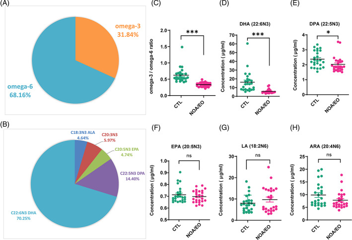
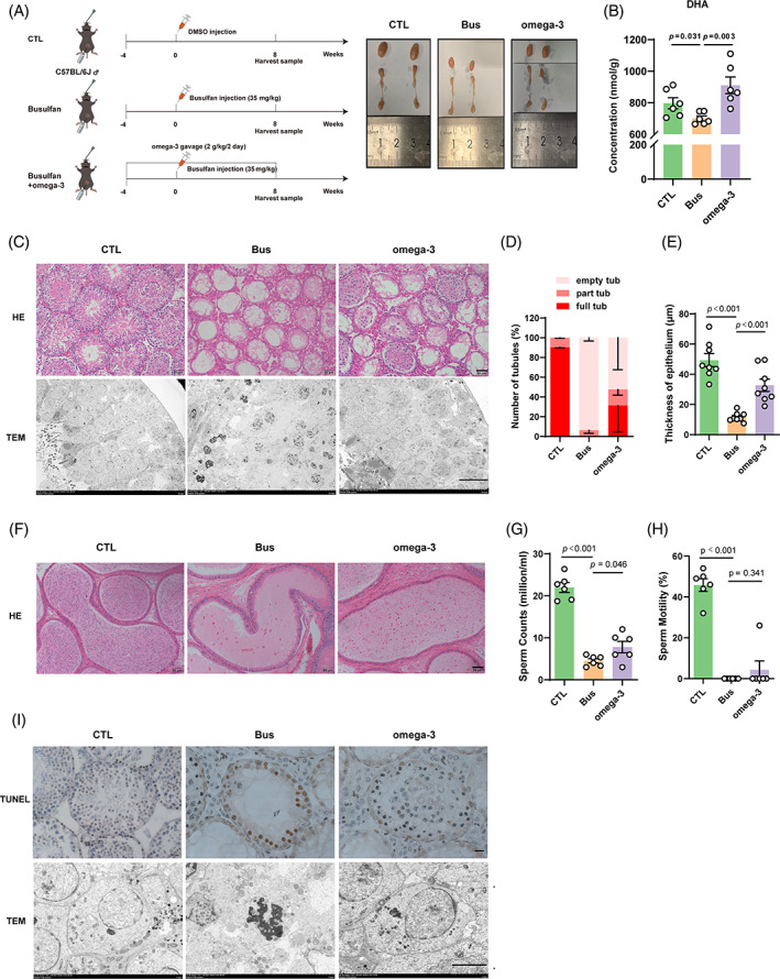
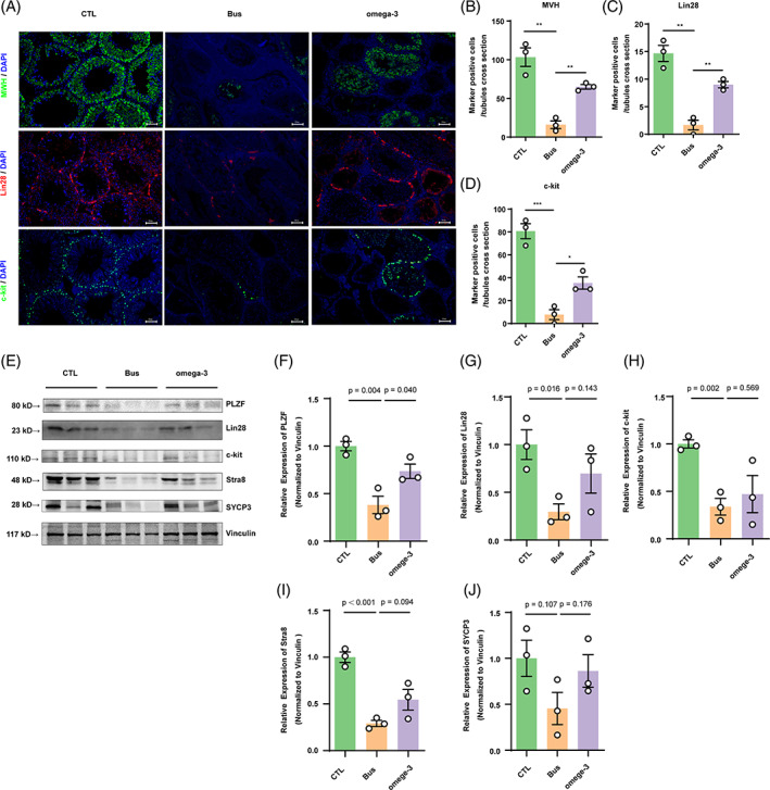
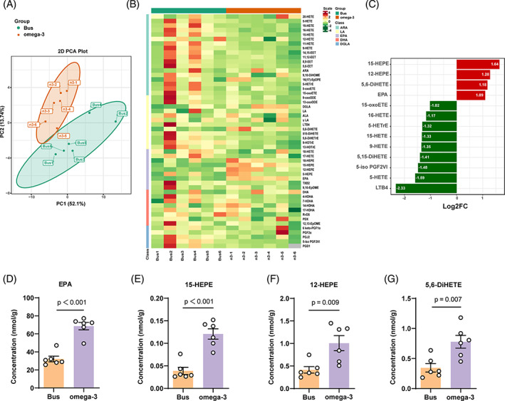
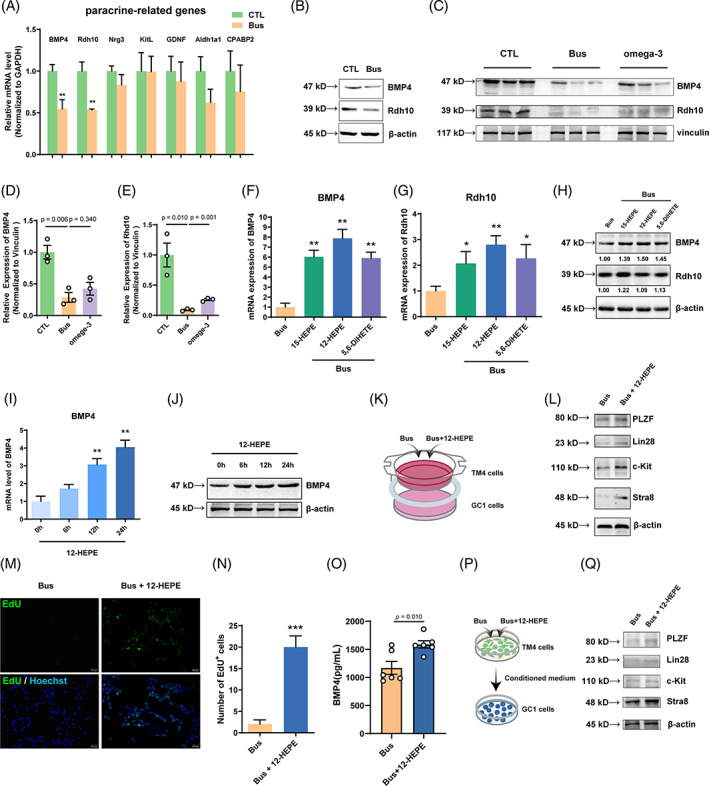
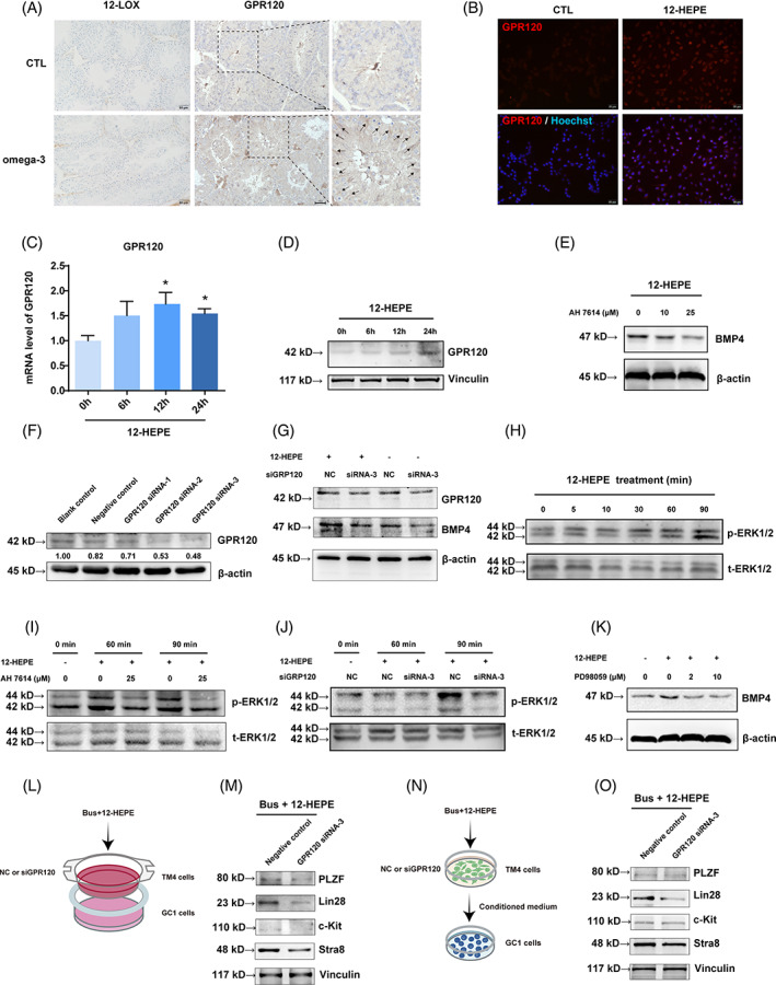
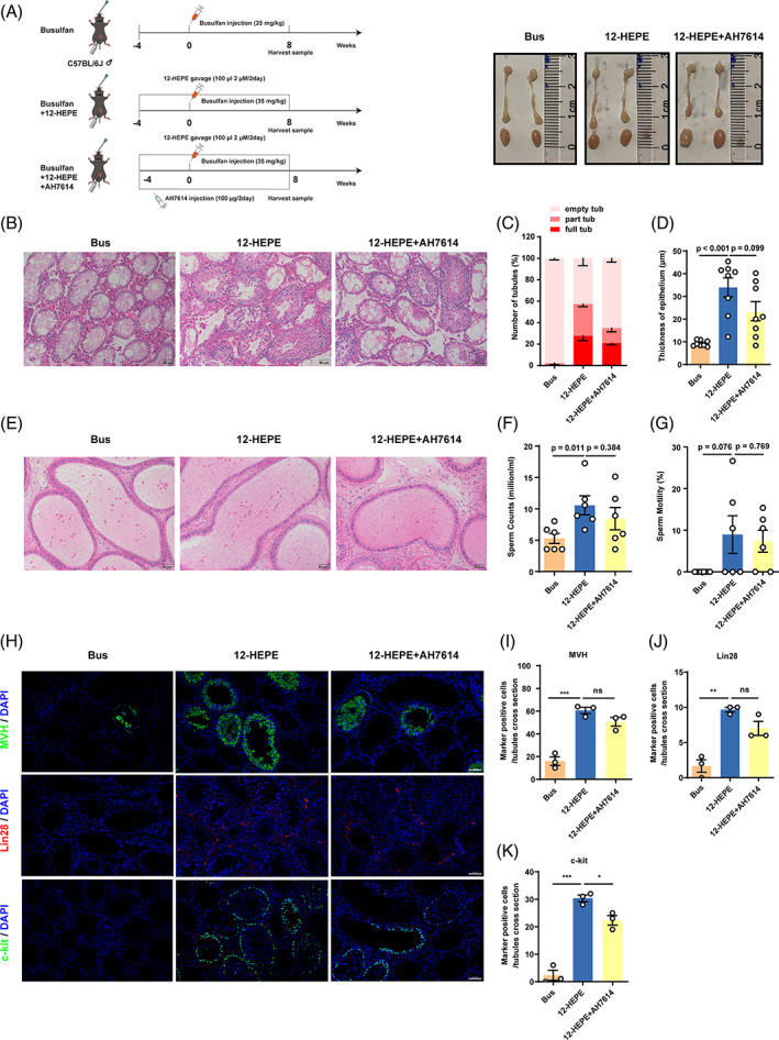
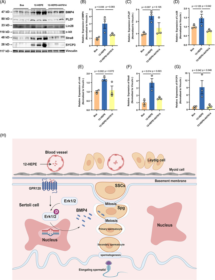
Busulfan is an antineoplastic, which is always accompanied with the abnormal of spermatogonia self-renewal and differentiation. It has been demonstrated that the omega-3 polyunsaturated fatty acids (PUFAs) benefits mature spermatozoa. However, whether omega-3 can protect endogenous spermatogonia and the detailed mechanisms are still unclear. Evaluate of spermatogenesis function (in vivo) were examined by histopathological analysis, immunofluorescence staining, and western blotting. The levels of lipid metabolites in testicular tissue were determined via liquid chromatography. We investigated the effect of lipid metabolites on Sertoli cells provided paracrine factors to regulate spermatogonia proliferation and differentiation using co-culture system. In our study, we showed that omega-3 PUFAs significantly improved the process of sperm production and elevated the quantity of both undifferentiated Lin28+ spermatogonia and differentiated c-kit+ spermatogonia in a mouse model where spermatogenic function was disrupted by busulfan. Mass spectrometry revealed an increase in the levels of several omega-3 metabolites in the testes of mice fed with omega-3 PUFAs. The eicosapentaenoic acid metabolite 12-hydroxyeicosapentaenoic acid (12-HEPE) up-regulated bone morphogenic protein 4 (BMP4) expression through GPR120-ERK1/2 pathway activation in Sertoli cells and restored spermatogonia proliferation and differentiation. Our study provides evidence that omega-3 PUFAs metabolite 12-HEPE effectively protects spermatogonia and reveals that GPR120 might be a tractable pharmacological target for fertility in men received chemotherapy or severe spermatogenesis dysfunction.


Conflict of interest statement

The authors declare that they have no competing interests.

Figures

FIGURE 1
FIGURE 1
The omega‐3/omega‐6 ratio in seminal plasma is significantly lower in patients with severe dyszoospermia. The proportions of polyunsaturated fatty acid (PUFA) constitution (A) and different components of omega‐3 (B) were measured. The omega‐3/omega‐6 ratio (F), docosahexaenoic acid docosahexaenoic acid (DHA) (C), docosapentaenoic acid (DPA) (D), eicosapentaenoic acid (EPA) (E), linoleic acid (LA) (G), and arachidonic acid (ARA) (H) levels in the seminal plasma were measured by gas chromatography coupled with mass spectrometry. The study included a group of healthy controls (n = 25) and two groups of patients: one with non‐obstructive azoospermia (NOA, n = 21) and the other with extreme oligospermia (EO, n = 4). Compared with the healthy control group: *p < 0.05; **p < 0.01; ***p < 0.001; ns, p > 0.05.
FIGURE 2
FIGURE 2
Dietary omega‐3 protects busulfan disrupted spermatogenesis in vivo. (A) Diagram of the animal experiments and images of testis morphology. (B) The docosahexaenoic acid (DHA) levels in testicular tissue from each group (n = 6). (C) Representative hematoxylin and eosin (H&E) staining images and transmission electron microscopy images of testicular cross‐sections from each group (n = 8 per group; scale bar, 50 μm). Testicular cross‐sections were examined using representative H&E staining and transmission electron microscopy from every group (n = 8; scale bar, 50 micrometres). Comparison between the model mice and control mice involves assessing the quantity of seminiferous tubules exhibiting empty, partial or full spermatogenesis (D) and tubule thickness (E). (F) Representative H&E staining images of caudal epididymis cross‐sections from each group (n = 8; scale bar, 50 μm). The sperm count (G) and motility (H) of the model mice were assessed and compared with those of control mice. I TUNEL staining of testicular spermatogenic tubules from each group and the morphology of spermatogonial cells under projective electron microscopy (n = 8; scale bar, 20 μm).
FIGURE 3
FIGURE 3
Dietary omega‐3 increases busulfan disrupted spermatogonia proliferation and differentiation in vivo. (A) Immunohistochemical staining indicated that Lin28 (red) was expressed in undifferentiated spermatogonia, and c‐kit (green) was found in differentiated spermatogonia in all groups (scale bar, 50 μm). (B–D) The count of MVH+, Lin28+, or c‐kit+ cells per cross‐section of tubules (n = 3). Compared with the control group: *p < 0.05; **p < 0.01; ***p < 0.001. (E) The protein expression of PLZF, Lin28, c‐kit, Stra8 and SYCP3 in each group (n = 3) was assessed by western blotting and compared them to the protein levels in control mice. (F–J) The quantification of germ cell markers' protein expression was quantified by normalizing the levels of these proteins to the level of the internal control vinculin. The data are displayed as the mean ± SEM from a minimum of three separate trials.
FIGURE 4
FIGURE 4
Dietary omega‐3 alters lipid metabolite levels in testicular tissue. (A) Principal component analysis (PCA) of metabonomic data from testicular tissue samples from the two groups (n = 6 per group). (B) Heatmap showing eicosanoid levels in testicular tissue samples from mice treated with busulfan and omega‐3 PUFAs (n = 6 per group). (C) Chart showing the fold change (FC) in the levels of metabolic products that showed an FC (log2) of at least two between the busulfan‐ and omega‐3‐treated groups. The red bars indicate compounds that were found at higher concentrations in omega‐3‐treated group, while the green bars indicate compounds that were found at lower concentrations in the omega‐3‐treated group. (D‐G) Quantitation of lipid metabolite levels in testicular tissue. The levels of eicosapentaenoic acid (EPA), 15‐hydroxyeicosapentaenoic acid (15‐HEPE), 12‐hydroxyeicosapentaenoic acid (12‐HEPE) and 5,6‐dihydroxy‐eicosatrienoic acid (5,6‐DiHETE) in testicular tissue from the omega‐3‐treated group were markedly elevated compared with the busulfan‐treated groups (n = 6). The data are displayed as the mean ± SEM.
FIGURE 5
FIGURE 5
12‐Hydroxyeicosapentaenoic acid (12‐HEPE) enhances bone morphogenic protein 4 (BMP4) expression in Sertoli cells to rescue spermatogonia proliferation and differentiation. (A,B) After subjecting TM4 cells to a 24‐h treatment of 10−4 M busulfan, the expression of paracrine factors was assessed using reverse transcription‐quantitative polymerase chain reaction (RT‐qPCR) for mRNA and western blotting for protein. Compared with the control group: **p < 0.01. (C–E) In omega‐3‐treated mice, BMP4 and Rdh10 protein expression was quantified by western blotting. (F–H) TM4 cells were treated with 15‐hydroxyeicosapentaenoic acid (15‐HEPE), 12‐HEPE, or 5‐(6)‐DiHETE and then with busulfan for 24 h, and the mRNA and protein levels of BMP4 and Rdh10 were examined by RT‐qPCR and western blotting, respectively. TM4 cells were exposed to 15‐HEPE, 12‐HEPE, or 5,6‐dihydroxy‐eicosatrienoic acid (5,6‐DiHETE) followed by busulfan treatment for 24 h. The expression of BMP4 and Rdh10 at mRNA and protein levels was assessed using RT‐qPCR and western blotting. (I,J) TM4 cells were treated with 12‐HEPE at various time points and the expression of BMP4 and Rdh10 at mRNA and protein levels were assessed using qRT‐PCR and western blotting. (K) Schematic of the co‐culture system including GC‐1 cells and TM4 cells treated with busulfan or Bus+12‐HEPE. (L) The protein expression of germ cell markers were assessed using western blotting in GC‐1 cells. (M) The 5‐ethynyl‐2‐deoxyuridine (EdU) incorporation assay was used to assess GC‐1 cell proliferation (scale bar, 50 μm). (N) The count of cells labelled with EdU in each group. In comparison to the group that received only busulfan: ***p < 0.001. (O) TM4 cells were exposed to 10−4 M busulfan alone or in combination with 12‐HEPE. The ELISA method was used to evaluate the secretion of BMP4 in the culture medium. (P) Schematic of the protocol used to treat GC‐1 cells treated with conditioned medium from TM4 cells pre‐treated with treated with busulfan or Bus+12‐HEPE for 24 h. (Q) The protein levels of germ cell markers were assessed using western blotting in GC‐1 cells. The data are displayed as the mean ± SEM from a minimum of three separate trials.
FIGURE 6
FIGURE 6
12‐Hydroxyeicosapentaenoic acid (12‐HEPE) regulates bone morphogenic protein 4 (BMP4) expression in Sertoli cells via the G protein‐coupled receptor 120 (GPR120)‐ERK signalling pathway. (A) GPR120 and 12‐lipoxygenase (12‐LOX) protein expression was assessed by immunohistochemistry. The arrows indicate positive staining (scale bar, 50 μm). (B) After a 24‐h treatment of TM4 cells with 12‐HEPE, the immunofluorescence analysis was conducted to the protein expression of GPR120 (red) (scale bar, 50 μm). (C,D) TM4 cells were exposed to 12‐HEPE for 0, 6, 12, or 24 h. The expression of GPR120 was assessed using reverse transcription‐quantitative polymerase chain reaction for mRNA and western blotting for protein levels. (E) TM4 cells were pretreated with different concentrations of AH7614 for a duration of 2 h to assess the expression of BMP4 protein. (F) After transfecting with three distinct siRNAs that targeted GPR120, blank control, or negative control (scrambled) siRNA for a duration of 24 h, and GPR120 protein expression was measured. (G) The cells were transfected with siRNA‐GPR120‐3 or scrambled siRNA and subsequently exposed to 12‐HEPE for 24 h. GPR120 and BMP4 protein expression was assessed. (H) TM4 cells were treated with 12‐HEPE for different durations, and the phosphorylated ERK1/2 levels were assessed. (I,J) TM4 cells were pretreated with AH7614 (a GPR120 inhibitor) or transfected with siRNA‐GPR120‐3 and then treated with 12‐HEPE for different durations. The phosphorylated ERK1/2 levels were assessed. (K) TM4 cells were pretreated with various concentrations of PD98059 (a p‐ERK inhibitor) for 2 h and then treated with 12‐HEPE for another 24 h. Then, BMP4 protein expression was evaluated by western blotting. TM4 cells were pre‐exposed to different doses of PD98059, an inhibitor of p‐ERK, for a duration of 2 h. Subsequently, BMP4 protein expression was assessed. (L) Schematic of the co‐culture system including GC‐1 and TM4 cells transfected with siRNA‐GPR120‐3 or scrambled siRNA. Afterward, they were treated with busulfan and 12‐HEPE for 24 h. (M) The germ cell markers protein expression was evaluated and compared with negative control group. (N) Schematic of the protocol used to treat GC‐1 cells with conditioned medium from TM4 cells transfected with siRNA‐GPR120‐3 or scrambled siRNA and then treated with busulfan and 12‐HEPE for a duration of 24 h. (O) The germ cell markers protein expression was evaluated and compared with negative control group. The data are displayed as the mean ± SEM from a minimum of three separate trials.
FIGURE 7
FIGURE 7
Dietary 12‐hydroxyeicosapentaenoic acid (12‐HEPE) restores spermatogenesis through the G protein‐coupled receptor 120 (GPR120) pathway. (A) Diagram of the animal experiments and images of testis morphology in all groups. (B) Representative hematoxylin and eosin (H&E) staining images of testicular cross‐sections from each group (n = 8; scale bar, 50 μm). Quantification of the quantity of seminiferous tubules exhibiting empty, partial or full spermatogenesis tubules (C) and tubule thickness (D) in relation to the control group. (E) Representative H&E staining images of caudal epididymis cross‐sections from each group (n = 8; scale bar, 50 μm). The sperm count (F) and motility (G) of model mice were assessed and compared with those of control mice. (H) Immunohistochemical staining indicated that MVH (green) was detected in newborn germ cells, Lin28 (red) was expressed in undifferentiated spermatogonia, and c‐kit (green) was found in differentiated spermatogonia in all groups (scale bar, 50 μm). (I–K) The count of MVH+, Lin28+, or c‐kit+ cells per cross‐section of tubules (n = 3). Compared with the control group: *p < 0.05; **p < 0.01; ***p < 0.001. The data are presented as the mean ± SEM.
FIGURE 8
FIGURE 8
Dietary 12‐hydroxyeicosapentaenoic acid (12‐HEPE) restores spermatogonia proliferation and differentiation through the G protein‐coupled receptor 120 (GPR120) pathway. (A) The protein expression of bone morphogenic protein 4 (BMP4), PLZF, Lin28, c‐kit, Stra8, and SYCP3 in each group was assessed by western blotting (n = 3). (B–G) BMP4, PLZF, Lin28, c‐kit, Stra8, and SYCP3 protein expression was quantified by normalizing the levels of these proteins to the level of the internal control vinculin. The information is displayed as the mean ± SEM from a minimum of three separate trials. (H) Diagram illustrates how omega‐3 fatty acids and their metabolite 12‐HEPE restore spermatogenesis in busulfan‐treated mice by enhancing BMP4 secretion from Sertoli cells.

Similar articles

Cited by

References

    1. Ogawa T, Dobrinski I, Brinster RL. Recipient preparation is critical for spermatogonial transplantation in the rat. Tissue Cell. 1999;31(5):461‐472. - PubMed
    1. Ganjalikhan Hakemi S, Sharififar F, Haghpanah T, Babaee A, Eftekhar‐Vaghefi SH. The effects of olive leaf extract on the testis, sperm quality and testicular germ cell apoptosis in male rats exposed to busulfan. Int J Fertil Steril. 2019;13(1):57‐65. - PMC - PubMed
    1. Bishop JB, Wassom JS. Toxicological review of busulfan (Myleran). Mutat Res. 1986;168(1):15‐45. - PubMed
    1. Zhou R, Wu J, Liu B, et al. The roles and mechanisms of Leydig cells and myoid cells in regulating spermatogenesis. Cell Mol Life Sci. 2019;76(14):2681‐2695. - PMC - PubMed
    1. de Rooij DG. The spermatogonial stem cell niche. Microsc Res Tech. 2009;72(8):580‐585. - PubMed