Review
. 2023 Nov 2;42(21):e113891.
doi: 10.15252/embj.2023113891. Epub 2023 Sep 25.

Emerging principles of primary cilia dynamics in controlling tissue organization and function

Affiliations
Review

Emerging principles of primary cilia dynamics in controlling tissue organization and function

Jay Gopalakrishnan et al. EMBO J. .

Abstract

Primary cilia project from the surface of most vertebrate cells and are key in sensing extracellular signals and locally transducing this information into a cellular response. Recent findings show that primary cilia are not merely static organelles with a distinct lipid and protein composition. Instead, the function of primary cilia relies on the dynamic composition of molecules within the cilium, the context-dependent sensing and processing of extracellular stimuli, and cycles of assembly and disassembly in a cell- and tissue-specific manner. Thereby, primary cilia dynamically integrate different cellular inputs and control cell fate and function during tissue development. Here, we review the recently emerging concept of primary cilia dynamics in tissue development, organization, remodeling, and function.

Keywords: cilia dynamics; ciliary signaling; cilium disassembly; primary cilia; tissue organization.


Conflict of interest statement

The authors declare that they have no conflict of interest.

Figures

Figure 1
Figure 1. Primary cilia: structure and dynamics
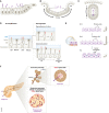
(A) The core structure of the primary cilium is the microtubule‐based axoneme. The primary cilium originates from a modified centriole called the basal body. Protein transport in and out of the cilium is passively controlled by the transition zone (TZ) and the transition fibers, and actively by the intraflagellar transport (IFT) machinery, which transports proteins anterogradely in a kinesin‐2‐dependent manner using IFT‐B trains and retrogradely in a dynein‐2‐depentend manner using IFT‐A trains. The BBSome is assembled with the help of the BBS chaperonin complex and functions as a cargo adaptor complex for the retrograde IFT transport of transmembrane proteins, in particular G protein‐coupled receptors (GPCRs). (B) Primary cilia dynamics can be observed on three different levels: 1. Compositional dynamics, determined by the active and passive gating structures that determine the compartmentalization of the primary cilium; 2. Signaling dynamics, referring to the signal‐dependent fine tuning to adapt the distinct molecular ciliary makeup appropriate to the signal status across time, space, and cellular state; 3. Assembly/disassembly dynamics, which are tightly coupled to the cell cycle. Figure has been created using Biorender.
Figure 2
Figure 2. Primary cilium assembly/disassembly dynamics in a cycling cell in vitro
The mature mother centriole (purple) templates the axoneme for cilium formation when the cell exits mitosis to the G1 phase. The daughter centriole at this stage is indicated in yellow. The cilium begins to disassemble at the onset of the S‐phase. Failure of or delay in cilium disassembly acts as a brake (“cilium checkpoint”) in cell cycle progression (stop sign). Cilium disassembly at G2 triggers cells to continue with mitotic progression.
Figure 3
Figure 3. Primary cilia dynamics during tissue development
(A) Primary cilia dynamics in tissue morphogenesis. From left to right: Cilium retraction and assembly (purple arrows) during proliferation (purple curved arrow), apical constriction, and cell delamination. (B, C) Primary cilia dynamics during neural tube closure. (B) Transverse view of the neuroepithelium (dark gray cells) flanked by non‐neural ectoderm (light cells); short primary cilium retraction and assembly during cell proliferation (purple and purple curved arrows) occurs differentially along medial‐to‐lateral (med./lat.) axis, which becomes the ventral‐to‐dorsal (V/D) axis during neural fold elevation. (C) Upon neural tube closure, cilia on the ventral (V) floor plate elongate dynamically, while cilia on lateral and dorsal (D) aspects of the neural tube remain short. (D) Primary cilia dynamics during brain development. Left: Apical progenitors' (AP) cilia are located at the apical surface of the neural epithelium (NE) in direct contact with embryonic cerebrospinal fluid (CSF). Right: Two different examples of ciliary dynamics upon neuronal differentiation. Top: The cilium is re‐assembled within the neuronal tissue following cell division, away from embryonic CSF. Bottom: Apical abscission removes the cilium from the differentiating neuron. The cilium will eventually be re‐assembled away from the apical surface of the NE. This leads to a transition from canonical to non‐canonical Hh signaling. (E) Primary cilia dynamics in the retinal pigment epithelium (RPE). Left: Cross‐section of the vertebrate eye. The apical processes of pigmented RPE cells (purple) engulf the outer segments of the light‐sensitive photoreceptors (beige). Middle/Right: A short primary cilium (red) can be detected from E14 onwards. Primary cilia are longest and most abundant at E16, before disassembling around birth. Disassembly of the primary cilium precedes extension of actin‐based apical processes. (F) Primary cilia dynamics in the pancreas. An adult mouse pancreas (left) comprises a network of ducts (brown) terminated by acini (top), forming the exocrine pancreas. A section of a large duct displayed on the right shows cilia on ductal cells (red), pointing towards the lumen. The islets of Langerhans (bottom) are located close to ducts and assemble multiple endocrine cells, which harbor a cilium on their lateral surface (red). The direction of fluid flow and the direction of cilia movement are indicated with blue arrows. Parts of the figure have been created using Biorender.

References

    1. Abraham SP, Nita A, Krejci P, Bosakova M (2022) Cilia kinases in skeletal development and homeostasis. Dev Dyn 251: 577–608 - PubMed
    1. Adachi C, Kakinuma N, Jo SH, Ishii T, Arai Y, Arai S, Kitaguchi T, Takeda S, Inoue T (2019) Sonic hedgehog enhances calcium oscillations in hippocampal astrocytes. J Biol Chem 294: 16034–16048 - PMC - PubMed
    1. Akella JS, Carter SP, Nguyen K, Tsiropoulou S, Moran AL, Silva M, Rizvi F, Kennedy BN, Hall DH, Barr MM et al (2020) Ciliary Rab28 and the BBSome negatively regulate extracellular vesicle shedding. Elife 9: e50580 - PMC - PubMed
    1. Alhassen W, Kobayashi Y, Su J, Robbins B, Nguyen H, Myint T, Yu M, Nauli SM, Saito Y, Alachkar A (2022) Regulation of brain primary cilia length by MCH signaling: evidence from pharmacological, genetic, optogenetic, and chemogenic manipulations. Mol Neurobiol 59: 245–265 - PMC - PubMed
    1. Al‐Jassar C, Andreeva A, Barnabas DD, McLaughlin SH, Johnson CM, Yu M, van Breugel M (2017) The ciliopathy‐associated Cep104 protein interacts with tubulin and Nek1 kinase. Structure 25: 146–156 - PMC - PubMed

Publication types