Case Reports
. 2023 Sep 22;7(9):ytad454.
doi: 10.1093/ehjcr/ytad454. eCollection 2023 Sep.

A multimodality approach to a rare case of ruptured sinus of Valsalva aneurysm with tricuspid valve involvement: a case report

Affiliations
Case Reports

A multimodality approach to a rare case of ruptured sinus of Valsalva aneurysm with tricuspid valve involvement: a case report

Vindhya C Wilson et al. Eur Heart J Case Rep. .

Abstract

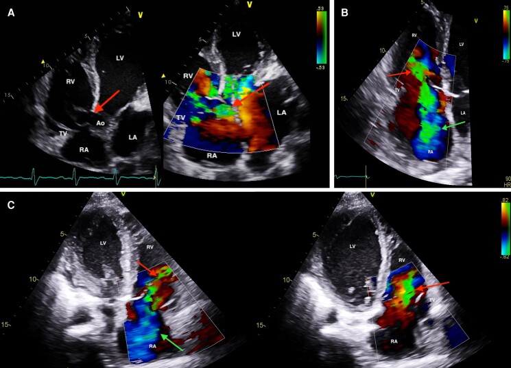
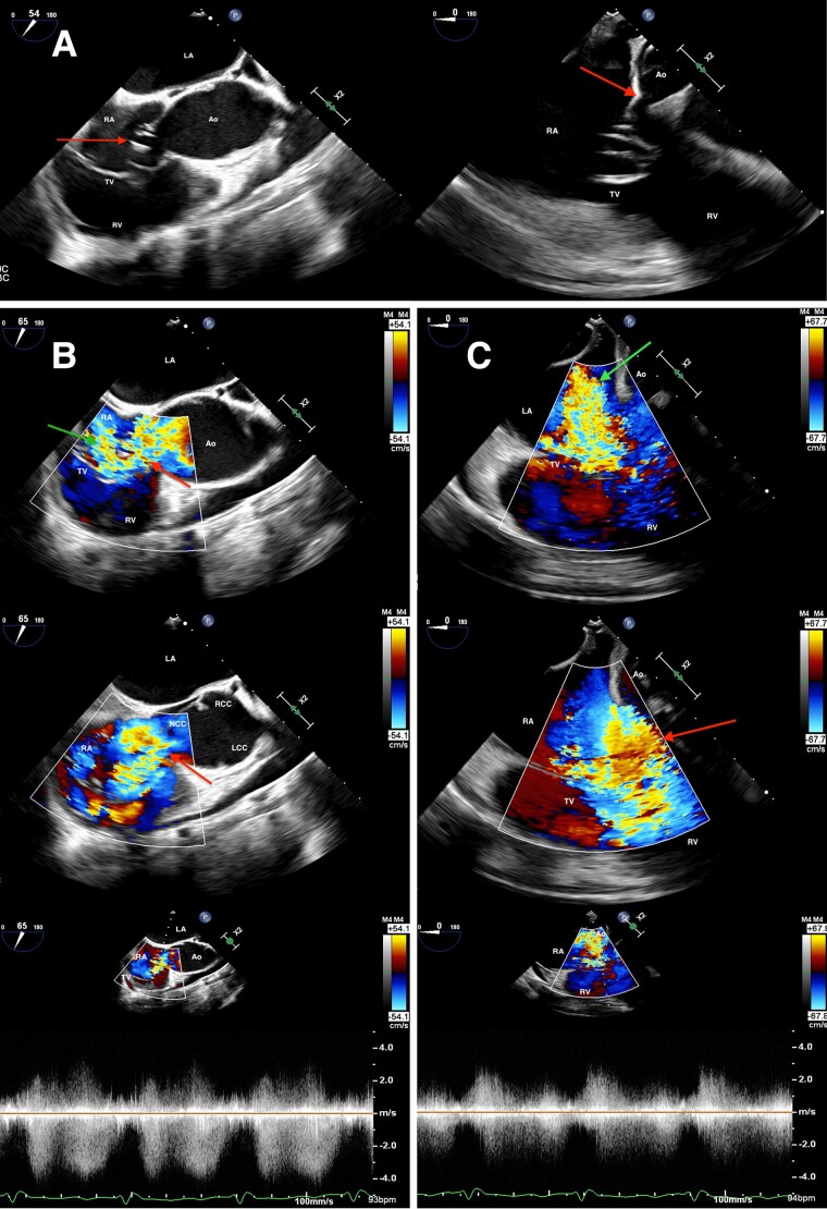
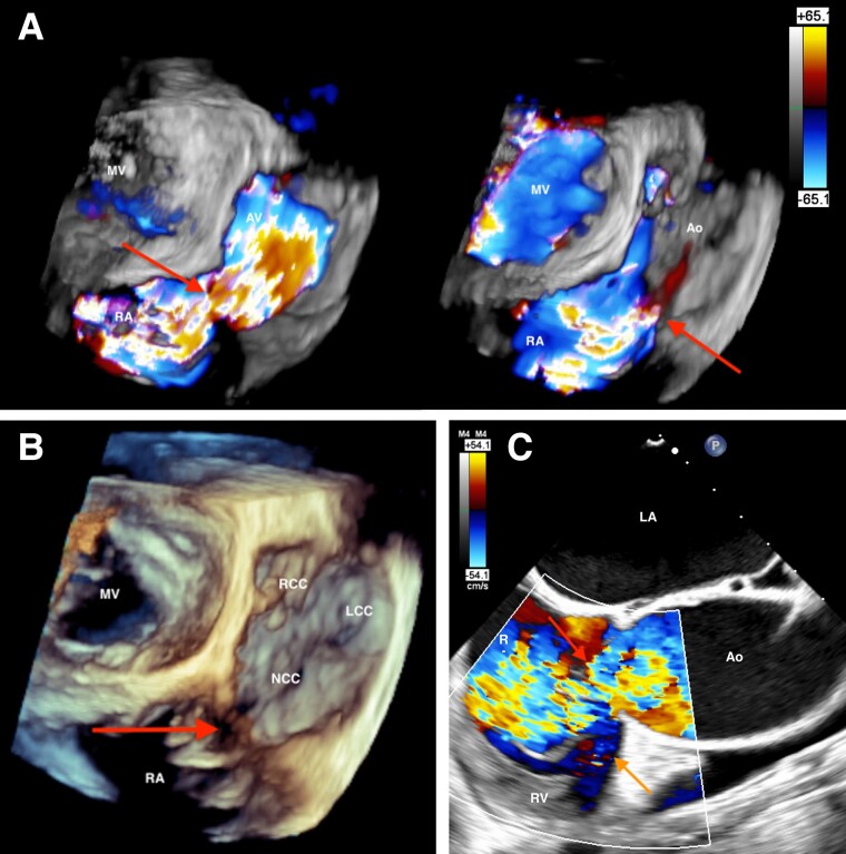
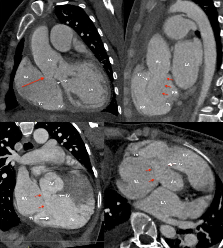
Background: Ruptured sinus of Valsalva aneurysm is a rare disease entity that is potentially life-threatening if left untreated. While imaging is the mainstay of diagnosis, resultant tricuspid valve involvement may mask typical findings providing a diagnostic challenge. Disruption of the tricuspid valve during ruptured sinus of Valsalva aneurysm with consequent tricuspid regurgitation is rare and infrequently described in the literature. Description of the utility and limitations of multimodality imaging in this scenario is equally scarce.

Case summary: We review the case of a young patient presenting with acute ruptured sinus of Valsalva aneurysm and involvement of the tricuspid valve on a background of severe aortic regurgitation requiring multimodality imaging for diagnostic and pre-surgical assessment.

Discussion: In young patients presenting with acute decompensation and pre-existing bicuspid aortic valve regurgitation, an increased clinical suspicion of a sinus of Valsalva aneurysm rupture is imperative. Doppler and 3D transoesophageal echocardiographic assessment should be pursued to characterize abnormal flows and clarify aetiology in the context of tricuspid involvement and resultant tricuspid regurgitation. A large-volume left-right shunt in proximity to the tricuspid annulus may result in disproportionately severe tricuspid regurgitation in the absence of annular disruption due to forced systolic opening of the leaflets by shunt flow and 'windsock' prolapse. Multimodality imaging can be essential in these cases to adequately assess the extent of the ruptured sinus of Valsalva aneurysm and overcome limitations of single modality imaging.

Keywords: Case report; Congenital heart defect; Echocardiography; Multimodality imaging; Tricuspid valve.


Conflict of interest statement

Conflict of interest: None declared.

Figures

Figure 1
Figure 1
Transthoracic echocardiographic images. Apical five-chamber view (A) demonstrating a ruptured sinus of Valsalva aneurysm in close proximity to septal tricuspid valve leaflet insertion (left) with colour flow through the defect into the tricuspid valve (right). Apical four-chamber view (B) exhibiting bidirectional systolic flow through the tricuspid valve with moderate–severe tricuspid regurgitation. Reverse apical four-chamber view (C) shows bidirectional systolic flow (left) and high-velocity diastolic forward flow through the tricuspid valve (right). RV, right ventricle; LV, left ventricle; TV, tricuspid valve; Ao, aorta; RA, right atrium; LA, left atrium; red arrow, RSOVA and/or RSOVA flow; green arrow, tricuspid regurgitation; RSOVA, ruptured sinus of Valsalva aneurysm; TR, tricuspid regurgitation.
Figure 2
Figure 2
Transoesophageal echocardiographic images. Modified mid-oesophageal aortic valve short-axis view (A, left) and mid-oesophageal five-chamber view (A, right) revealing disruption of the aortic sinus and prolapse of the ‘windsock’ into the tricuspid valve. Modified mid-oesophageal aortic valve short-axis views (B) differentiating two individual systolic jets representing tricuspid regurgitation and ruptured sinus of Valsalva aneurysm flow (top), high-velocity diastolic ruptured sinus of Valsalva aneurysm flow (mid), and Doppler profile describing continuous flow through the ruptured sinus of Valsalva aneurysm (bottom). Mid-oesophageal five-chamber views (C) demonstrating moderate–severe systolic tricuspid regurgitation (top) and high-velocity diastolic ruptured sinus of Valsalva aneurysm flow through the TV (mid). Doppler profile demonstrates continuous forward flow and bidirectional systolic flow through the tricuspid valve (C, bottom). Red arrow, RSOVA and/or RSOVA flow; green arrow, TR. Other abbreviations as in Figure 1 legend.
Figure 3
Figure 3
Transoesophageal 3D mid-oesophageal tilted enface left atrium view (A, B) demonstrating a ruptured sinus of Valsalva aneurysm with systolic (A, left) and diastolic (A, right) forward flow into the right atrium. Modified mid-oesophageal aortic valve short-axis view showing potential systolic flow apical to the septal tricuspid annulus concerning annular disruption (C, orange arrow). 3D, 3 dimensional.
Figure 4
Figure 4
Contrast computed tomography angiogram images with pan-cardiac enhancement despite electrocardiographic gating. Standard coronal (top left) and sagittal (top right) reconstruction views and oblique coronal (bottom left) and oblique axial reconstruction views demonstrate the proximity of the ruptured sinus of Valsalva aneurysm to the septal tricuspid valve leaflet insertion with suboptimal annular definition impairing assessment of annular involvement. Prolapse of the ‘windsock’ through the tricuspid valve is demonstrated. Abbreviations as in Figure 1 and 2 legends.
Figure 5
Figure 5
Cardiac magnetic resonance imaging oblique aortic sagittal views (A) demonstrating a non-coronary cusp ruptured sinus of Valsalva aneurysm with high-velocity flow through the right atrium, along the anterior tricuspid valve leaflet into the right ventricle. Cardiac magnetic resonance imaging TRUFI retro four-chamber view depicting systolic (left) and diastolic (right) high-velocity forward flow through the tricuspid valve. No direct flow into the right ventricle is demonstrated.

Similar articles

Cited by

References

    1. Weinreich M, Yu PJ, Trost B. Sinus of Valsalva aneurysms: review of the literature and an update on management. Clin Cardiol 2015;38:185–189. - PMC - PubMed
    1. Bricker AO, Avutu B, Mohammed TL, Williamson EE, Syed IS, Julsrud PR, et al. . Valsalva sinus aneurysms: findings at CT and MR imaging. Radiographics 2010;30:99–110. - PubMed
    1. Kuroda M, Takahashi K, Matsumoto S, Oshima M. Impact of intraoperative transesophageal echocardiography for noncoronary sinus of Valsalva aneurysm with severe tricuspid regurgitation. J Cardiothorac Vasc Anesth 2020;34:2169–2173. - PubMed
    1. Plambeck C, Eiseman M, Iqbal Z, Pagel P. A small circular structure in the right atrium: a cause for right atrial and ventricular dilatation? J Cardiothorac Vasc Anesth 2013;27:628–630. - PubMed
    1. Doost A, Craig JA, Soh SY. Acute rupture of a sinus of Valsalva aneurysm into the right atrium: a case report and a narrative review. BMC Cardiovasc Disord 2020;20:84. - PMC - PubMed

Publication types

LinkOut - more resources