. 2023 Sep 5:34:102024.
doi: 10.1016/j.omtn.2023.09.001. eCollection 2023 Dec 12.

Peptide-conjugated antimiRs improve myotonic dystrophy type 1 phenotypes by promoting endogenous MBNL1 expression

Affiliations

Peptide-conjugated antimiRs improve myotonic dystrophy type 1 phenotypes by promoting endogenous MBNL1 expression

Irene González-Martínez et al. Mol Ther Nucleic Acids. .

Abstract

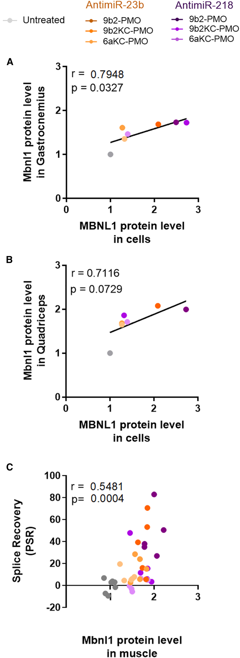
Myotonic dystrophy type 1 (DM1) is a rare neuromuscular disease caused by a CTG repeat expansion in the DMPK gene that generates toxic RNA with a myriad of downstream alterations in RNA metabolism. A key consequence is the sequestration of alternative splicing regulatory proteins MBNL1/2 by expanded transcripts in the affected tissues. MBNL1/2 depletion interferes with a developmental alternative splicing switch that causes the expression of fetal isoforms in adults. Boosting the endogenous expression of MBNL proteins by inhibiting the natural translational repressors miR-23b and miR-218 has previously been shown to be a promising therapeutic approach. We designed antimiRs against both miRNAs with a phosphorodiamidate morpholino oligonucleotide (PMO) chemistry conjugated to cell-penetrating peptides (CPPs) to improve delivery to affected tissues. In DM1 cells, CPP-PMOs significantly increased MBNL1 levels. In some candidates, this was achieved using concentrations less than two orders of magnitude below the median toxic concentration, with up to 5.38-fold better therapeutic window than previous antagomiRs. In HSALR mice, intravenous injections of CPP-PMOs improve molecular, histopathological, and functional phenotypes, without signs of toxicity. Our findings place CPP-PMOs as promising antimiR candidates to overcome the treatment delivery challenge in DM1 therapy.

Keywords: MBNL proteins; MT: Oligonucleotides: Therapies and Applications; PMO chemistry; alternative splicing; antisense oligonucleotides; biodistribution; cell-penetrating peptides; microRNAs; muscle dysfunction; myotonic dystrophy.


Conflict of interest statement

B.L. and R.A. are founders, and CEO and scientific consultant of Arthex Biotech, respectively. Arthex Biotech is developing antimiRs for use in the treatment of DM1.

Figures

None
Graphical abstract
Figure 1
Figure 1
Activity effect of CPP-PMOs and previous antagomiRs in DM1 cells Data show the quantification by QDB of MBNL1 protein levels after treatment with different ONs in DM1 cells at the indicated concentrations: (A) 9b2-23b, (B) 9b2KC-23b, (C) 6aKC-23b, (D) 9b2-218, (E) 9b2KC-218, (F) 6aKC-218, (G) AntagomiR-23b, (H) AntagomiR-218, and (I) scrambled control (SC). The highest value of each was used as the Emax of the corresponding ON. Black bars represent MBNL1 levels of unaffected control (CNT) myotubes and light gray bars represent MBNL1 levels in DM1 mock-treated cells. Fold change of (J) MBNL1 protein levels and percentage of (K) cell growth inhibition of DM1 cells using transfection reagent (black columns) or gymnosis delivery (gray columns) with ONs against miR-23b or miR-218 at their corresponding EC50 concentration. Dotted line indicates MBNL1 levels in DM1 cells without treatment. ∗p < 0.05, ∗∗p < 0.01, ∗∗∗p < 0.001, ∗∗∗∗p < 0.0001 according to one-way ANOVA compared with DM1 non-treated cells (black stars in A–I, white stars in J) or according to Student’s t test between delivery strategies (black stars in J and K). Each concentration was tested in triplicate. Individual values are indicated as datapoints. Error bars indicate mean ± SEM.
Figure 2
Figure 2
CPP-PMO antimiR treatment results in sustained improvement of MBNL1- and MBNL2-dependent molecular functions in HSALR mice After 45 days of treatment with CPP-PMOs, gastrocnemius and quadriceps muscles were dissected to measure functional (A) miR-23b and (B) miR-218 expression relative to U1 and U6 snRNA endogenous controls, (C) Mbnl1 transcript levels relative to Gapdh endogenous control on gastrocnemius and quadriceps, (D) Mbnl1 protein levels relative to endogenous tubulin control using quantitative dot blot. Statistical comparisons were performed in each case against PBS-treated HSALR values (indicated by a black dashed line with ANOVA one-way test. ∗p < 0.05, ∗∗p < 0.01, ∗∗∗p < 0.001, ∗∗∗∗p < 0.0001). PBS n = 7, SC n = 5, 9b2-218 n = 5, 9b2-23b n = 5, 9b2KC-218 n = 5, 9b2KC-23b n = 5, 6aKC-218 n = 5, and 6aKC-23b n = 5. Individual values are indicated as datapoints. Error bars indicate mean ± SEM.
Figure 3
Figure 3
CPP-PMOs rescue Mbnl1 splicing alterations in mice (A–F) Quantification of the percentage of exon inclusion (Ψ) of (A and B) Nfix exon 7, (C and D) Clcn1 exon 7a, and (E and F) Atp2a1 exon 22 in gastrocnemius and quadriceps muscles of HSALR mice upon treatment with PBS, SC, or CPP-PMOs antimiRs against (A, C, and E) miR-23b or (B, D, and F) miR-218. (G and H) Splice recovery of Nfix ex7, Clcn1 ex7a, and Atp2a1 ex22. Reference non-DM1 Ψ values were estimated from FVB controls. Individual values are indicated as datapoints. Error bars indicate mean ± SEM. Data were analyzed by unpaired Student’s t tests compared with PBS-treated HSALR mice. ∗p < 0.05, ∗∗p < 0.01, ∗∗∗p < 0.001, ∗∗∗∗p < 0.0001.
Figure 4
Figure 4
Activity of CPP-PMOs shows a strong correlation between in vitro and in vivo models Positive correlation of Pearson (r > 0.5) between MBNL1 protein levels in DM1 cells and (A) gastrocnemius or (B) quadriceps tissue or (C) between average of Mbnl1 protein levels in muscle and percentage splice recovery (PSR) in individual mouse, after treatment with CPP-PMOs antimiRs. Gray dots indicate untreated samples (DM1 cells in A and B, PBS-treated HSALR mice in C).
Figure 5
Figure 5
Recursive administration of CPP-PMO antimiRs improved functional and histological alterations in DM1 mice (A) Relative change in myotonia grade at 45 days compared with the first injection day. (B) Quantification of the percentage of muscle fibers with central nuclei in quadriceps of mice from each treatment group. Individual values are indicated as datapoints. (C–K) Images from hematoxylin and eosin staining of quadriceps muscles from representative mice of each treatment group. Black arrows point to examples of centrally located nuclei in muscle fibers. Error bars indicate mean ± SEM. The data were analyzed by ANOVA one-way test or Kruskal-Wallis test when it was necessary, compared with untreated HSALR mice. ∗p < 0.05, ∗∗p < 0.01, ∗∗∗p < 0.001, ∗∗∗∗p < 0.0001. PBS n = 7, SC n = 5, 9b2-218 n = 5, 9b2-23b n = 5, 9b2KC-218 n = 5, 9b2KC-23b n = 5, 6aKC-218 n = 5, and 6aKC-23b n = 5.
Figure 6
Figure 6
Levels of CPP-PMO antimiRs and Mbnl1 in selected target tissues After 45 days of treatment with the CPP-PMOs, different tissues were dissected to measure by ELISA levels of each antimiR: (A) 9b2-23b, (B) 9b2-218, (C) 9b2KC-23b, (D) 9b2KC-218, (E) 6aKC-23b, and (F) 6aKC-218. The selected target tissues were kidney, liver, heart, quadriceps, gastrocnemius, and brain. (G) Mbnl1 protein levels in kidney relative to endogenous tubulin control using quantitative dot blot. Individual values are indicated as datapoints. Error bars indicate mean ± SEM. The data were analyzed by Kruskal-Wallis test, compared with untreated HSALR mice ∗∗p < 0.01.

References

    1. Brook J.D., McCurrach M.E., Harley H.G., Buckler A.J., Church D., Aburatani H., Hunter K., Stanton V.P., Thirion J.P., Hudson T. Molecular basis of myotonic dystrophy: expansion of a trinucleotide (CTG) repeat at the 3' end of a transcript encoding a protein kinase family member. Cell. 1992;68:799–808. - PubMed
    1. Ozimski L.L., Sabater-Arcis M., Bargiela A., Artero R. The hallmarks of myotonic dystrophy type 1 muscle dysfunction. Biol. Rev. Camb. Phil. Soc. 2021;96:716–730. - PubMed
    1. Ashizawa T., Gagnon C., Groh W.J., Gutmann L., Johnson N.E., Meola G., Moxley R., 3rd, Pandya S., Rogers M.T., Simpson E., et al. Consensus-based care recommendations for adults with myotonic dystrophy type 1. Neurol. Clin. Pract. 2018;8:507–520. - PMC - PubMed
    1. Goers E.S., Purcell J., Voelker R.B., Gates D.P., Berglund J.A. MBNL1 binds GC motifs embedded in pyrimidines to regulate alternative splicing. Nucleic Acids Res. 2010;38:2467–2484. - PMC - PubMed
    1. Batra R., Charizanis K., Manchanda M., Mohan A., Li M., Finn D.J., Goodwin M., Zhang C., Sobczak K., Thornton C.A., Swanson M.S. Loss of MBNL leads to disruption of developmentally regulated alternative polyadenylation in RNA-mediated disease. Mol. Cell. 2014;56:311–322. - PMC - PubMed

LinkOut - more resources